Review
. 2016 Aug;14(8):535-542.
doi: 10.1038/nrmicro.2016.85. Epub 2016 Jun 27.

Sporulation, bacterial cell envelopes and the origin of life

Affiliations
Review

Sporulation, bacterial cell envelopes and the origin of life

Elitza I Tocheva et al. Nat Rev Microbiol. 2016 Aug.

Abstract

Electron cryotomography (ECT) enables the 3D reconstruction of intact cells in a near-native state. Images produced by ECT have led to the proposal that an ancient sporulation-like event gave rise to the second membrane in diderm bacteria. Tomograms of sporulating monoderm and diderm bacterial cells show how sporulation can lead to the generation of diderm cells. Tomograms of Gram-negative and Gram-positive cell walls and purified sacculi suggest that they are more closely related than previously thought and support the hypothesis that they share a common origin. Mapping the distribution of cell envelope architectures onto a recent phylogenetic tree of life indicates that the diderm cell plan, and therefore the sporulation-like event that gave rise to it, must be very ancient. One explanation for this model is that during the cataclysmic transitions of the early Earth, cellular evolution may have gone through a bottleneck in which only spores survived, which implies that the last bacterial common ancestor was a spore.


Conflict of interest statement

Competing interests statement

The authors declare no competing interests.

Figures
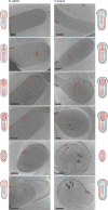
Figure 1
Figure 1. Sporulation in Gram-positive and Gram-negative bacteria
Overview of bacterial sporulation in Bacillus subtilis (left) and Acetonema longum (right) by electron cryotomography (ECT). Each panel represents a tomographic slice through a bacterial cell at a different stage of sporulation. Part a shows vegetative cells, part b shows the formation of the septum, part c shows engulfment, part d shows forespores, part e shows mature spores and part f shows germinating cells. The inner membrane and cytoplasmic membrane are depicted in red and the outer membrane of A. longum is depicted in blue. Schematic representations of all sporulation stages are shown next to the tomographic slices. Scale bar 200 nm. Images in the left panels of parts df are adapted with permission from REF. , Wiley. Images in the right panels of parts a, b, d, e and f are adapted with permission from REF., Elsevier.
Figure 2
Figure 2. Model for how the outer membrane arose as a byproduct of sporulation and how losses then led to the diversity of modern bacterial cell plans
At some point during early evolution, the cell division and nutrient-uptake processes of a primordial cell were combined into a sporulation-like process. Retention of the second spore membrane led to sporulating diderm species. Losses of the outer membrane and/or the ability to sporulate in various lineages can explain the distribution of cell envelope architectures that is observed in modern bacteria. An alternative hypothesis (not shown) is that some monoderms evolved directly from a monoderm last common ancestor.
Figure 3
Figure 3. Peptidoglycan remodelling during sporulation
Peptidoglycan and cortex remodelling during sporulation in Bacillus subtilis and Acetonema longum show that both Gram-positive and Gram-negative bacterial cells can synthesize thick and thin peptidoglycan and can remodel one into the other, which supports the notion that all bacterial cell walls have the same basic architecture that is inherited from a common ancestor. Green lines represent the placement and thickness of peptidoglycan. Scale bar 100 nm. Adapted with permission from REF. , Wiley.
Figure 4
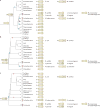
Figure 4. Rooted phylogenetic trees that represent relationships between bacterial phyla
Three different schematic reproductions are shown of a recent phylogenetic tree of life based on 42 gene markers that are conserved throughout Bacteria and Archaea rooted in three different ways, following leading proposals in the field,,,–. Cell envelope structures that are present in members of these phyla are shown on the right. Reported abilities to sporulate are represented by small circles within the cells (for endospores) or next to them (for exospores). The presence of the conserved Omp85 or Omp87 is denoted by the ‘β’ symbol and the presence of other conserved outer membrane proteins (OMPs) is denoted by the ‘β*’ symbol. The deduced basic cell plans of ancient ancestors are depicted on early branches. Losses of sporulation (red) and the outer membrane (blue) are marked, which together with the hypothesized cell plans of ancient ancestors minimize the number of evolutionary events that are required to explain the modern distributions of cell plans and sporulation abilities. In all cases, an endospore-forming diderm preceded all or at most one major branch point. A. longum, Acetonema longum; B. subtilis, Bacillus subtilis; E. coli, Escherichia coli; L. monocytogenes, Listeria monocytogenes; M. pneumoniae, Mycoplasma pneumoniae; M. tuberculosis, Mycobacterium tuberculosis; M. xanthus, Myxococcus xanthus; PVC, Planctomycetes–Verrucomicrobia–Chlamydiae; S. coelicolor, Streptomyces coelicolor; V. parvula, Veillonella parvula.

Comment in

Similar articles

Cited by

References

    1. Gram C. Ueber die isolirte firbung der schizomyceten iu schnitt-und trockenpriparate. Fortschitte Med. 1884;2:185–189. (in German)
    1. Sutcliffe IC. A phylum level perspective on bacterial cell envelope architecture. Trends Microbiol. 2010;18:464–470. - PubMed
    1. Gupta RS. Protein phylogenies and signature sequences: a reappraisal of evolutionary relationships among archaebacteria, eubacteria, and eukaryotes. Microbiol Mol Biol Rev. 1998;62:1435–1491. - PMC - PubMed
    1. Silhavy TJ, Kahne D, Walker S. The bacterial cell envelope. Cold Spring Harb Perspect Biol. 2010;2:a000414. - PMC - PubMed
    1. Martin W, Baross J, Kelley D, Russell MJ. Hydrothermal vents and the origin of life. Nat Rev Microbiol. 2008;6:805–814. - PubMed

Publication types

MeSH terms

LinkOut - more resources