. 2017 Feb 23;8(1):5.
doi: 10.1038/s41467-016-0011-z.

Genomic RNA folding mediates assembly of human parechovirus

Affiliations

Genomic RNA folding mediates assembly of human parechovirus

Shabih Shakeel et al. Nat Commun. .

Erratum in

Abstract

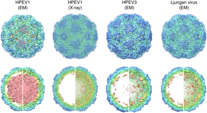
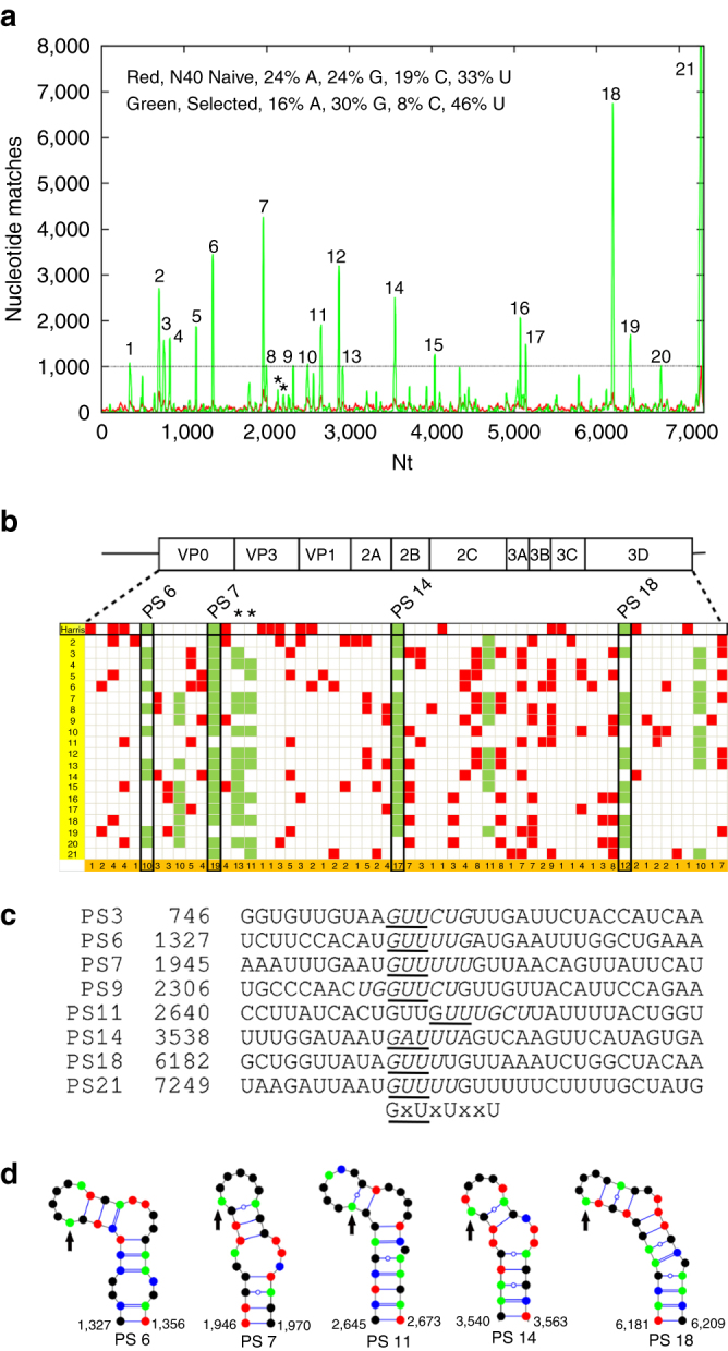
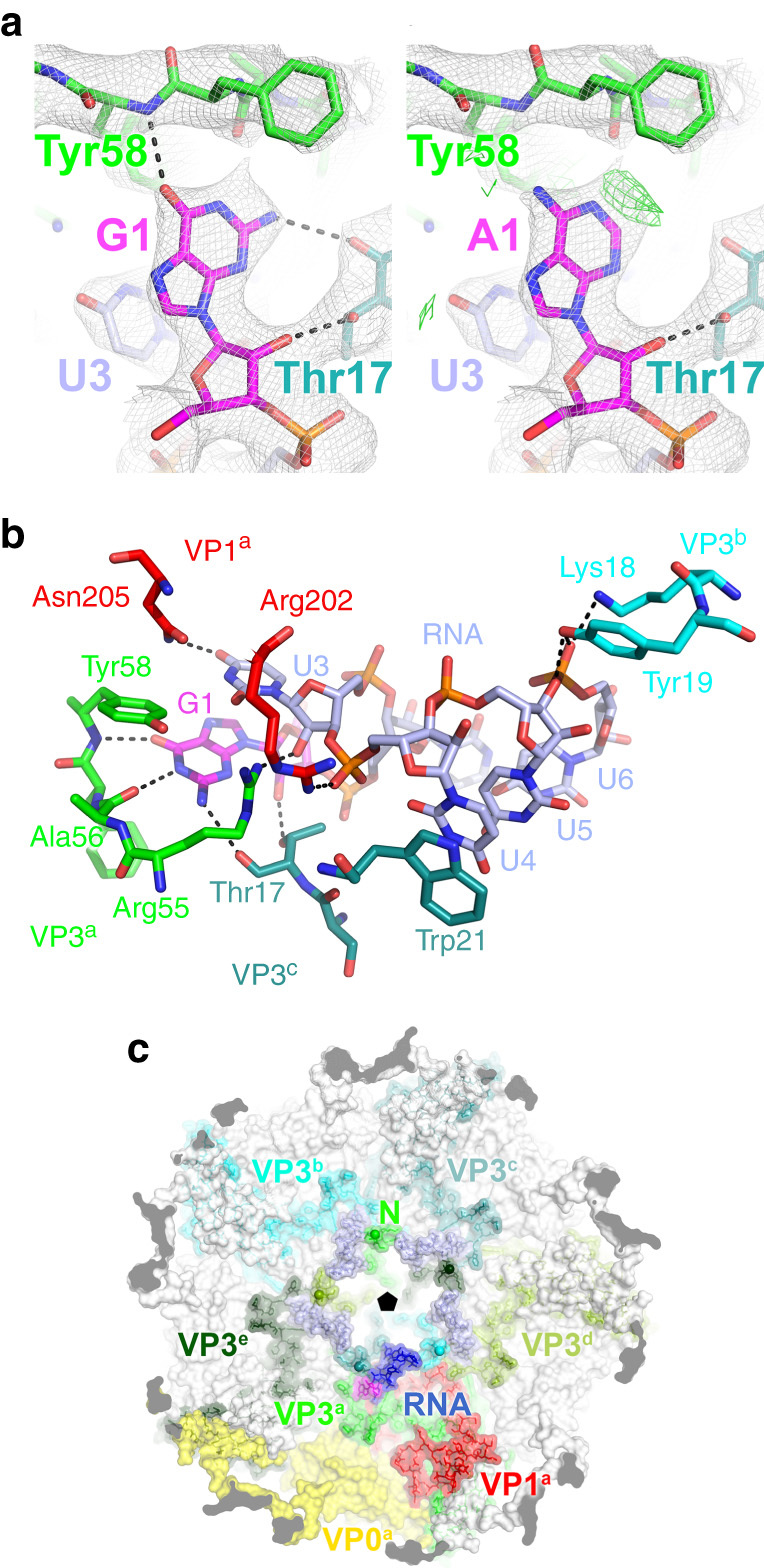
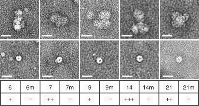
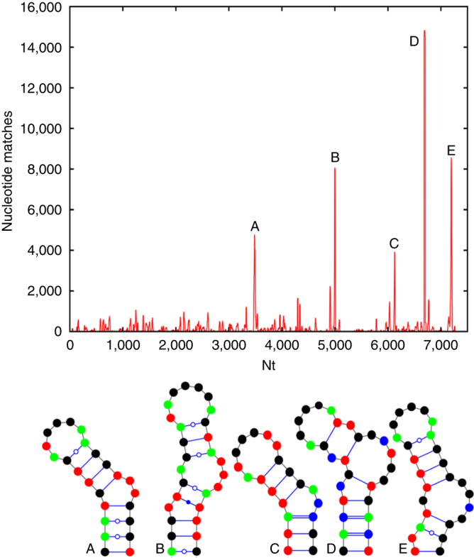
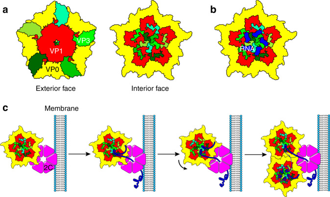
Assembly of the major viral pathogens of the Picornaviridae family is poorly understood. Human parechovirus 1 is an example of such viruses that contains 60 short regions of ordered RNA density making identical contacts with the protein shell. We show here via a combination of RNA-based systematic evolution of ligands by exponential enrichment, bioinformatics analysis and reverse genetics that these RNA segments are bound to the coat proteins in a sequence-specific manner. Disruption of either the RNA coat protein recognition motif or its contact amino acid residues is deleterious for viral assembly. The data are consistent with RNA packaging signals playing essential roles in virion assembly. Their binding sites on the coat proteins are evolutionarily conserved across the Parechovirus genus, suggesting that they represent potential broad-spectrum anti-viral targets.The mechanism underlying packaging of genomic RNA into viral particles is not well understood for human parechoviruses. Here the authors identify short RNA motifs in the parechovirus genome that bind capsid proteins, providing approximately 60 specific interactions for virion assembly.


Conflict of interest statement

The authors declare no competing financial interests.

Figures

Fig. 1
Fig. 1
Ordered RNA segments in the structures of picornaviruses. Four icosahedrally-symmetric structures for parechovirus virions are currently available. These are HPeV1 (Cryo-EM, 8.5 Å resolution, EMD-1690), HPeV1 (X-ray crystallography, 3.1 Å resolution, PDB 4Z92), HPeV3 (Cryo-EM, 4.3 Å resolution, EMD-3137), and Ljungan virus (Cryo-EM, 4.5 Å resolution, EMD-6395). On the top row, an exterior view of each capsid is shown, viewed perpendicular to an icosahedral two-fold axis. On the bottom row, each virus is shown in the same orientation, as a 60 Å thick central section on the left hand side, and the rear half of the capsid on the right hand side. All panels are coloured with an identical radial colour scheme (red: 98 Å, yellow: 111 Å, green: 124 Å, cyan: 137 Å, blue: 151 Å). The diameters of all capsids are very similar, with density ascribed to RNA shown in yellow/red in each case. More RNAs can be seen in the lower resolution HPeV1 EM reconstruction at 8.5 Å, than in the X-ray density at 3.1 Å resolution, suggesting that only a few nucleotides are identical in all 60 positions, but similar stem-loops occupy all positions
Fig. 2
Fig. 2
Identification of putative PS positions within the HPeV1 Harris genome. a Histogram plot showing alignments of aptamer sequences with Bernoulli scores ≥ 12 to the HPeV1 genome for the enriched SELEX pool (green) and the N40 naïve library (red), base compositions of both pools shown inset. The dashed line, corresponding to the highest peak in the match of the naïve library to the Harris genome, indicates the initial threshold used to identify areas with statistically significant sequence similarities. b Alignment of the results of similar plots for the other 20 complete genome sequences of HPeV1 strain variants are shown with the data from the Harris strain (vertical axis, Harris strain = 1; see Supplementary Table 1 for the Genbank IDs). The positions of statistically significant peaks, i.e. peaks above the level of the highest background peak, within the coding regions of each genome are shown as filled cells in the Table. These were aligned within the polyprotein coding region using Clustal. Peaks within the Harris strain are termed PSs and are aligned with equivalent sites in the other strains, provided the peak nucleotides lie within 10 nucleotides of the Harris PSs. The last row of the table shows the number of such matches from all the strains. Co-localised peaks in more than 47% of the strains are shown in green, the others are in red. The asterisks in a, b indicate two peaks, PS8A and PS8B, which are below the statistical cut-off defined by the naïve library but are highly conserved in >47% of the non-Harris strains. They have similar predicted secondary structures, suggesting that they are also PSs (Supplementary Fig. 2b). c Alignments of a subgroup of Harris PS secondary structures, some of which are shown as VARNA representations in d with G—green; C—blue; A—red and U—black. Sequences that appear in the terminal loops of these structures are italicised in c while occurrences of the triplet GxU (see Fig. 3) are underlined. d Mfold structures for four PSs with the arrows indicating the start of the GxU motifs that occur at the 5′ end of the single-stranded loops (see Supplementary Fig. 2)
Fig. 3
Fig. 3
HPeV1 protein:RNA interactions. A recently published crystal structure by Kalnych et al., PDB: 4Z92, had a new model (PDB: 5MJV) built into the electron density. a Comparison of the refined crystal structures of the virus with guanosine (left), and adenosine (right) modelled in position 1 of the RNA. Sigma-weighted 2mFo–DFc maps are contoured in grey at 1.0σ. In the right panel, the mFo–DFc difference map is contoured in green at 2.5σ. b Close-up showing a single copy of the viral RNA and its interactions with the surrounding amino acid residues from the viral CPs. a, b Protein and RNA are shown in stick representation with the following colours: oxygen, coral; nitrogen, dark blue; phosphorus, orange. Carbon atoms are coloured by subunit as shown in b, c; additionally, carbon atoms in the RNA base G1 are shown in magenta. Putative hydrogen bonds shown as dashed lines. c View of the viral capsid from inside the virus showing the arrangement of proteins and viral RNA around the icosahedral five-fold. VP1, VP3 and VP0 from one asymmetric unit (denoted VP1a, VP3a and VP0a) are shown in red, green and yellow respectively. VP3 contains a long N-terminal extension that reaches around the icosahedral five-fold. The N-terminal residue of each copy of VP3 in the crystal structure (Met15) is marked with a ball on the nitrogen atom and labelled N for VP3a. The RNA associated with VP1a and VP3a is shown in dark blue, with the 5′ guanosine in magenta. Five-fold related copies of the RNA are shown in light blue. Five-fold related copies of VP3a (VP3b through VP3e) are also coloured in different shades of green and labelled as shown. Note how the VP3 N-terminal regions interdigitate between adjacent RNA copies around the five-fold, so that each RNA is recognised by VP1 and 3 copies of VP3 (VP3a, VP3b and VP3c for the RNA molecule shown in dark blue, see Table 1)
Fig. 4
Fig. 4
In vitro pentamer binding to the predicted PSs. In vitro analysis of PS: pentamer interactions by MST, HPeV1 pentamers were titrated into fixed amounts of 5ʹ-Alexa 488-labelled short RNAs encompassing PSs 6, 7, 9, 14 and 21 and their variants (labelled m). The binding data were assessed semi-quantitatively (+/−) noting the amplitude of the signal change over the range of pentamer concentrations and where significant binding began to occur. These were compared to a known PS-CP interaction, that of the 19 nt bacteriophage MS2 TR (translational operator) stem-loop and its CP (K d~1–4 nM, depending on binding assay used), which was designated as +++++, i.e. low nano-molar affinity. The highest protein concentration samples from these assays were recovered, negatively-stained and examined by electron microscopy (top row shows the PS and the bottom row shows its mutant). Scale bar is 25 nm
Fig. 5
Fig. 5
Conservation across the HPeV genus. We used the selected HPeV1 library to derive a Bernoulli plot for a representative of the HPeV3 group (GQ183026), which is most distal from the HPeV1 group from the point of view of VP1-VP4 conservation (Supplementary Fig. 6). The number of nucleotide matches of the HPeV3 genome to the selected HPeV1 library is remarkable: with up to 15,000 nucleotide matches for higher peaks and smaller peaks between 1000 and 2000, as opposed to a maximum of 8000 for the higher peaks and 1000–3000 for the lower peaks in HPeV1, indicating that a similar recognition motif is likely to occur also in HPeV3. Consistent with this, sequence fragments ±20 nucleotides around the peak nucleotides can fold into secondary structures that reveal the characteristic GxU motif in the loop of a stem-loop
Fig. 6
Fig. 6
Cartoon of a plausible virion assembly mechanism. The crystal structure (PDB: 4Z92) was used to create a simplified cartoon of the pentameric capsomere. a left, shows a view along the five-fold axis into the virion, i.e. the outer surface. The CP subunits are coloured, yellow for VP0; red for VP1 and various shades of green for the different VP3 subunits. The right hand panel shows the view along the same axis from the centre of the virus, highlighting the extension of the N-terminal arms of the VP3s around the symmetry axis. b shows a similar view with the ordered RNA segments in dark blue. c A cartoon of a hypothesised model of capsid assembly based on known aspects of picornavirus morphogenesis. Pentamers of the viral CPs are bound by the 2CATPase, associated with a replication factory at the cell membrane via sequence-specific protein-protein contacts to VP1 or VP3. Here they contact newly replicated genomic RNA. The 2CATPase is known to play multiple roles in virion replication and morphogenesis. Its RNA helicase activity would allow it to bias RNA folding to short-range contacts, favouring the appearance of the PSs in HPeV1. These could then associate with their binding sites on a pentamer. As each protomer binds RNA, the genome would form a loop until the next PS sequence appeared. This could attach to a neighbouring binding site by chance, or pentamers could rotate relative to 2C to orientate the RNA at unbound sites. Once a pentamer’s RNA sites are full, a second pentamer may become associated forming the CP-CP contacts seen in the virion. Such a model accounts both for the data described here and previous models of picornavirus assembly

References

    1. Coleman JR, et al. Virus attenuation by genome-scale changes in codon pair bias. Science. 2008;320:1784–1787. doi: 10.1126/science.1155761. - DOI - PMC - PubMed
    1. Jiang P, Liu Y, Ma HC, Paul AV, Wimmer E. Picornavirus morphogenesis. Microbiol. Mol. Biol. Rev. 2014;78:418–437. doi: 10.1128/MMBR.00012-14. - DOI - PMC - PubMed
    1. Nugent CI, Kirkegaard K. RNA binding properties of poliovirus subviral particles. J. Virol. 1995;69:13–22. - PMC - PubMed
    1. Kalynych S, Palkova L, Plevka P. The structure of Human Parechovirus-1 reveals an association of the RNA genome with the capsid. J. Virol. 2015;90:1377–1386. doi: 10.1128/JVI.02346-15. - DOI - PMC - PubMed
    1. Zhu L, et al. Structure of Ljungan virus provides insight into genome packaging of this picornavirus. Nat. Commun. 2015;6:8316. doi: 10.1038/ncomms9316. - DOI - PMC - PubMed

Publication types

MeSH terms