Review
. 2017 Jan;8(1):4-23.
doi: 10.1159/000452082. Epub 2016 Nov 5.

Bloom's Syndrome: Clinical Spectrum, Molecular Pathogenesis, and Cancer Predisposition

Affiliations
Review

Bloom's Syndrome: Clinical Spectrum, Molecular Pathogenesis, and Cancer Predisposition

Christopher Cunniff et al. Mol Syndromol. 2017 Jan.

Abstract

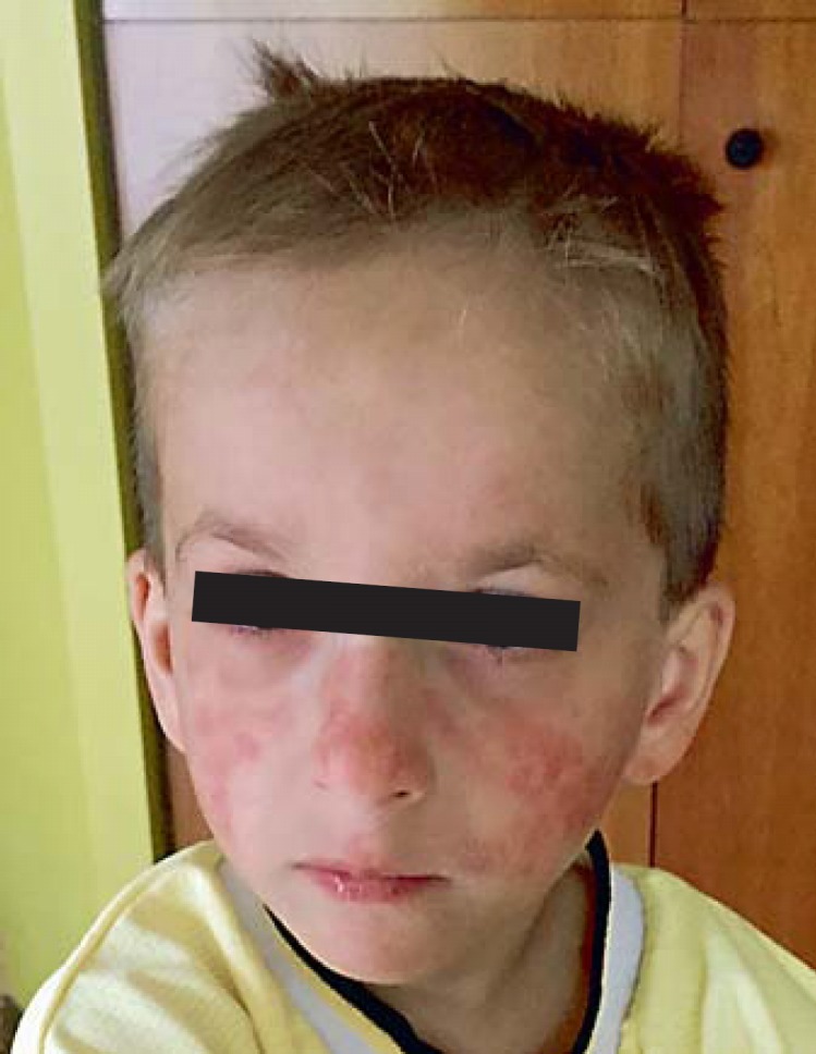
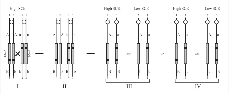
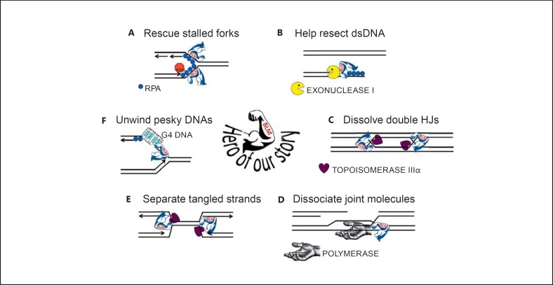
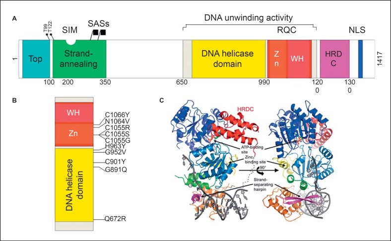
Bloom's syndrome is an autosomal recessive disorder characterized by prenatal and postnatal growth deficiency, photosensitive skin changes, immune deficiency, insulin resistance, and a greatly increased risk of early onset of cancer and for the development of multiple cancers. Loss-of-function mutations of BLM, which codes for a RecQ helicase, cause Bloom's syndrome. The absence of a functional BLM protein causes chromosome instability, excessive homologous recombination, and a greatly increased number of sister chromatid exchanges that are pathognomonic of the syndrome. A common founder mutation designated blmAsh is present in about 1 in 100 persons of Eastern European Jewish ancestry, and there are additional recurrent founder mutations among other populations. Missense, nonsense, and frameshift mutations as well as multiexonic deletions have all been observed. Bloom's syndrome is a prototypical chromosomal instability syndrome, and the somatic mutations that occur as a result of that instability are responsible for the increased cancer risk. Although there is currently no treatment aimed at the underlying genetic abnormality, persons with Bloom's syndrome benefit from sun protection, aggressive treatment of infections, surveillance for insulin resistance, and early identification of cancer.

Keywords: BLM; Bloom's syndrome; Cancer.


Figures

Fig. 1
Fig. 1
Characteristic sun-sensitive facial rash in a young boy with BS.
Fig. 2
Fig. 2
Model to generate a functionally normal BLM locus by somatic intergenic recombination. I The pairs of sister chromatids of the homologous chromosome 15 after the chromosomes have been duplicated in S phase in a somatic cell of a BS compound heterozygote (blm1/blm2) are numbered 1-1 to 4-4. Each of the 2 mutations in BLM (grey rectangle), represented by black dots, one inherited from each parent, is at a different site in the gene. Flanking markers proximal to and distal to the mutated loci are heterozygous A/a and B/b. II After homologous interchange between chromatids 2-2 and 3-3 at a point between the sites of mutation within BLM (the x in I), a non-mutant gene is reconstituted on chromatid 2-3 that is capable of correcting the high-SCE phenotype of BS cells. Simultaneously, the distal marker b becomes associated with the non-mutant gene on chromatid 2-3. III, IV By segregational events at mitosis, 2 pairs of daughter cells are possible. If chromatids 2-3 and 4-4 cosegregate, the distal marker becomes homozygous b/b (the diagram on the right side of III). On the other hand, if chromatids 2-3 and 3-2 cosegregate, the distal marker remains heterozygous b/B (the diagram on the right side of IV). The proximal marker remains heterozygous A/a in both cases. In the sister cells, segregation of chromatids 1-1 and 4-4 (the diagram on the left side of IV) or of chromatids 1-1 and 3-2 (the diagram on the left side of III) do not give rise to a low-SCE phenotype. Note that cells of heterozygous carriers of a mutation in BLM, namely, blm+ parents of persons with BS, display a low-SCE rate [Ellis et al., 1995a].
Fig. 3
Fig. 3
BLM's heroic fight against topological disaster and genome chaos. A BLM is recruited to stalled replications to stabilize the structure, regulate the accumulations of ssDNA-binding protein RPA and RAD51 recombinase to prevent premature or illegitimate engagement of homologous recombination, and to assist in replication restart. B BLM is active in the resection of dsDNA to create ssDNA for loading of RAD51 recombinase. C BLM in conjunction with topoisomerase IIIα will disentangle DNA molecules with 2 closely apposed double HJs, sliding the 2 HJs together and dissolving them without creating crossover products. D BLM is active in a DSB repair pathway referred to as synthesis-dependent strand annealing, in which an intact donor DNA molecule allows extension of the recipient DNA molecule past the break site. BLM may dissociate the extended DNA molecule from the donor template whereupon it may anneal with DNA from the other broken end. E BLM disentangles topological constrained molecules; such as might be found at approaching replication forks and underreplicated regions where the approaching forks failed to successfully terminate DNA replication. F BLM can unwind stable ssDNA structures that interfere with DNA replication, such as G4. DNA that forms these structures is broadly distributed around the genome - in the promoter regions of many genes, at telomere, in the rDNA.
Fig. 4
Fig. 4
Domain structure of human BLM. A Schematic representation of BLM domains. The functional domains depicted in the diagram are (from left to right) the topoisomerase IIIα-binding region (marked Top, blue block), the ssDNA strand-annealing and strand exchange domain (green block), the DNA helicase domain (yellow block), the RecQ C-terminal domain (RQC, red block) containing the Zn2+-binding motif (Zn, orange block) and winged helix (WH, fuchsia block), the helicase and ribonuclease D C-terminal (HRDC) domain (purple block), and the nuclear localization signal (NLS, blue block). The grey block indicates the region required for the DNA unwinding activity which contains the DNA helicase and RQC domains. Also represented in the diagram are the sites for phosphorylation by ATR and ATR protein kinases Thr99 and Thr122, the SUMO-interaction motif (SIM) which binds the small ubiquitin-related modifiers SUMO-1 and SUMO-2 as well as the SUMO-acceptor sites (depicted by the callout buttons), and the major sites being the lysines at amino acid residues 317 and 331. B A blow-up of the region containing the DNA unwinding activity, depicting the highly conserved amino acids residues that are mutated in persons with BS - missense mutations that constitute catalytic nulls. C Representation of the protein-folding structure of BLM640-1298 in complex with ADP and a DNA duplex with a 3′-overhang. The structure is rotated 90° around the vertical axis and the colors are different from the above-mentioned colors (A). The helicase domain is blue, the Zn2+-binding motif is green and yellow, the winged-helix (WH) domain is orange, with the β-hairpin that forms the DNA scalpel in pink, and the HRDC domain is red. The ADP is shown in stick form and the phosphate backbone of the DNA with bases is dark grey. The calcium and zinc ions are shown as light grey spheres. Reproduced with permission from the International Union of Crystallography [Swan et al., 2014].

Similar articles

Cited by

References

    1. Adams MD, McVey M, Sekelsky JJ. Drosophila BLM in double-strand break repair by synthesis-dependent strand annealing. Science. 2003;299:265–267. - PubMed
    1. Antczak A, Kluźniak A, Kashyap A, Jakubowska A, et al. A common nonsense mutation of the BLM gene and prostate cancer risk and survival. Gene. 2013;532:173–1766. - PubMed
    1. Arora H, Chacon AH, Choudhary S, McLeod MP, Meshkov L, et al. Bloom syndrome. Int J Dermatol. 2014;53:798–802. - PubMed
    1. Babbe H, Chester N, Leder P, Reizis B. The Bloom's syndrome helicase is critical for development and function of the αβ T-cell lineage. Mol Cell Biol. 2007;27:1947–1959. - PMC - PubMed
    1. Babbe H, McMenamin J, Hobeika E, Wang J, Rodig SJ, et al. Genomic instability resulting from Blm-deficiency compromises development, maintenance, and function of the B cell lineage. J Immunol. 2009;182:347–360. - PMC - PubMed