Nicotine Elicits Convulsive Seizures by Activating Amygdalar Neurons
- PMID: 28232801
- PMCID: PMC5298991
- DOI: 10.3389/fphar.2017.00057
Nicotine Elicits Convulsive Seizures by Activating Amygdalar Neurons
Abstract
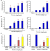
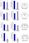
Nicotinic acetylcholine (nACh) receptors are implicated in the pathogenesis of epileptic disorders; however, the mechanisms of nACh receptors in seizure generation remain unknown. Here, we performed behavioral and immunohistochemical studies in mice and rats to clarify the mechanisms underlying nicotine-induced seizures. Treatment of animals with nicotine (1-4 mg/kg, i.p.) produced motor excitement in a dose-dependent manner and elicited convulsive seizures at 3 and 4 mg/kg. The nicotine-induced seizures were abolished by a subtype non-selective nACh antagonist, mecamylamine (MEC). An α7 nACh antagonist, methyllycaconitine, also significantly inhibited nicotine-induced seizures whereas an α4β2 nACh antagonist, dihydro-β-erythroidine, affected only weakly. Topographical analysis of Fos protein expression, a biological marker of neural excitation, revealed that a convulsive dose (4 mg/kg) of nicotine region-specifically activated neurons in the piriform cortex, amygdala, medial habenula, paratenial thalamus, anterior hypothalamus and solitary nucleus among 48 brain regions examined, and this was also suppressed by MEC. In addition, electric lesioning of the amygdala, but not the piriform cortex, medial habenula and thalamus, specifically inhibited nicotine-induced seizures. Furthermore, microinjection of nicotine (100 and 300 μg/side) into the amygdala elicited convulsive seizures in a dose-related manner. The present results suggest that nicotine elicits convulsive seizures by activating amygdalar neurons mainly via α7 nACh receptors.
Keywords: Fos expression; amygdala; convulsive seizures; nicotine; nicotinic acetylcholine receptors.
Figures


Similar articles
-
Nicotine evokes kinetic tremor by activating the inferior olive via α7 nicotinic acetylcholine receptors.Behav Brain Res. 2016 Nov 1;314:173-80. doi: 10.1016/j.bbr.2016.08.013. Epub 2016 Aug 6. Behav Brain Res. 2016. PMID: 27506652
-
Mechanism Underlying Organophosphate Paraoxon-Induced Kinetic Tremor.Neurotox Res. 2019 Apr;35(3):575-583. doi: 10.1007/s12640-019-0007-7. Epub 2019 Feb 7. Neurotox Res. 2019. PMID: 30729450
-
Pharmacological characterization of nicotine-induced seizures in mice.J Pharmacol Exp Ther. 1999 Dec;291(3):1284-91. J Pharmacol Exp Ther. 1999. PMID: 10565853
-
Involvement of alpha7- and alpha4beta2-type postsynaptic nicotinic acetylcholine receptors in nicotine-induced excitation of dopaminergic neurons in the substantia nigra: a patch clamp and single-cell PCR study using acutely dissociated nigral neurons.Brain Res Mol Brain Res. 2004 Oct 22;129(1-2):1-7. doi: 10.1016/j.molbrainres.2004.06.040. Brain Res Mol Brain Res. 2004. PMID: 15469877
-
Isocortical hyperemia and allocortical inflammation and atrophy following generalized convulsive seizures of thalamic origin in the rat.Cell Mol Neurobiol. 2003 Oct;23(4-5):773-91. doi: 10.1023/a:1025057004447. Cell Mol Neurobiol. 2003. PMID: 14514031 Free PMC article. Review.
Cited by
-
Three Seizures Provoked by E-cigarette Use in a Five-Year Period: A Case Report.Cureus. 2022 Aug 2;14(8):e27616. doi: 10.7759/cureus.27616. eCollection 2022 Aug. Cureus. 2022. PMID: 36059307 Free PMC article.
-
Nicotinic Acetylcholine Receptor Dysfunction in Addiction and in Some Neurodegenerative and Neuropsychiatric Diseases.Cells. 2023 Aug 11;12(16):2051. doi: 10.3390/cells12162051. Cells. 2023. PMID: 37626860 Free PMC article. Review.
-
Molecular Recognition of Nerve Agents and Their Organophosphorus Surrogates: Toward Supramolecular Scavengers and Catalysts.Chemistry. 2021 Sep 20;27(53):13280-13305. doi: 10.1002/chem.202101532. Epub 2021 Aug 10. Chemistry. 2021. PMID: 34185362 Free PMC article. Review.
-
Nicotine Neurotoxicity Involves Low Wnt1 Signaling in Spinal Locomotor Networks of the Postnatal Rodent Spinal Cord.Int J Mol Sci. 2021 Sep 3;22(17):9572. doi: 10.3390/ijms22179572. Int J Mol Sci. 2021. PMID: 34502498 Free PMC article.
-
Altered neuronal activity in the ventromedial prefrontal cortex drives nicotine intake escalation.Neuropsychopharmacology. 2023 May;48(6):887-896. doi: 10.1038/s41386-022-01428-9. Epub 2022 Aug 30. Neuropsychopharmacology. 2023. PMID: 36042320 Free PMC article.
References
-
- Besson M., David V., Baudonnat M., Cazala P., Guilloux J. P., Reperant C., et al. (2012). Alpha7-nicotinic receptors modulate nicotine-induced reinforcement and extracellular dopamine outflow in the mesolimbic system in mice. Psychopharmacology (Berl) 220 1–14. 10.1007/s00213-011-2422-1 - DOI - PubMed
LinkOut - more resources
Full Text Sources
Other Literature Sources

