. 2017 Feb 9:8:38.
doi: 10.3389/fphys.2017.00038. eCollection 2017.

On Biophysical Properties and Sensitivity to Gap Junction Blockers of Connexin 39 Hemichannels Expressed in HeLa Cells

Affiliations

On Biophysical Properties and Sensitivity to Gap Junction Blockers of Connexin 39 Hemichannels Expressed in HeLa Cells

Anibal A Vargas et al. Front Physiol. .

Abstract

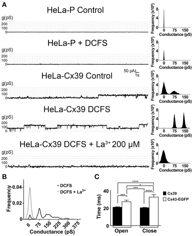
Although connexins (Cxs) are broadly expressed by cells of mammalian organisms, Cx39 has a very restricted pattern of expression and the biophysical properties of Cx39-based channels [hemichannels (HCs) and gap junction channels (GJCs)] remain largely unknown. Here, we used HeLa cells transfected with Cx39 (HeLa-Cx39 cells) in which intercellular electrical coupling was not detected, indicating the absence of GJCs. However, functional HCs were found on the surface of cells exposed to conditions known to increase the open probability of other Cx HCs (e.g., extracellular divalent cationic-free solution (DCFS), extracellular alkaline pH, mechanical stimulus and depolarization to positive membrane potentials). Cx39 HCs were blocked by some traditional Cx HC blockers, but not by others or a pannexin1 channel blocker. HeLa-Cx39 cells showed similar resting membrane potentials (RMPs) to those of parental cells, and exposure to DCFS reduced RMPs in Cx39 transfectants, but not in parental cells. Under these conditions, unitary events of ~75 pS were frequent in HeLa-Cx39 cells and absent in parental cells. Real-time cellular uptake experiments of dyes with different physicochemical features, as well as the application of a machine-learning approach revealed that Cx39 HCs are preferentially permeable to molecules characterized by six categories of descriptors, namely: (1) electronegativity, (2) ionization potential, (3) polarizability, (4) size and geometry, (5) topological flexibility and (6) valence. However, Cx39 HCs opened by mechanical stimulation or alkaline pH were impermeable to Ca2+. Molecular modeling of Cx39-based channels suggest that a constriction present at the intracellular portion of the para helix region co-localizes with an electronegative patch, imposing an energetic and steric barrier, which in the case of GJCs may hinder channel function. Results reported here demonstrate that Cx39 form HCs and add to our understanding of the functional roles of Cx39 HCs under physiological and pathological conditions in cells that express them.

Keywords: Cx39; dye-uptake; electrical coupling; gap junction; membrane potential; permeability; unitary conductance.


Figures

Figure 1
Figure 1
HeLa-Cx39 cells do not form functional gap junctions. (A,B) Phase contrast view (left) and immunofluorescence of Cx39 (A, right) and Cx43-EGFP fluorescence (Right) in HeLa-Cx39 and fluorescence of HeLa-Cx43EGFP cells (B, right). Bar scale: 20 μM. Arrows denote small Cx39 gap junctions plaques. (C) Junctional current (Ij) at different voltages (left) and normalized junctional conductance (gj) at different voltages (right) using dual voltage-clamp measurements in HeLa-Cx39 and HeLa-Cx43EGFP cells. Values of Ij for HeLa-Cx39 cells were zero at all voltages studied. Records of intercellular current transfer from cell1 (C1) to cell2 (C2) and vice versa in HeLa-Cx39 cells (Below cells shown in A) and in HeLa-Cx43EGFP (Below cells shown in B) upon voltage steps (increasing in 20 mV from −100 to 0 mV with duration of 200 ms, left bottom traces) applied to C1 and C2, respectively (n = 9).
Figure 2
Figure 2
DCFS reduces the resting membrane potential and increases the macroscopic membrane current of HeLa Cx39 cells. (A) The RMP of cells was measured using conventional microelectrodes. The graph shows the RMP of HeLa-P and HeLa-Cx39 cells bathed with normal saline solution or DCFS. In parallel experiments cells bathed with normal saline or DCFS were incubated for 30 min with 200 μM La3+ or 100 nM ouabain and the RMP was evaluated (****p < 0.0001 and ***p < 0.0005 compared only between HeLa-Cx39 cells under control conditions v/s all conditions and δδδδp < 0.0001 compared only between HeLa-P cells under control conditions v/s all conditions) n = 40 cells. (B) Representative membrane I/V curve obtained under application of a voltage ramp from −80 to +80 mV to HeLa-Cx39 cells under control conditions (Control) or DCFS or to DCFS-La3+ (200 μM), a HCs blocker. The macroscopic current at V = 0 mV was zero in all conditions. Right inset shows a unitary current event (Rectangle of discontinuous lines) of approximately 75 pS, recorded in the membrane of a HeLa-Cx39 cell. Left inset shows the slope of macroscopic currents under different conditions (n = 10, ****p < 0.0001).
Figure 3
Figure 3
DCFS increases the open probability of Cx39 HCs. (A) Unitary current events recorded under voltage clamp at +60 mV in HeLa-P (two top traces) or HeLa-Cx39 cells under the indicated conditions. The frequency of events recorded under each condition is shown in the right of each trace. (B) Conductance frequency chart obtained in HeLa-Cx39 cells in DCFS. The most frequent unitary conductance event recorded was 75 ± 5 pS in cells bathed with DCFS (n = 40 cells), and was blocked by La3+ (n = 10). (C) Delay times of aperture and closure of Cx39 and Cx43-EGFP HCs. (*P < 0.05; ***P < 0.0005; ****P < 0.0001, n = 30).
Figure 4
Figure 4
The dye uptake of HeLa-Cx39 cells is increased by DCFS. Uptake rate of the different molecules with different molecular mass and net charge (Bottom list) of HeLa-Cx39 cells under each condition indicated (n = 6 for each dye). Inset shows representative real time ethidium (Etd+) and propidium (Pro) uptake, HeLa-Cx39 cells respectively, and Etd+ in HeLa-P. After 5 min recording under control conditions, the extracellular solution was quickly changed by DCFS and dye uptake was recorded for 8 min followed by additional 5 min recording in the presence of 200 μM La3+. (****P < 0.0001 and ***p < 0.0005 compared to basal value in each case; n = 4, with 30 cells recorded in each case). AU, arbitrary Units.
Figure 5
Figure 5
Alkaline pH opens Cx39 HCs, which are permeable to ethidium but not to Ca2+. HeLa-P, HeLa-Cx39, or HeLa-Cx43 cells were exposed to extracellular saline solution with pH 7.4 or pH 8.5. (A,B) Etd+ uptake rate and Ca2+ signal, respectively, were evaluated over time. (B) Inset. Real time of Ca2+ signal increase in HeLa-P, HeLa-Cx39 and HeLa-Cx43 cells, bathed with alkaline solution after register in control conditions. ****p < 0.0001 and ***p < 0.0005 respectively, n = 3 experiments and 20 cells were recorded in each experiment. AU, Arbitrary Units.
Figure 6
Figure 6
Cx39 HCs of cells exposed to mechanical stress are permeable to ethidium but not to Ca2+. (A) Dye (Etd+) uptake rate of HeLa-P and HeLa-Cx39 cells mechanical stimulated with different volumes of saline solution from 15 cm high under control conditions. HeLa-Cx39 cells were also mechanically stimulated with 8 ml in the presence of 200 μM La3+ (8+La3+). HeLa-P cells were also stimulated with 8 ml of saline solution. (****p < 0.0001, n = 5 with 30 cells recorder in each case). (B) Intracellular Ca2+ signal of HeLa-Cx39 cells under control conditions (first 2.5 min), after mechanical stimulation (8 ml from 15 cm high) and after the addition of 2.5 μM of 4-Bromo calcium ionophore A23187 to increase calcium in the cells. MS, mechanical stress.
Figure 7
Figure 7
Cx39 HCs are inhibited by some conventional Cx HCs blockers. The graph shows normalized data with respect to the basal condition of Etd+ uptake rate induced by DCFS in HeLa-Cx43 EGFP and HeLa Cx39 cells. Heptanol (350 μM), octanol (310 μM), 18β-glycyrrhetinic acid (50 μM, β-GA), oleamide (100 μM) or carbenoxolone (5 to 50 μM, CBX) was applied together with DCFS (n = 10, &&&&p < 0.0001, &&&p < 0.0005 and ****p < 0.0001 compared only between HeLa-Cx39 cells and HeLa-Cx43 EGFP respectively, under increased dye uptake induced by DCFS v/s all dye uptake blockers; ####p < 0.0001 compared only between HeLa-Panx1 cells).
Figure 8
Figure 8
mCx39 resemble a Cx based channel but exhibit structural differences in PH region compared with mCx43 an hCx26. (A) Top row, structural representations of the hCx26 crystal structure of the HC used as a template, and the molecular models of mCx39 and mCx43. Each HC shows two monomers that are depicted by ribbons and color coded to represent different regions of the protein (NTH = magenta, TM1 = red, PH = cyan, TM2 = orange, TM3 = yellow, and TM4 = green). Two other monomers are depicted in a gray solid surface, while the other two at the front were omitted for clarity. (A) Bottom row, structural representations of the hCx26 GJC used as template, and the molecular models of mCx39 and mCx43. Each HC shows three monomers that are depicted by cartoons (cylinders = alpha helices, arrows = beta sheets, tubes = coils and loops) and color coded to differentiate from each other. (B) Pore radius along the z-axis of the Cx channels. Left, pore radius along the z-axis calculated for the hCx26 crystal structure (black) of the GJCs and the mCx39 (red) and Cx43 (blue) molecular model. Right, pore radius along the z-axis calculated for the hCx26 crystal structure (black) of the HC and the mCx39 (red) and Cx43 (blue) molecular models. The NTH region is depicted in magenta while the PH regions appears in cyan.
Figure 9
Figure 9
Molecular surface along the pore of Cx-based channels colored by electrostatic potential. Top row, the electrostatic potential mapped onto the molecular surface of the hCx26 crystal structure of the HC used as a template, and the mCx39 and mCx43 molecular models. Bottom row, the electrostatic potential mapped onto the molecular surface of the hCx26 crystal structure of the GJC used as a template, and the mCx39 and mCx43 molecular models. All structural representations depict 4 monomers per HC while the other two at the front were omitted for clarity. Color coding corresponds to the value of the electrostatic potential according to the spectrum shown at the right (in kT/e units).

Similar articles

Cited by

References

    1. Abascal F., Zardoya R. (2013). Evolutionary analyses of gap junction protein families. Biochim. Biophys. Acta 1828, 4–14. 10.1016/j.bbamem.2012.02.007 - DOI - PubMed
    1. Altevogt B. M., Kleopa K. A., Postma F. R., Scherer S. S., Paul D. L. (2002). Connexin29 is uniquely distributed within myelinating glial cells of the central and peripheral nervous systems. J. Neurosci. 22, 6458–6470. - PMC - PubMed
    1. Araya-Secchi R., Lagos C. F., Abarca F., Martinez A. D., Perez-Acle T. (2012). Molecular dynamics simulations of Cx26-Wt and deafness related mutants M34A, A40G and V37I. Biophys. J. 102, 106a–107a. 10.1016/j.bpj.2011.11.600 - DOI
    1. Araya-Secchi R., Perez-Acle T., Kang S. G., Huynh T., Bernardin A., Escalona Y., et al. . (2014). Characterization of a novel water pocket inside the human Cx26 hemichannel structure. Biophys. J. 107, 599–612. 10.1016/j.bpj.2014.05.037 - DOI - PMC - PubMed
    1. Bao L., Locovei S., Dahl G. (2004). Pannexin membrane channels are mechanosensitive conduits for ATP. FEBS Lett. 572, 65–68. 10.1016/j.febslet.2004.07.009 - DOI - PubMed

LinkOut - more resources