

γδ T Cell-Mediated Immunity to Cytomegalovirus Infection
- PMID: 28232834
- PMCID: PMC5298998
- DOI: 10.3389/fimmu.2017.00105
γδ T Cell-Mediated Immunity to Cytomegalovirus Infection
Abstract
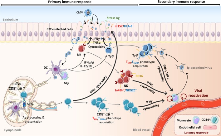
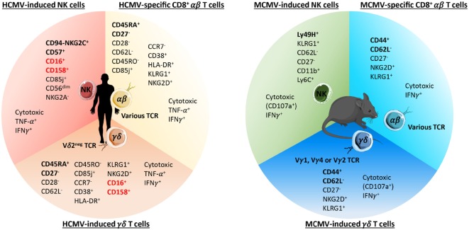
γδ T lymphocytes are unconventional immune cells, which have both innate- and adaptive-like features allowing them to respond to a wide spectrum of pathogens. For many years, we and others have reported on the role of these cells in the immune response to human cytomegalovirus in transplant patients, pregnant women, neonates, immunodeficient children, and healthy people. Indeed, and as described for CD8+ T cells, CMV infection leaves a specific imprint on the γδ T cell compartment: (i) driving a long-lasting expansion of oligoclonal γδ T cells in the blood of seropositive individuals, (ii) inducing their differentiation into effector/memory cells expressing a TEMRA phenotype, and (iii) enhancing their antiviral effector functions (i.e., cytotoxicity and IFNγ production). Recently, two studies using murine CMV (MCMV) have corroborated and extended these observations. In particular, they have illustrated the ability of adoptively transferred MCMV-induced γδ T cells to protect immune-deficient mice against virus-induced death. In vivo, expansion of γδ T cells is associated with the clearance of CMV infection as well as with reduced cancer occurrence or leukemia relapse risk in kidney transplant patients and allogeneic stem cell recipients, respectively. Taken together, all these studies show that γδ T cells are important immune effectors against CMV and cancer, which are life-threatening diseases affecting transplant recipients. The ability of CMV-induced γδ T cells to act independently of other immune cells opens the door to the development of novel cellular immunotherapies that could be particularly beneficial for immunocompromised transplant recipients.
Keywords: antiviral immunity; bone marrow and organ transplantation; cytomegalovirus; memory T cells; γδ T cells.
Figures
Similar articles
-
Direct and Indirect Effects of Cytomegalovirus-Induced γδ T Cells after Kidney Transplantation.Front Immunol. 2015 Jan 21;6:3. doi: 10.3389/fimmu.2015.00003. eCollection 2015. Front Immunol. 2015. PMID: 25653652 Free PMC article. Review.
-
Control of murine cytomegalovirus infection by γδ T cells.PLoS Pathog. 2015 Feb 6;11(2):e1004481. doi: 10.1371/journal.ppat.1004481. eCollection 2015 Feb. PLoS Pathog. 2015. PMID: 25658831 Free PMC article.
-
γδ T cells confer protection against murine cytomegalovirus (MCMV).PLoS Pathog. 2015 Mar 6;11(3):e1004702. doi: 10.1371/journal.ppat.1004702. eCollection 2015 Mar. PLoS Pathog. 2015. PMID: 25747674 Free PMC article.
-
Cytomegalovirus infection alters phenotypes of different γδ T-cell subsets in renal transplant recipients with long-term stable graft function.J Med Virol. 2017 Aug;89(8):1442-1452. doi: 10.1002/jmv.24784. Epub 2017 Mar 6. J Med Virol. 2017. PMID: 28198539
-
Antigen-specific γδ T cells contribute to cytomegalovirus control after stem cell transplantation.Curr Opin Immunol. 2023 Jun;82:102303. doi: 10.1016/j.coi.2023.102303. Epub 2023 Mar 20. Curr Opin Immunol. 2023. PMID: 36947903 Review.
Cited by
-
γδ T cells: Major advances in basic and clinical research in tumor immunotherapy.Chin Med J (Engl). 2024 Jan 5;137(1):21-33. doi: 10.1097/CM9.0000000000002781. Epub 2023 Aug 18. Chin Med J (Engl). 2024. PMID: 37592858 Free PMC article. Review.
-
Robust Identification of Suitable T-Cell Subsets for Personalized CMV-Specific T-Cell Immunotherapy Using CD45RA and CD62L Microbeads.Int J Mol Sci. 2019 Mar 20;20(6):1415. doi: 10.3390/ijms20061415. Int J Mol Sci. 2019. PMID: 30897843 Free PMC article.
-
Human γδ T Cell Receptor Repertoires in Peripheral Blood Remain Stable Despite Clearance of Persistent Hepatitis C Virus Infection by Direct-Acting Antiviral Drug Therapy.Front Immunol. 2018 Mar 16;9:510. doi: 10.3389/fimmu.2018.00510. eCollection 2018. Front Immunol. 2018. PMID: 29616028 Free PMC article. Clinical Trial.
-
Characterization of the γδ T-cell compartment during infancy reveals clear differences between the early neonatal period and 2 years of age.Immunol Cell Biol. 2020 Jan;98(1):79-87. doi: 10.1111/imcb.12303. Epub 2019 Dec 1. Immunol Cell Biol. 2020. PMID: 31680329 Free PMC article.
-
Mapping and Characterization of HCMV-Specific Unconventional HLA-E-Restricted CD8 T Cell Populations and Associated NK and T Cell Responses Using HLA/Peptide Tetramers and Spectral Flow Cytometry.Int J Mol Sci. 2021 Dec 27;23(1):263. doi: 10.3390/ijms23010263. Int J Mol Sci. 2021. PMID: 35008688 Free PMC article.
References
Publication types
LinkOut - more resources
Full Text Sources
Other Literature Sources
Research Materials