. 2017 Feb 9:8:106.
doi: 10.3389/fimmu.2017.00106. eCollection 2017.

Mesenchymal Stem Cells Support Survival and Proliferation of Primary Human Acute Myeloid Leukemia Cells through Heterogeneous Molecular Mechanisms

Affiliations

Mesenchymal Stem Cells Support Survival and Proliferation of Primary Human Acute Myeloid Leukemia Cells through Heterogeneous Molecular Mechanisms

Annette K Brenner et al. Front Immunol. .

Abstract

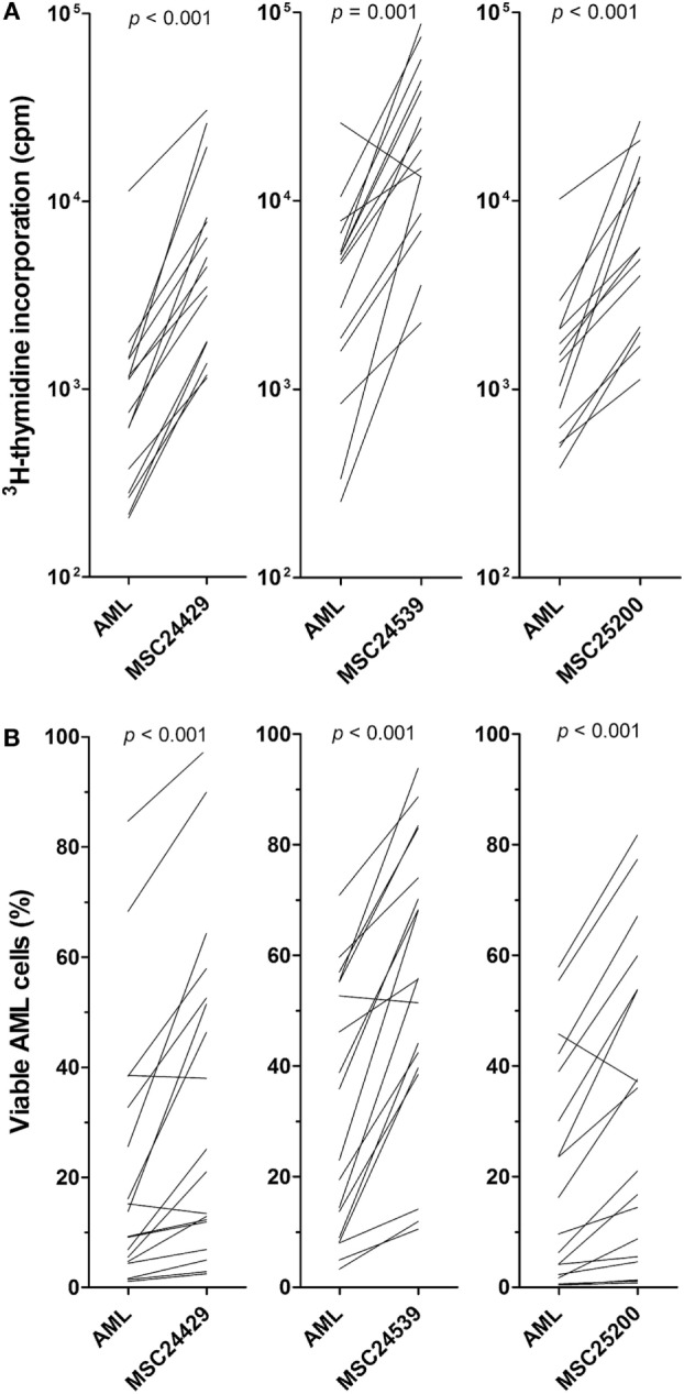
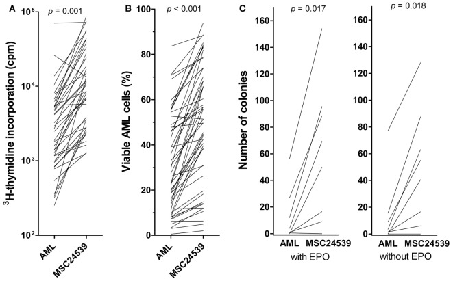
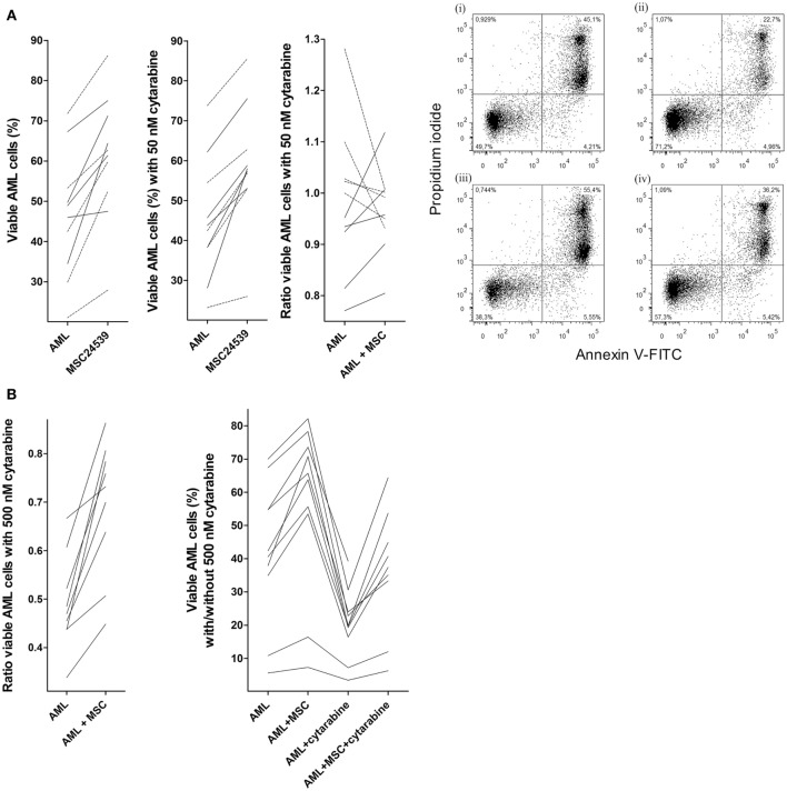
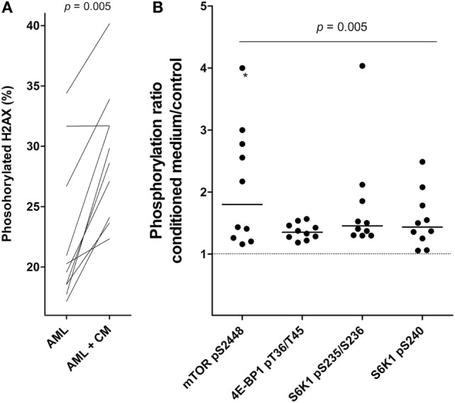
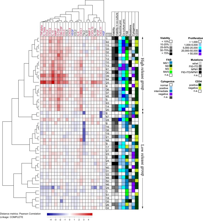
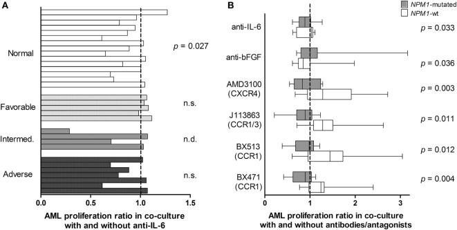
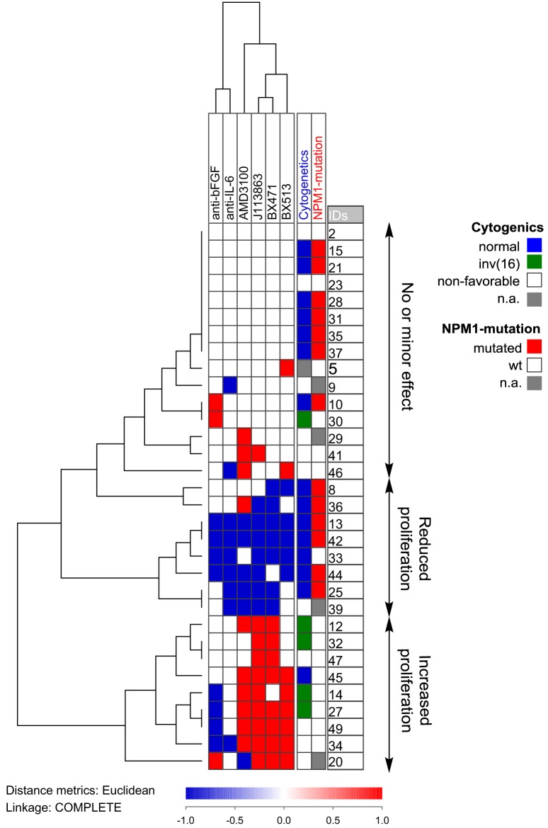
Acute myeloid leukemia (AML) is a bone marrow malignancy, and various bone marrow stromal cells seem to support leukemogenesis, including osteoblasts and endothelial cells. We have investigated how normal bone marrow mesenchymal stem cells (MSCs) support the in vitro proliferation of primary human AML cells. Both MSCs and primary AML cells show constitutive release of several soluble mediators, and the mediator repertoires of the two cell types are partly overlapping. The two cell populations were cocultured on transwell plates, and MSC effects on AML cells mediated through the local cytokine/soluble mediator network could thus be evaluated. The presence of normal MSCs had an antiapoptotic and growth-enhancing effect on primary human AML cells when investigating a group of 51 unselected AML patients; this was associated with increased phosphorylation of mTOR and its downstream targets, and the effect was independent of cytogenetic or molecular-genetic abnormalities. The MSCs also supported the long-term proliferation of the AML cells. A subset of the patients also showed an altered cytokine network with supra-additive levels for several cytokines. The presence of cytokine-neutralizing antibodies or receptor inhibitors demonstrated that AML cells derived from different patients were heterogeneous with regard to effects of various cytokines on AML cell proliferation or regulation of apoptosis. We conclude that even though the effects of single cytokines derived from bone marrow MSCs on human AML cells differ among patients, the final cytokine-mediated effects of the MSCs during coculture is growth enhancement and inhibition of apoptosis.

Keywords: acute myeloid leukemia; apoptosis; chemokines; cytokines; mesenchymal stem cells; proliferation.


Figures

Figure 1
Figure 1
The effect of normal bone marrow mesenchymal stem cells (MSCs) on in vitro proliferation and viability of primary human acute myeloid leukemia (AML) cells; a comparison of MSCs derived from three healthy donors (MSC24429, MSC24539, MSC25200). Primary human AML cells derived from 18 consecutive/unselected leukemia patients were cocultured with normal MSCs from the three donors. The MSCs were initially cultured for 3 days to allow them to establish adherent in vitro proliferation; primary human AML cells were then added and both leukemia cell proliferation and viability were assayed after additional 3 days of culture. [(A), upper part] Proliferation was assayed as 3H-thymidine incorporation. The results for each patient are presented as nuclear thymidine incorporation (counts per minute, cpm). The p-value for the statistical comparison of the overall results is given at the top of the figure for each of the individual MSC. Each line represents the results for one patient. [(B), lower part] Leukemia cell viability was assayed by the Annexin V-PI flow cytometric assay. The p-value for the statistical comparison of the overall results for each of the MSCs is given at the top of the figure for the MSCs. Each line represents the results for one patient.
Figure 2
Figure 2
The effects of mesenchymal stem cells (MSCs) on acute myeloid leukemia (AML) cell proliferation/viability in suspension cultures and AML cells tested in a clonogenic assay. The effect of MSCs on the in vitro proliferation [(A), left] and viability [(B), middle] of primary human AML cells derived from 51 consecutive patients was examined. The AML cells were then cocultured with normal MSC24539. The MSCs were initially cultured for 3 days to allow them to establish adherent in vitro proliferation; primary human AML cells were thereafter added and (A) leukemia cell proliferation was assayed as 3H-thymidine incorporation after additional 3 days of culture; additionally, (B) AML cell viability was assayed by the Annexin V-PI flow cytometric assay. The p-values for the statistical comparison of the overall results are given at the top of the figure. Each line represents the results for one patient. [(C), right] The effects of MSCs on clonogenic AML cells were also investigated. The leukemic cells were cultured either in medium alone or in transwell cocultures together with MSC24593 for 3 weeks; the number of clonogenic cells were thereafter compared for AML cells precultured in medium alone and together with MSCs. The total culture period was thus 5 weeks. The MSCs significantly increased the number of colony-forming units (p-values are given on top of the figure) in growth media both with and without erythropoietin. The results are presented as mean of duplicate samples (average deviation from mean corresponding to 1.3 colonies and 8.7% of the total colony number). Each line represents the results for one patient.
Figure 3
Figure 3
Effects of mesenchymal stem cells (MSCs) on primary human acute myeloid leukemia (AML) cells treated with cytarabine; (A) effect of 50 nM cytarabine-exposure on AML cell viability in transwell cocultures and (B) effect of 500 nM cytarabine tested in direct-contact cocultures. Primary AML cells derived from the same 10 patients were included in all these studies, and MSC24539 was used in all experiments. The AML cell viability was analyzed by flow cytometry. [(A), transwell cocultures] For each patient, we compared cultures containing AML cells alone (AML) or AML cells plus MSCs (AML + MSC). The three figures from left to the middle right show (i) AML cell viability for cells cultured in medium alone with or without MSCs; (ii) AML cell viability in the presence of 50 nM cytarabine for leukemic cells cultured with and without MSCs; and (iii) a comparison of the medium culture ratio (i.e., viable cells in cytarabine-containing cultures versus drug-free controls; medium alone) and MSC culture ratio (i.e., viable cells in cytarabine-containing cultures versus drug-free controls; MSCs being added to both cultures). The results for the five patients (solid lines) for which 50 nM cytarabine exhibited a proapoptotic effect in the presence of MSCs are indicated. The results for one responding patient are presented in detail (right part of the figure; i/ii: drug-free control, iii/iv: 50 nM cytarabine without and with MSCs, respectively); the percentage of viable cells (population at the lower left) is indicated in the figures for each of the four cultures. [(B), direct-contact cultures] The left part of the figure compares the medium culture ratio (i.e., viable cells in cytarabine-containing cultures versus drug-free controls; medium alone) and MSC culture ratio (i.e., viable cells in cytarabine-containing cultures versus drug-free controls; MSCs being added to both cultures) when testing 500 nM cytarabine. The right part of the figure shows the percentage of viable primary AML cells (10 patients tested, one patient not tested for AML cells + cytarabine + MSC) cultured in medium alone, together with MSCs, in medium containing 500 nM cytarabine, and together with 500 nM cytarabine and MSCs.
Figure 4
Figure 4
Effects of mesenchymal stem cell (MSC)-conditioned medium on primary human acute myeloid leukemia (AML) cells; a study of H2AX phosphorylation and mTOR signaling. (A) The change in percentage of phosphorylated H2AX for AML cells cultured in MSC-conditioned medium (CM, 50%) compared to medium alone. (B) The effect of MSC-conditioned medium on the phosphorylation of mTOR and its downstream targets. For each patient, we compared the level of phosphorylation for AML cells cultured with MSC-conditioned medium versus control cells cultured in medium alone. The results are presented as the relative level, i.e., the levels for MSC-conditioned cultures versus control cultures. The value marked with an asterisk represents a value that was undetectable in the control; for simplicity, the ratio was set to the same value as the highest observed ratio in the data series.
Figure 5
Figure 5
Hierarchical clustering analysis based on the ratio of cytokine levels in acute myeloid leukemia (AML)-mesenchymal stem cell (MSC) cocultures divided by the concentration sums of AML cells and MSCs cultured alone; primary AML cells were derived from 51 patients and cocultured with MSC24539. Each horizontal row in the figure represents the observation for one patient, and the vertical columns represent the observations for the soluble mediators. Red indicates supra-additive effects in coculture. Likewise cytokines marked in red showed supra-additive effect on the total patient cohort, whereas mediators marked in blue showed higher levels in MSC cultures alone than in coculture. The patients clustered into two main groups (see right part of the figure) indicating high and low relative coculture cytokine levels, respectively. The figure also shows the distribution of various biological characteristics between the patient subsets (AML cell viability after 72 h of culture in medium alone and in coculture, proliferative capacity in medium alone and in coculture, FAB classification, genetic abnormalities, and expression of the CD34 stem cell marker).
Figure 6
Figure 6
Cytokine-targeted treatment during coculture of primary human acute myeloid leukemia (AML) cells and bone marrow mesenchymal stem cells (MSCs); a comparison of patients with different karyotype (A) and patients with and without NPM1 mutations. (A) Proliferative AML cell responses were compared for cocultures prepared with an IL-6 specific antibody and control antibody. The study included patients with normal AML cell karyotype (n = 15), favorable (n = 5), intermediate (n = 4), and adverse (n = 7) cytogenetics. The results for individual patients are presented as the proliferative response in cultures with anti-IL-6 relative to the proliferation in control cultures. The IL-6-specific antibody decreased the proliferation only for patients with normal cytogenetics but not for the other patients (Wilcoxon’s test for paired samples). (B) AML cell proliferation was also compared for patients with (gray columns) and without NPM1 mutations (white columns). Decreased AML cell proliferation was seen for several cytokine-directed treatment, especially chemokine-directed interventions (Wilcoxon’s test for paired samples for IL-6/bFGF, χ2 likelihood analyses for the chemokines). The box-plots show the median value, the 25 and 75% quartiles and the variation range (minimum to maximum value).
Figure 7
Figure 7
Hierarchical clustering of the effects of anti-cytokine treatment (neutralizing antibodies or receptor blocking) on acute myeloid leukemia (AML) cell proliferation during coculture with normal mesenchymal stem cells. AML cells derived from 32 AML patients were examined. Each horizontal row in the figure represents the observations for one patient, and the vertical columns represent the observations for each of the antibodies and receptor antagonists. Red indicates increased proliferation corresponding to an increase in the absolute value of >2,000 cpm and >20% proliferation increase compared to the control cultures, whereas blue indicates reduced proliferation according to the same definitions. The patients clustered into three main groups (see right part of the figure): one group (above) showing only minor effects, a second group (middle) showing reduced proliferation in the presence of both antibodies and receptor antagonists, and a last group (below) that showed increased proliferation especially for chemokine inhibition.

Similar articles

Cited by

References

    1. Estey EH. Acute myeloid leukemia: 2014 update on risk-stratification and management. Am J Hematol (2014) 89:1063–81.10.1002/ajh.23834 - DOI - PubMed
    1. Arber DA, Orazi A, Hasserjian R, Thiele J, Borowitz MJ, Le Beau MM, et al. The 2016 revision to the World Health Organization classification of myeloid neoplasms and acute leukemia. Blood (2016) 127:2391–405.10.1182/blood-2016-03-643544 - DOI - PubMed
    1. Burnett A, Wetzler M, Löwenberg B. Therapeutic advances in acute myeloid leukemia. J Clin Oncol (2011) 29:487–94.10.1200/JCO.2010.30.1820 - DOI - PubMed
    1. Falini B, Nicoletti I, Bolli N, Martelli MP, Liso A, Gorello P, et al. Translocations and mutations involving the nucleophosmin (NPM1) gene in lymphomas and leukemias. Haematologica (2007) 92:519–32.10.3324/haematol.11007 - DOI - PubMed
    1. Deans RJ, Moseley AB. Mesenchymal stem cells: biology and potential clinical uses. Exp Hematol (2000) 28:875–84.10.1016/S0301-472X(00)00482-3 - DOI - PubMed