. 2014 Dec 12;2(1):e970499.
doi: 10.4161/21690693.2014.970499. eCollection 2014.

Unreported intrinsic disorder in proteins: Building connections to the literature on IDPs

Affiliations

Unreported intrinsic disorder in proteins: Building connections to the literature on IDPs

Vladimir N Uversky. Intrinsically Disord Proteins. .

Abstract

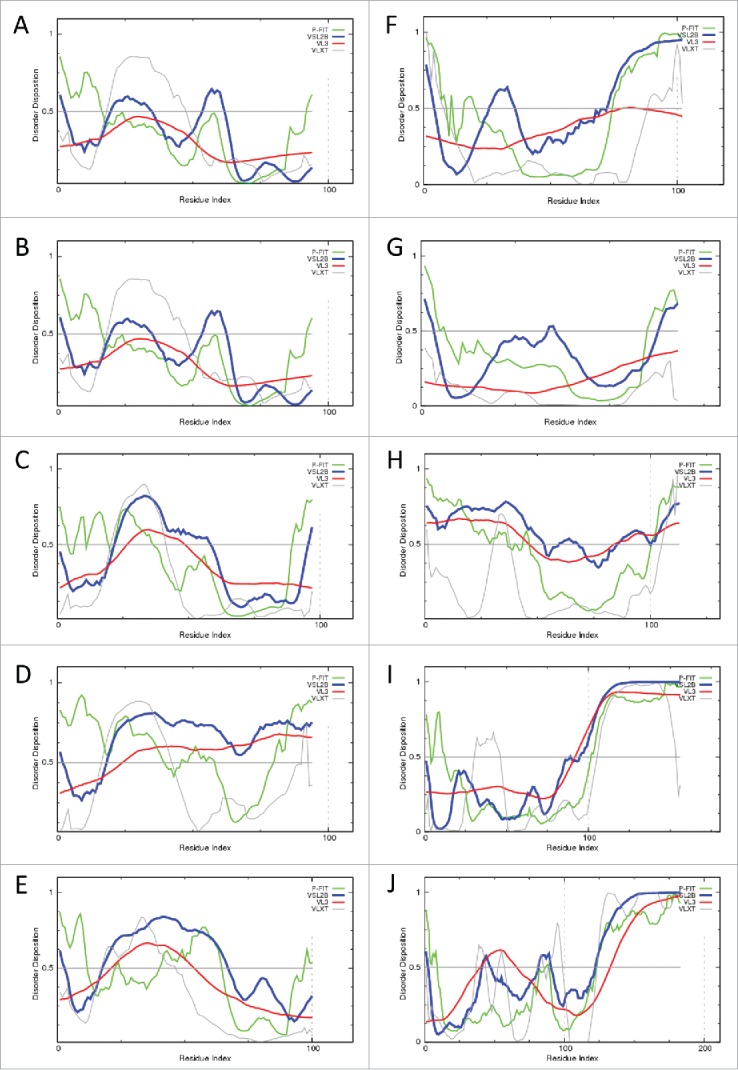
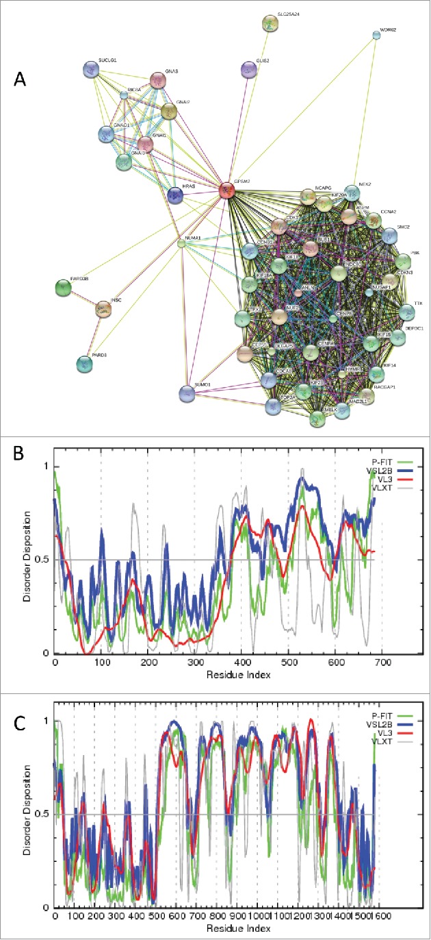
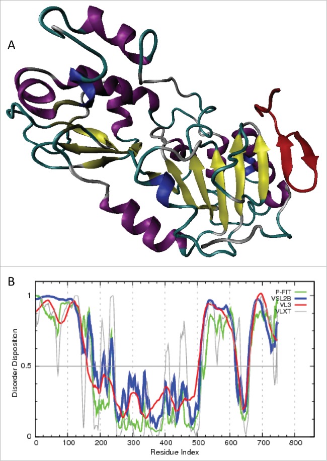
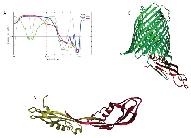
This review opens a new series entitled "Unreported intrinsic disorder in proteins." The goal of this series is to bring attention of researchers to an interesting phenomenon of missed (or overlooked, or ignored, or unreported) disorder. This series serves as a companion to "Digested Disorder" which provides a quarterly review of papers on intrinsically disordered proteins (IDPs) found by standard literature searches. The need for this alternative series results from the observation that there are numerous publications that describe IDPs (or hybrid proteins with ordered and disordered regions) yet fail to recognize many of the key discoveries and publications in the IDP field. By ignoring the body of work on IDPs, such publications often fail to relate their findings to prior discoveries or fail to explore the obvious implications of their work. Thus, the goal of this series is not only to review these very interesting and important papers, but also to point out how each paper relates to the IDP field and show how common tools in the IDP field can readily take the findings in new directions or provide a broader context for the reported findings.

Keywords: intrinsically disordered protein; molecular recognition; posttranslational modification; protein function; protein-protein interaction.


Figures

Figure 1.
Figure 1.
Intrinsic disorder in the eIF4A (UniProt ID: P10081, A) eIF4B (UniProt ID: P34167, C), and eIF4G (UniProt ID: P39935, D) evaluated by PONDR® FIT (red lines), PONDR® VLXT (blue lines), PONDR® VSL2B (green lines), and PONDR® VL3 (cyan lines). (B) Crystal structure (PDB ID: 2VSO) of the complex between the eIF4A (red) and the middle domain of eIF4G (residues 572–853, blue).
Figure 2.
Figure 2.
(A) Intrinsic disorder in human plasminogen activator inhibitor 1 (PAI-1, UniProt ID: P05121). (B) Intrinsic disorder propensities of SAMHD1 (UniProt ID: Q9Y3Z3). (C). Intrinsic disorder in CspC from E. coli (UniProt ID: P0A9Y6). Intrinsic disorder propensities are evaluated by PONDR® FIT (green lines), PONDR® VLXT (gray lines), PONDR® VSL2B (blue lines), and PONDR® VL3 (red lines).
Figure 3.
Figure 3.
(A) Evaluation of the disorder propensity of the UvsW helicase (UniProt ID: P20703) from the bacteriophage T4. Disorder propensity was evaluated by a set of predictors from the PONDR family, PONDR® FIT, VSL2B, VL3, and VLXT. Scores above 0.5 correspond to disordered residues/regions. (B) Crystal structure of the bacteriophage T4 UvsW helicase (PDB ID: 2OCA). (C). Crystal structure of gp32 (PDB ID: 1GPC). (D) Disorder propensity in the gp32 protein (UniProt ID: P03695) evaluated by a set of predictors from the PONDR family, PONDR-FIT, VSL2B, VL3, and VLXT.
Figure 4.
Figure 4.
Intrinsic disorder in the type 4 fimbrial biogenesis protein PilF (A, UniProt ID: Q9HXJ2); the fimbrial assembly protein PilQ (B, UniProt ID: P34750). Intrinsic disorder propensities are evaluated by PONDR® FIT (green lines), PONDR® VLXT (gray lines), PONDR® VSL2B (blue lines), and PONDR® VL3 (red lines).
Figure 5.
Figure 5.
For figure legend, see page 11.Figure 5 (see previous page). (A) A crystal structure (PDB ID: 2V7D) of the complex between the 14–3–3ζ protein (blue cloud) and a phosphorylated peptide from the β2 integrin tail (red chain). (B) Interactome of the β2 integrin as evaluated by STRING. Here, settings were chosen to find binding partners with the highest confidence (0.9). (C). Disorder-based alignments of the C-terminal tails of several human integrins: residues 1001–1032 of human integrin α4 (black line, UniProt ID: P13612), residues 752–801 of human integrin β1 (black line, UniProt ID: P05556), residues 724–769 of human integrin β2 (black line, UniProt ID: P05107), residues 742–788 of human integrin β3 (black line, UniProt ID: P05106), and residues 747–798 of human integrin β7 (black line, UniProt ID: P26010).
Figure 6.
Figure 6.
For figure legend, see page 13.Figure 6 (see previous page). (A) Evaluation of the interactivity of the human viperin (UniProt ID: Q8WXG1) by STRING, which is the online database resource Search Tool for the Retrieval of Interacting Genes that provides both experimental and predicted interaction information. STRING produces the network of predicted associations for a particular group of proteins. The network nodes are proteins. The edges represent the predicted functional associations. An edge may be drawn with up to 7 differently colored lines - these lines represent the existence of the 7 types of evidence used in predicting the associations. A red line indicates the presence of fusion evidence; a green line - neighborhood evidence; a blue line – co-occurrence evidence; a purple line - experimental evidence; a yellow line – text mining evidence; a light blue line - database evidence; a black line – co-expression evidence. (B) Intrinsic disorder propensity of the human viperin. Disorder propensity was evaluated by a set of predictors from the PONDR family, PONDR® FIT, VSL2B, VL3, and VLXT. Scores above 0.5 correspond to disordered residues/regions.
Figure 7.
Figure 7.
(A) Crystal structure of the Pukovnik Xis pentamer (PDB ID: 4J2N) and 4 pairs of different dimers found within this pentameric filament. (B) Intrinsic disorder propensity of Pukovnik Xis (UniProt ID: B3VGI6) evaluated by by a set of predictors from the PONDR family, PONDR® FIT, VSL2B, VL3, and VLXT. Scores above 0.5 correspond to disordered residues/regions.
Figure 8.
Figure 8.
Evaluation of intrinsic disorder in Human aryl-hydrocarbon-interacting protein-like 1 (A, AIPL1, UniProt ID: Q9NZN9); Human AIP (B, UniProt ID: O00170); Human APC (C, UniProt ID: P04070, residues 16–266); E.coli DnaT protein (D, UniProt ID: P0A8J2); Human PECAM-1 (E, UniProt ID: P16284); Mouse Serine/arginine-rich splicing factor 1 (F, UniProt ID: Q6PDM2). Intrinsic disorder propensities are evaluated by PONDR® FIT (green lines), PONDR® VLXT (gray lines), PONDR® VSL2B (blue lines), and PONDR® VL3 (red lines).
Figure 9.
Figure 9.
Intrinsic disorder in: (A) Human IFITM1 (UniProt ID: P13164); (B) Human IFITM2 (UniProt ID: Q01629); (C) Human IFITM3 (UniProt ID: Q01628); (D) Human IFITM5 (UniProt ID: A6NNB3); (E) Human IFITM10 (UniProt ID: A6NMD0); (F) Human semaphorin-3 (UniProt ID: Q13275). Intrinsic disorder propensities are evaluated by PONDR® FIT (green lines), PONDR® VLXT (gray lines), PONDR® VSL2B (blue lines), and PONDR® VL3 (red lines).
Figure 10.
Figure 10.
(A) Crystal structure of the isolated ε subunit of the F0F1 ATP synthase from the thermophile Bacillus PS3 (Tε) in a compact structure in the non-bound form (PDB ID: 1AQT). (B) Crystal structure of the Tε subunit in a highly extended open form in a complex with other subunits of the bacterial F1 complex (PDB ID: 3OAA). (C). Disorder propensity of the Tε (UniProt ID: P0A6E6) evaluated by PONDR® FIT (green lines), PONDR® VLXT (gray lines), PONDR® VSL2B (blue lines), and PONDR® VL3 (red lines).
Figure 11.
Figure 11.
Intrinsic disorder in the CaPcl5 evaluated by PONDR® FIT (red lines), PONDR® VLXT (blue line), PONDR® VSL2B (green line), and PONDR® VL3 (cyan line). Light pink shadow around the corresponding PONDR® FIT curve represents distribution of statistical errors.
Figure 12.
Figure 12.
Intrinsic disorder propensities of the isoforms-1 (A) and the alternatively spliced isoform-5 (B) of human siruin-2 evaluated by PONDR® FIT, PONDR® VSL2B, PONDR® VL3, and PONDR® VLXT.
Figure 13.
Figure 13.
The multiparametric evaluation of intrinsic disorder propensities of HSF-1 (A, UniProt ID: Q00613), CREB (B, UniProt ID: P16220), NF-YA (C, UniProt ID: P23511), NF-YB (D, UniProt ID: P25208), NF-YC (E, UniProt ID: Q13952), and NF-kB (F, UniProt ID: P19838). Intrinsic disorder propensities are evaluated by PONDR® FIT (green lines), PONDR® VLXT (gray lines), PONDR® VSL2B (blue lines), and PONDR® VL3 (red lines).
Figure 14.
Figure 14.
Intrinsic disorder propensities of mouse melanopsin (A. UniProt ID: Q9QXZ9); human retinoblastoma-associated protein (B, UniProt ID: P06400); human AMSH (C, UniProt ID: O95630); mouse transcriptional activator protein Purβ (D, UniProt ID: O35295). Intrinsic disorder propensities are evaluated by PONDR® FIT (green lines), PONDR® VLXT (gray lines), PONDR® VSL2B (blue lines), and PONDR® VL3 (red lines).
Figure 15.
Figure 15.
Intrinsic disorder propensity of human defensins evaluated by PONDR® FIT, PONDR® VSL2B, PONDR® VL3, and PONDR® VLXT. (A) Neutrophil defensin 1 (P59665); (B) Neutrophil defensin 3 (P59666), (C) Neutrophil defensin 4 (P12838); (D) Defensin-5 (Q01523); (E) Defensin-6 (Q01524); (F) Beta-defensin 116 (Q30KQ4); (G) Beta-defensin 1 (P60022); (H) Beta-defensin 112 (Q30KQ8); (I) Beta-defensin 125 (Q8N687); (J) Beta-defensin 129 (Q9H1M3).
Figure 16.
Figure 16.
(A) Evaluation of the interactivity of the human LGN by STRING, which is the online database resource Search Tool for the Retrieval of Interacting Genes that provides both experimental and predicted interaction information. STRING produces the network of predicted associations for a particular group of proteins. The network nodes are proteins. The edges represent the predicted functional associations. An edge may be drawn with up to 7 differently colored lines - these lines represent the existence of the 7 types of evidence used in predicting the associations. A red line indicates the presence of fusion evidence; a green line - neighborhood evidence; a blue line – co-occurrence evidence; a purple line - experimental evidence; a yellow line – text mining evidence; a light blue line - database evidence; a black line – co-expression evidence. (B) Intrinsic disorder propensity of the LGN protein. (C) Per-residue disorder distribution in the Frmpd1 protein. Disorder propensity was evaluated by a set of predictors from the PONDR family, PONDR® FIT, VSL2B, VL3, and VLXT. Scores above 0.5 correspond to disordered residues/regions.
Figure 17.
Figure 17.
(A) Crystal structure of a complex between the catalytic domain of the human sirtuin-1 (residues 234–510) and the C-terminal regulatory segment of this protein (CTR, residues 641–665). In this structure (PDB ID: 4IG9) the catalytic domain is colored based on its secondary structure, whereas CTR is shown as a red β-hairpin. (B) Disorder predictions of the human Sirt1 (UniProt ID: Q96EB6) evaluated by PONDR® FIT, PONDR® VSL2B, PONDR® VL3, and PONDR® VLXT.
Figure 18.
Figure 18.
(A) Intrinsic disorder in the periplasmic domain of the TolB protein (residues 33–239, UniProt ID: P02929). (B) Crystal structure of the dimer containing C-terminal fragment of the E.coli TolB protein (residues 150–239, PDB ID: 1U07). (C) Crystal structure (PDB ID: 2GRX) of the complex between the TolB (residues 31–239, red structure) and FhuA complex (green structure). Intrinsic disorder propensities are evaluated by PONDR® FIT, PONDR® VSL2B, PONDR® VL3, and PONDR® VLXT.
Figure 19.
Figure 19.
(A) NMR solution structure of the human DNAJA1 J-domain (PDB ID: 2M6Y). Intrinsic disorder propensity of the human DNAJA1 (UniProt ID: P31689) evaluated by PONDR® FIT (green lines), PONDR® VLXT (gray lines), PONDR® VSL2B (blue lines), and PONDR® VL3 (red lines).
Figure 20.
Figure 20.
Intrinsic disorder propensities of human bone marrow stromal antigen 2 or tetherin (A, UniProt ID: Q10589); human cyclin-dependent kinase inhibitor 1 (B, UniProt ID: P38936); human proline- and glutamine-rich splicing factor (C, UniProt ID: P23246).
Figure 21.
Figure 21.
Intrinsic disorder propensities of human (A, UniProt ID: P02647) and mouse (B, UniProt ID: Q00623) ApoA-I proteins evaluated by by PONDR® FIT (red lines), PONDR® VLXT (blue line), PONDR® VSL2B (green line), and PONDR® VL3 (cyan line). (C). A model structure of the dimeric complex of human ApoA-I with lipids (PDB ID: 3K2S).
Figure 22.
Figure 22.
(A) Evaluating interactome of human MAVS with STRING. (B) Intrinsic disorder propensitof the human MAVS (UniProt ID: Q7Z434) evaluated by PONDR® FIT (green lines), PONDR® VLXT (gray lines), PONDR® VSL2B (blue lines), and PONDR® VL3 (red lines).
Figure 23.
Figure 23.
(A) Evaluating interactome of human MYPT1 with STRING. (B) Intrinsic disorder propensity of the human MYPT1 (UniProt ID: O14974) evaluated by PONDR® FIT (green lines), PONDR® VLXT (gray lines), PONDR® VSL2B (blue lines), and PONDR® VL3 (red lines).
Figure 24.
Figure 24.
(A) Crystal structure of the Geobacillus stearothermophilus GerD protein (PDB ID: 4O8W). Top panel represents crystal structure of the GerD trimer as a molecular surface, middle plot represents structure of this trimer as a ribbon diagram, and bottom panel shows the highly extended structure of one of the GerD monomers. (B) Intrinsic disorder propensity of the of the Geobacillus stearothermophilus GerD protein (UniProt ID: Q5L3Q1) evaluated by PONDR® FIT (green line), PONDR® VLXT (gray line), PONDR® VSL2B (blue line), and PONDR® VL3 (red line).
Figure 25.
Figure 25.
(A) Crystal structure of 3 Pup molecules bound to the Mpa hexamer (PDB ID: 3M9D) and enlarged structure of a complex between the Pup and Mpa dimer. (B) The results of disorder prediction for the Mycobacterium tuberculosis Pup (UniProt ID: P9WHN5) by PONDR® FIT (green line), PONDR® VLXT (gray line), PONDR® VSL2B (blue line), and PONDR® VL3 (red line).
Figure 26.
Figure 26.
(A) Evaluation of the interactivity of the human PICT-1/GLTSCR2 (UniProt ID: Q9NZM5) by STRING. (B) Intrinsic disorder propensity of PICT-1/GLTSCR2 (UniProt ID: Q9NZM5) analyzed by PONDR® FIT (green line), PONDR® VLXT (gray line), PONDR® VSL2B (blue line), and PONDR® VL3 (red line).

Similar articles

Cited by

References

    1. Uversky VN. Digested disorder: quarterly intrinsic disorder digest (January/February/ March, 2013). Intrinsically Disord Proteins 2013; 1:e25496; http://dx.doi.org/ 10.4161/idp.25496 - DOI - PMC - PubMed
    1. DeForte S, Reddy KD, Uversky VN. Digested disorder, issue #2: quarterly intrinsic disorder digest (April/May/June, 2013). Intrinsically Disord Prot 2013; 1:e27454; http://dx.doi.org/ 10.4161/idp.27454 - DOI - PMC - PubMed
    1. Reddy KD, DeForte S, Uversky VN. Digested disorder, issue #3: quarterly intrinsic disorder digest (July-August-September, 2013). Intrinsically Disord Prot 2014; 2:e27833; http://dx.doi.org/ 10.4161/idp.27833 - DOI - PMC - PubMed
    1. Peng ZL, Kurgan L. Comprehensive comparative assessment of in-silico predictors of disordered regions. Curr Protein Pept Sci 2012; 13:6-18; PMID:22044149; http://dx.doi.org/ 10.2174/138920312799277938 - DOI - PubMed
    1. Andreou AZ, Klostermeier D. eIF4B and eIF4G jointly stimulate eIF4A ATPase and unwinding activities by modulation of the eIF4A conformational cycle. J Mol Biol 2014; 426:51-61; PMID:24080224; http://dx.doi.org/ 10.1016/j.jmb.2013.09.027 - DOI - PubMed

LinkOut - more resources