. 2015 Apr 22;3(1):e1010999.
doi: 10.1080/21690707.2015.1010999. eCollection 2015.

Unreported intrinsic disorder in proteins: Disorder emergency room

Affiliations

Unreported intrinsic disorder in proteins: Disorder emergency room

Vladimir N Uversky. Intrinsically Disord Proteins. .

Abstract

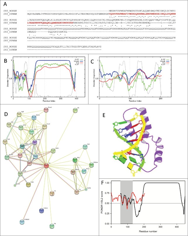
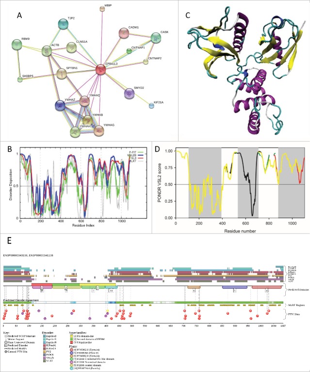
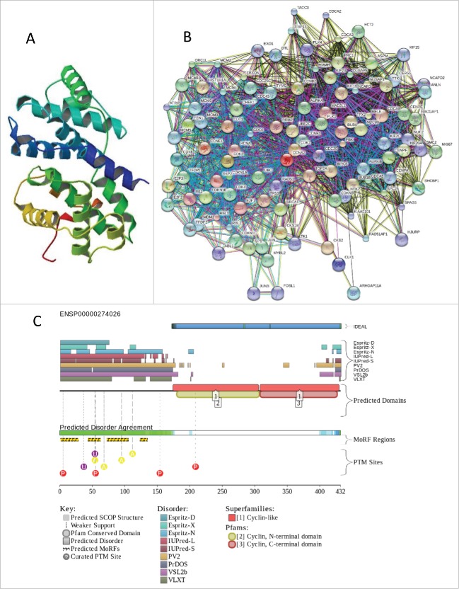
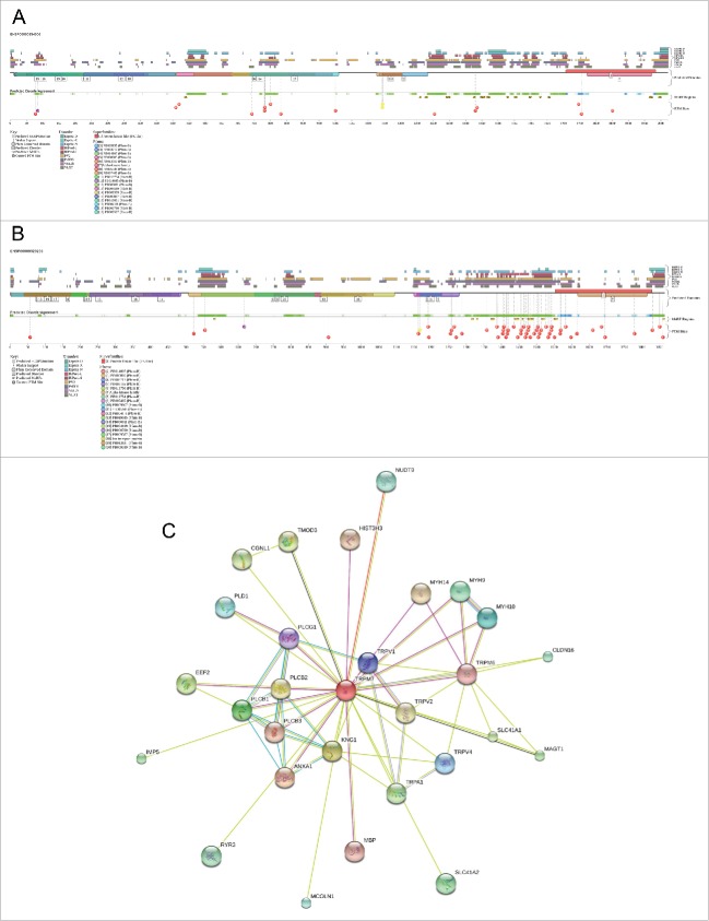
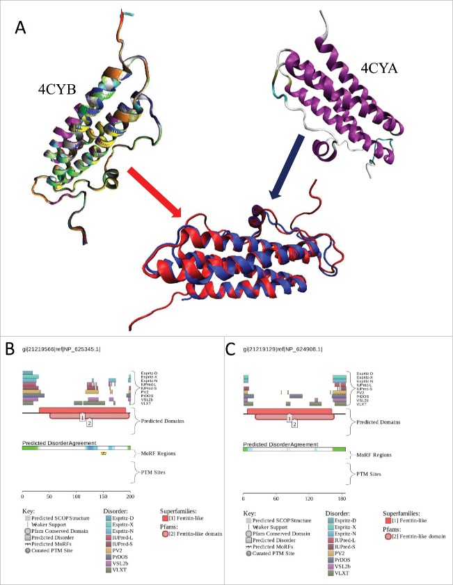
This article continues an "Unreported Intrinsic Disorder in Proteins" series, the goal of which is to expose some interesting cases of missed (or overlooked, or ignored) disorder in proteins. The need for this series is justified by the observation that despite the fact that protein intrinsic disorder is widely accepted by the scientific community, there are still numerous instances when appreciation of this phenomenon is absent. This results in the avalanche of research papers which are talking about intrinsically disordered proteins (or hybrid proteins with ordered and disordered regions) not recognizing that they are talking about such proteins. Articles in the "Unreported Intrinsic Disorder in Proteins" series provide a fast fix for some of the recent noticeable disorder overlooks.

Keywords: disorder prediction; intrinsically disordered protein; intrinsically disordered protein region; molecular recognition; posttranslational modifications; protein-protein interactions.


Figures

Figure 1 (See previous page).
(A). Sequence alignment of mouse (UniProt ID: Q05738) and human SRY proteins (UniProt ID: Q05066) by CLUSTAL W.106 Bold red characters show the HMG-box domain of human protein. (B). Per-residue disorder distribution in the mouse SRY protein. Disorder propensity was evaluated by a set of predictors from the PONDR family, PONDR® FIT, VSL2B, VL3, and VLXT. (C). Per-residue disorder distribution in the human SRY protein. Disorder propensity was evaluated by a set of predictors from the PONDR family, PONDR® FIT, VSL2B, VL3, and VLXT. Scores above 0.5 correspond to disordered residues/regions. (D). Evaluation of the interactivity of the mouse SRY protein (UniProt ID: Q05738) by STRING, which is the online database resource Search Tool for the Retrieval of Interacting Genes that provides both experimental and predicted interaction information. STRING produces the network of predicted associations for a particular group of proteins. The network nodes are proteins. The edges represent the predicted functional associations. An edge may be drawn with up to 7 differently colored lines - these lines represent the existence of the 7 types of evidence used in predicting the associations. A red line indicates the presence of fusion evidence; a green line - neighborhood evidence; a blue line – co-occurrence evidence; a purple line - experimental evidence; a yellow line – text mining evidence; a light blue line - database evidence; a black line – co-expression evidence. (E). Solution structure of the fragment of human SRY protein (residues 56–131) corresponding to the DNA binding HMG box domain in a complex with its DNA target site in the promoter of the Müllerian substance gene solved by the multidimensional NMR spectroscopy (PDB ID: 1HRZ). (F). Aligned PONDR® VSL2 disorder profiles of mouse (black line) and human (red line) SRY proteins. The localization of the HMG box domain of human protein is shown by gray shadow area.
Figure 2.
Figure 2.
(A). Analysis of the interactivity of the human 4.1B/DAL-1 protein (UniProt ID: Q9Y2J2) by STRING. (B). Per-residue disorder distribution in the 4.1B/DAL-1 protein. Disorder propensity was evaluated by a set of predictors from the PONDR family, PONDR® FIT, VSL2B, VL3, and VLXT. Scores above 0.5 correspond to disordered residues/regions. (C). Tertiary structure of the FERM domain (PDB ID: 2HE7; residues 108–390). (D). PONDR VSL2 intrinsic disorder profiles of the alternatively spliced isoforms of the human 4.1B/DAL-1 protein: black line – canonical form (UniProt ID: Q9Y2J2); red line – isoform 2 (UniProt ID: Q9Y2J2–2; differs from the canonical form as follows: 446–446: G → GASVNENHEIYMKDSMSAA; 503–689: missing; 708–719: missing; 784–824: missing); green line – isoform 3 (UniProt ID: Q9Y2J2–3; 446–446: G → GASVNENHEIYMKDSMSAA; 503–689: missing; 708–719: missing; 784–824: missing; 835–1087: missing); yellow line – isoform 4 (UniProt ID: Q9Y2J2–4; 446–446: G → GASVNENHEIYMKDSMSAA; 503–689: missing; 1052–1052: A → E; 1053–1087: missing). The locations of the FERM, SABD and CTD are shown by gray shadow areas. (E). Analysis of disorder propensity and disorder-based functionality of the human 4.1B/DAL-1 protein (UniProt ID: Q9Y2J2) by D2P2 database (http://d2p2.pro/).
Figure 3.
Figure 3.
(A). Crystal structure of the C-terminal half of human cyclin A2 (residues 175–432, PDB ID: 1VIN). (B). Analysis of the interactivity of the human cyclin A2 (UniProt ID: P20248) by STRING. (C). Evaluation of the functional intrinsic disorder propensity of the cyclin A2 (UniProt ID: P20248) by D2P2 database (http://d2p2.pro/).
Figure 4.
Figure 4.
(A). Evaluation of the functional intrinsic disorder propensity of the human TRPM6 (UniProt ID: Q9BX84) by D2P2 database (http://d2p2.pro/). (B). Evaluation of the functional intrinsic disorder propensity of TRPM7 (UniProt ID: Q96QT4) by D2P2 database (http://d2p2.pro/). (C). Analysis of the interactivity of the human TRPM6 (UniProt ID: Q9BX84) and TRPM7 (UniProt ID: Q96QT4) by STRING.
Figure 5.
Figure 5.
(A). Structural analysis of DpsC (PDB ID: 4CYB, residues 28–200) and DpsA (PDB ID: 4CYA, residues 4–163) Dps proteins from Streptomyces coelicolor. Since structural information on 12 chains constituting the DpsC dodecamer is available, the shown plot represents the results of structural alignment of these monomers using the MultiProt algorithm, where structures of different monomers are shown by different color. The MultiProt algorithm was also used to structurally align DpsC (shown as a red structure in the aligned plot) and DpsA (shown as a blue structure). (B). Evaluation of the functional intrinsic disorder propensity of DspC (UniProt ID: Q9K3L0) by D2P2 database (http://d2p2.pro/). (C). Evaluation of the functional intrinsic disorder propensity of DspA (UniProt ID: Q9R408) by D2P2 database (http://d2p2.pro/).

Similar articles

Cited by

References

    1. Uversky VN. Digested disorder: quarterly intrinsic disorder digest (january/february/ march, 2013). Intrinsically Disordered Proteins 2013; 1:e25496; http://dx.doi.org/10.4161/idp.25496 - DOI - PMC - PubMed
    1. DeForte S, Reddy KD, Uversky VN. Digested disorder, issue #2: quarterly intrinsic disorder digest (april/may/june, 2013). Intrinsically Disordered Proteins 2013; 1:e27454; http://dx.doi.org/10.4161/idp.27454 - DOI - PMC - PubMed
    1. Reddy KD, DeForte S, Uversky VN. Digested disorder, issue #3: quarterly intrinsic disorder digest (july-august-september, 2013). Intrinsically Disordered Proteins 2014; 2:e27833; http://dx.doi.org/10.4161/idp.27833 - DOI - PMC - PubMed
    1. Uversky VN. Unreported intrinsic disorder in proteins: building connections to the literature on IDPs. Intrinsically Disordered Proteins 2014; 2:e36409 - PMC - PubMed
    1. Obradovic Z, Peng K, Vucetic S, Radivojac P, Dunker AK. Exploiting heterogeneous sequence properties improves prediction of protein disorder. Proteins 2005; 61 7:176-82; PMID:16187360; http://dx.doi.org/10.1002/prot.20735 - DOI - PubMed

LinkOut - more resources