. 2017 Feb 24:7:43240.
doi: 10.1038/srep43240.

Caulobacter crescentus CdnL is a non-essential RNA polymerase-binding protein whose depletion impairs normal growth and rRNA transcription

Affiliations

Caulobacter crescentus CdnL is a non-essential RNA polymerase-binding protein whose depletion impairs normal growth and rRNA transcription

Aránzazu Gallego-García et al. Sci Rep. .

Abstract

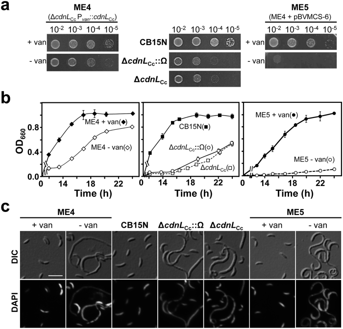
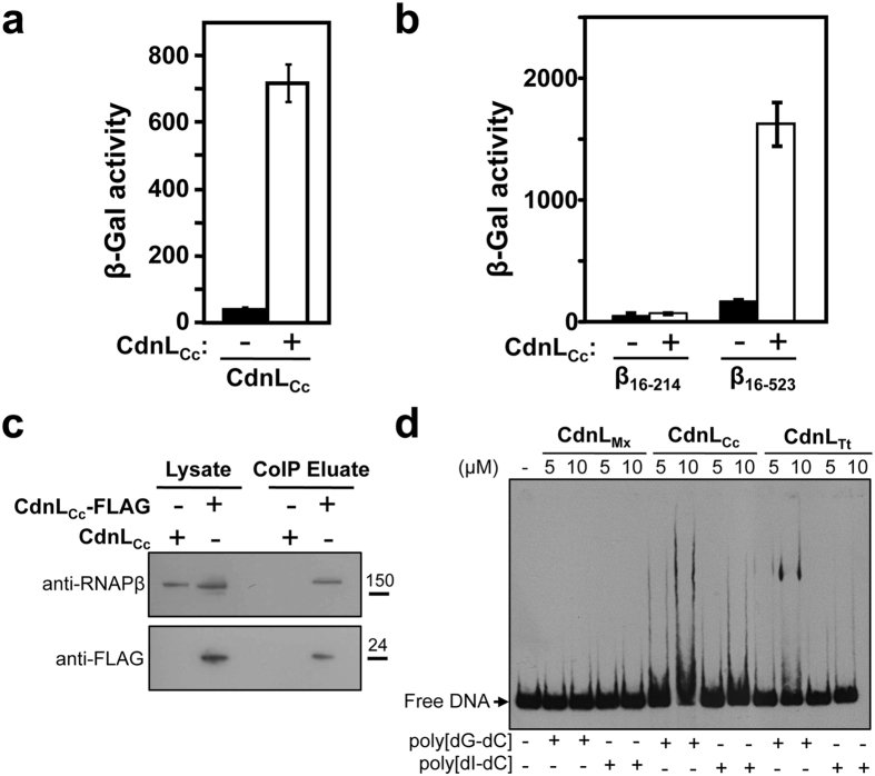
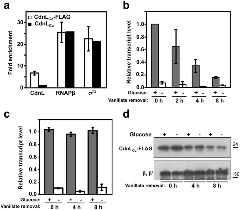
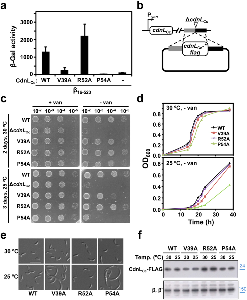
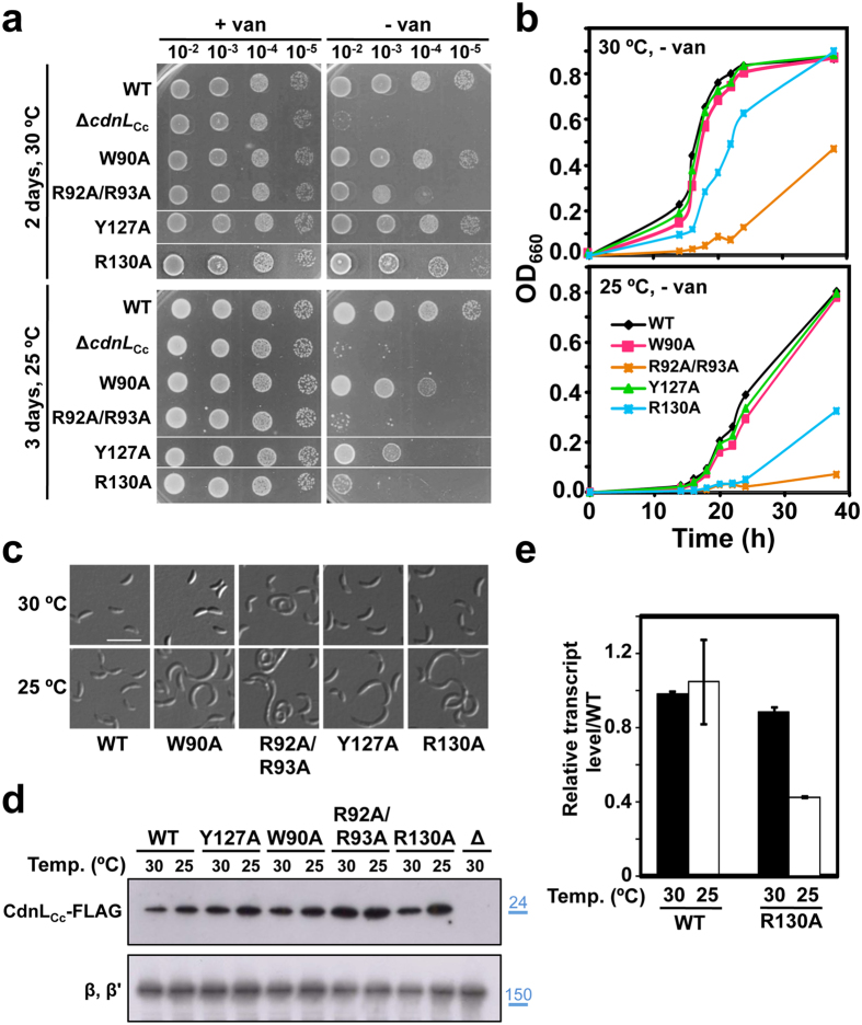
CdnL is an essential RNA polymerase (RNAP)-binding activator of rRNA transcription in mycobacteria and myxobacteria but reportedly not in Bacillus. Whether its function and mode of action are conserved in other bacteria thus remains unclear. Because virtually all alphaproteobacteria have a CdnL homolog and none of these have been characterized, we studied the homolog (CdnLCc) of the model alphaproteobacterium Caulobacter crescentus. We show that CdnLCc is not essential for viability but that its absence or depletion causes slow growth and cell filamentation. CdnLCc is degraded in vivo in a manner dependent on its C-terminus, yet excess CdnLCc resulting from its stabilization did not adversely affect growth. We find that CdnLCc interacts with itself and with the RNAP β subunit, and localizes to at least one rRNA promoter in vivo, whose activity diminishes upon depletion of CdnLCc. Interestingly, cells expressing CdnLCc mutants unable to interact with the RNAP were cold-sensitive, suggesting that CdnLCc interaction with RNAP is especially required at lower than standard growth temperatures in C. crescentus. Our study indicates that despite limited sequence similarities and regulatory differences compared to its myco/myxobacterial homologs, CdnLCc may share similar biological functions, since it affects rRNA synthesis, probably by stabilizing open promoter-RNAP complexes.


Conflict of interest statement

The authors declare no competing financial interests.

Figures

Figure 1
Figure 1. Growth and cell morphology upon CdnLCc depletion.
(a) Growth of the indicated C. crescentus strains. Liquid cultures (OD660 ∼ 0.5) were serially diluted, spotted (8 μl) on PYE plates with (+van) or without (−van) 0.5 mM vanillate, and incubated for 2 days at 30 °C. (b) Growth curves for the strains indicated (with symbols in parentheses). Freshly plated cells were innoculated into 10 ml of PYE (with 0.5 mM vanillate for ME4 and ME5) and grown at 30 °C to OD660 ∼ 0.8. 50 μl were aliquoted (for ME4 and ME5, after washing three times with PYE to remove the vanillate) into 10 ml of fresh PYE (for ME4 and ME5, one with 0.5 mM vanillate and one without). Growth was monitored at the indicated times, and the average and error of three independent measurements is shown. (c) Cellular morphology of cells from (b). Samples (concentrated ten-fold in the case of ME5 cultivated in the absence of vanillate) of each culture in (b) after ∼20 h of growth were DAPI-stained and examined by DIC (differential interference contrast; top panels) and fluorescence microscopy (bottom panels), as described in Methods. Scale bar: 5 μm.
Figure 2
Figure 2. Analysis of CdnLCc protein stability in vivo.
(a) Stability of FLAG-CdnLCc, CdnLCc-FLAG and FLAG-CdnLCc(DD) in vivo. C. crescentus strains ME27, ME28 and ME29 were grown in PYE with vanillate, and subjected to a previously described protocol (see Methods) prior to immunoblot analysis (−xyl: no xylose; +xyl: 0.3% xylose to induce clpX* expression). Total protein from 1 ml aliquots withdrawn every 20 min was detected in immunoblots using anti-FLAG antibodies (left). On the right is a semi-log plot of the relative band intensities (mean of three independent experiments) versus time for FLAG-CdnLCc (−xyl, circles; +xyl, diamonds), CdnLCc-FLAG (−xyl, triangles) and FLAG-CdnLCc(DD) (−xyl, squares). Slopes of the linear fits shown yield the decay rate constants used to estimate half-lives. (b) FLAG-CdnLCc levels during the cell cycle. Swarmer cells (SW) from strain ME24 grown in M2G with vanillate were isolated and used for synchronized cell cycle progression (∼150 min doubling time). Total protein from 1 ml aliquots taken every 20 min was subjected to immunoblot analysis using anti-FLAG antibodies. The control CtrA was probed on a separate blot (since its gel mobility is close to that of FLAG-CdnLCc) using anti-CtrA antibodies and equivalent samples from the same experiment processed in parallel. Samples at 150 min correspond to SW and ST (with a small proportion of PD) cells isolated from the culture remaining at the end of this assay. A plot of band intensities (% of the maximum value) versus time is shown (right). In (a,b), positions of molecular size markers (kDa) are shown in blue to the right of cropped immunoblots. Note that FLAG-tagged CdnLCc (∼19.5 kDa calculated Mw) migrates slower than expected. (c) Growth of C. crescentus strains (ME41, ME39, ME8) expressing CdnLCc, CdnLCc(DD) or CdnLCc-FLAG, respectively, at the endogenous site. Liquid cultures (OD660 ∼ 0.5) were serially diluted, spotted (8 μl) on PYE plates with or without vanillate, and incubated for 2 days at 30 °C. (d) Growth curves of strains in (c) expressing CdnLCc (squares, red), CdnLCc-FLAG (triangles, blue) or CdnLCc(DD) (circles, green) in PYE without vanillate at 30 °C.
Figure 3
Figure 3. CdnLCc interacts with itself, with RNAP, and with DNA.
(a) BACTH analysis of CdnLCc self-interaction in E. coli BTH101 transformed with plasmids pUT18-cdnLCc and pKT25-cdnLCc. (b) BACTH analysis of the interaction between CdnLCc (in pKT25) and C. crescentus RNAP β subunit fragments β16-214 and β16-523 (in pUT18C). In (a,b), the negative control (-) was pKT25 without insert. (c) Western blot of immunoprecipitated CdnLCc-FLAG probed for the presence of coprecipitating RNAPβ. Cells expressing CdnLCc-FLAG (strain ME17: ΔcdnLCc, Pvan::cdnLCc-flag, vanR) or, as the negative control, untagged CdnLCc (strain ME5: ΔcdnLCc, Pvan::cdnLCc, vanR) were immunoprecipitated with anti-FLAG agarose and processed in parallel, as described in Methods. Equal amounts of sample were then resolved by SDS-PAGE for immunoblot analysis. Monoclonal anti-RNAP β antibodies were used to detect RNAPβ (top) that coimmunoprecipitated with CdnLCc-FLAG, which was detected using anti-FLAG antibodies (bottom). Molecular size markers are shown to the right of the cropped immunoblots by the lines and corresponding values in kDa. (d) EMSA for the DNA binding of CdnLCc, CdnLTt, and CdnLMx. Reactions were performed as described in Methods with a 350-bp PrrnA DNA probe, and with 1 μg of poly[dG-dC] or poly[dI-dC] added as nonspecific competitor.
Figure 4
Figure 4. CdnLCc localizes at an rRNA promoter and affects its transcription in vivo.
(a) CdnLCc binds to PrrnA in vivo. ChIP-qPCR analysis using an anti-FLAG antibody on cells expressing CdnLCc-FLAG (strain ME17: ΔcdnLCc, Pvan::cdnLCc-flag, vanR; unfilled bars) or CdnLCc as negative control (strain ME5: ΔcdnLCc, Pvan::cdnLCc, vanR; black bars) showing CdnLCc-FLAG enrichment at PrrnA in vivo, relative to an intergenic region. As positive controls, ChIP-qPCR analysis was carried out using anti-RNAP β or anti-σA monoclonal antibodies for enrichment of RNAP or σ73, respectively, at PrrnA. (b) Effects of CdnLCc depletion on PrrnA promoter activity in vivo. CdnLCc was expressed from the Pvan promoter in strain ME42. Cells grown in M2G with vanillate to OD660~0.4 were washed and then grown in vanillate-free medium to block cdnLCc expression, and activity was measured by qRT-PCR at the times indicated. At each time point following vanillate withdrawal, one-half of the sample was washed and resuspended in medium without glucose and the other half remained untreated. Following 15 min incubation at 30 °C, the samples were collected for qRT-PCR analysis. (c) qRT-PCR analysis carried out with strain ME40, which expresses CdnLCc-FLAG from the Pvan promoter, using a procedure identical to that in (b). Data shown in (ac) correspond to the mean and standard error from three biological replicates. (d) Immunoblot analysis of CdnLCc-FLAG corresponding to samples in (c) with and without glucose deprivation and at the times indicated following vanillate withdrawal (top). As loading control, the same blot was probed using polyclonal anti-RNAP antibodies; the band corresponding to the RNAP β, β′ subunits is shown (bottom). Molecular size markers are shown to the right of the cropped immunoblots by lines and corresponding values in kDa.
Figure 5
Figure 5. Mutational analysis of CdnLCc-RNAP interaction in vivo.
(a) BACTH analysis of the interaction of CdnLCc mutants V39A, R52A and P54A (in pKT25) with C. crescentus RNAPβ fragment β16-523 (in pUT18C). The negative control (–) bears empty pKT25 and pUT18C-β16-523. (b) Schematic for the strategy employed to check for cdnLCc complementation in C. crescentus. A pMR3552 derivative with the required cdnLCc-flag allele (* indicates mutant) flanked by DNA segments upstream (grey) and downstream (black) of cdnLCc in the genome was introduced into strain ME5, which bears the ΔcdnLCc allele at the endogenous site and Pvan-cdnLCc at a heterologous site. Merodiploids resulting from plasmid integration by recombination express both CdnLCc*-FLAG and CdnLCc in the presence of vanillate (+van) and only the former in the absence of vanillate (−van). (c) Complementation analysis in C. crescentus of cells bearing the ΔcdnLCc allele or ones expressing at the endogenous site C-terminal FLAG-tagged wild-type CdnLCc (WT) or the indicated N-terminal CdnLCc variants. PYE plates with (+van) or without (−van) vanillate were spotted with 8 μl of liquid cultures (OD660 ∼ 0.5) at the dilutions indicated and incubated at 30 °C for two days or at 25 °C for three days. (d) Growth curves at 30 °C or 25 °C of C. crescentus expressing C-terminal FLAG-tagged CdnLCc (WT) or its indicated variants cultivated in liquid PYE without vanillate using the procedures described in Fig. 1b. (e) Cellular morphology examined by DIC microscopy of the wild-type (WT; scale bar: 5 μm) and the indicated mutant cells from (d) grown at 30 °C or 25 °C. (f) Immunoblot analysis to probe the stability of N-terminal CdnLCc variants. Cell extracts of strains expressing C-terminal FLAG-tagged CdnLCc (WT) or its indicated variants grown at 30 °C or 25 °C in PYE with vanillate were probed using anti-FLAG antibodies (top). As loading control, the same blot was probed using polyclonal anti-RNAP antibodies; the band corresponding to the RNAP β, β′ subunits is shown (bottom). Molecular size markers are shown to the right of the cropped immunoblots by lines and corresponding values in kDa (in blue).
Figure 6
Figure 6. Analysis of C-terminal CdnLCc mutations in vivo.
(a) Complementation analysis in C. crescentus of cells bearing the ΔcdnLCc allele or ones expressing at the endogenous site C-terminal FLAG-tagged wild-type CdnLCc (WT) or the indicated C-terminal CdnLCc variants. The analysis was carried out using the same procedures and conditions described in Fig. 5c. (b) Growth curves at 30 °C or 25 °C of C. crescentus strains expressing the C-terminal FLAG-tagged CdnLCc (WT) or its indicated variants cultivated in liquid PYE without vanillate using the procedures described in Fig. 1b. (c) Cellular morphology examined by DIC microscopy of the wild-type (WT; scale bar: 5 μm) and the indicated mutant cells from (b) grown at 30 °C or 25 °C. (d) Immunoblot analysis to probe the stability of C-terminal CdnLCc variants. Cell extracts of strains expressing C-terminal FLAG-tagged wild-type CdnLCc (WT) or the indicated CdnLCc mutants grown at 30 °C or 25 °C in PYE with vanillate were probed using anti-FLAG M2 antibodies (top). The negative control “Δ” corresponds to the strain (ME5) with the ΔcdnLCc allele at the endogenous site and expressing untagged CdnLCc under Pvan. As loading control, the same blot was probed using polyclonal anti-RNAP antibodies; the band corresponding to the RNAP β, β′ subunits is shown (bottom). Molecular size markers are shown to the right of the cropped immunoblots by lines and corresponding values in kDa (in blue). (e) PrrnA promoter activity in vivo at 30 °C or at 25 °C in cells expressing CdnLCc-FLAG (WT) or its variant with the R130A mutation under Pvan control (strains ME40 and ME38, respectively). Cells grown overnight at 30 °C in M2G with vanillate were diluted into the same medium to OD660 ∼ 0.1. One-half was grown at 30 °C and the other at 25 °C to OD660 of 0.3–0.4, and RNA was quantitated using qRT-PCR. Data shown correspond to the mean and standard error from three biological replicates.

Similar articles

Cited by

References

    1. Lee D. J., Minchin S. D. & Busby S. J. Activating transcription in bacteria. Annu. Rev. Microbiol. 66, 125–152 (2012). - PubMed
    1. Saecker R. M., Record M. T. Jr. & deHaseth P. L. Mechanism of bacterial transcription initiation: RNA polymerase - promoter binding, isomerization to initiation-competent open complexes, and initiation of RNA synthesis. J. Mol. Biol. 412, 754–771 (2011). - PMC - PubMed
    1. Ruff E. F., Record M. T. & Artsimovitch I. Initial events in bacterial transcription initiation. Biomolecules 5, 1035–1062 (2015). - PMC - PubMed
    1. Abellón-Ruiz J. et al. The CarD/CarG regulatory complex is required for the action of several members of the large set of Myxococcus xanthus extracytoplasmic function sigma factors. Environ. Microbiol. 16, 2475–2490 (2014). - PubMed
    1. Cayuela M. L., Elías-Arnanz M., Peñalver-Mellado M., Padmanabhan S. & Murillo F. J. The Stigmatella aurantiaca homolog of Myxococcus xanthus high-mobility-group A-type transcription factor CarD: insights into the functional modules of CarD and their distribution in bacteria. J. Bacteriol. 185, 3527–3537 (2003). - PMC - PubMed

Publication types

MeSH terms

LinkOut - more resources