. 2017 Feb 24:7:43198.
doi: 10.1038/srep43198.

Increased 4R tau expression and behavioural changes in a novel MAPT-N296H genomic mouse model of tauopathy

Affiliations

Increased 4R tau expression and behavioural changes in a novel MAPT-N296H genomic mouse model of tauopathy

Heike J Wobst et al. Sci Rep. .

Abstract

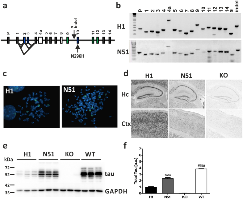
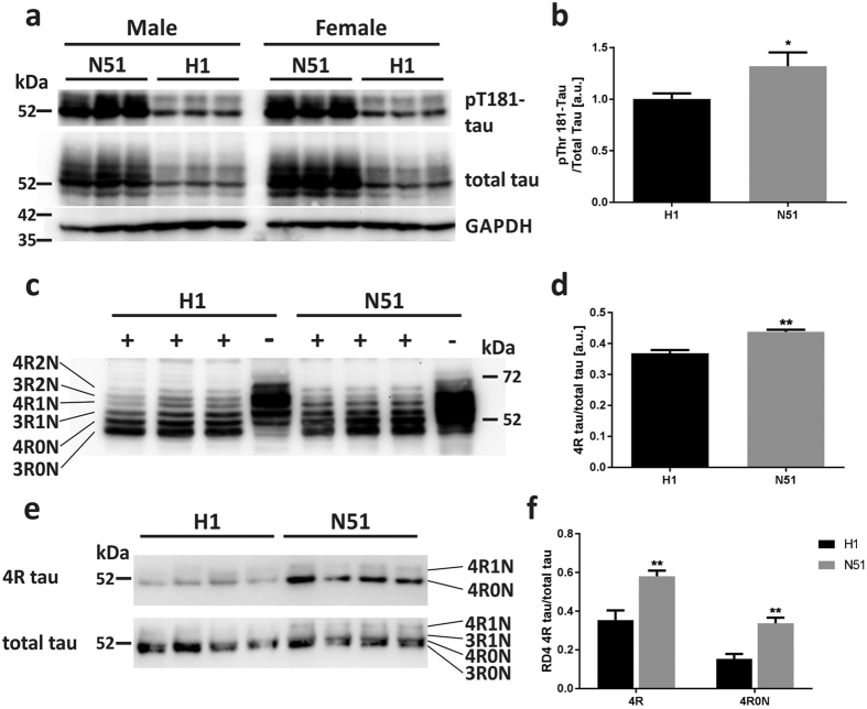
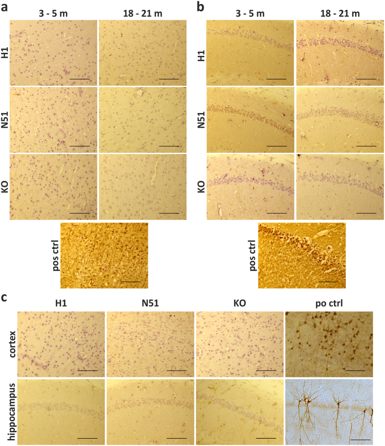
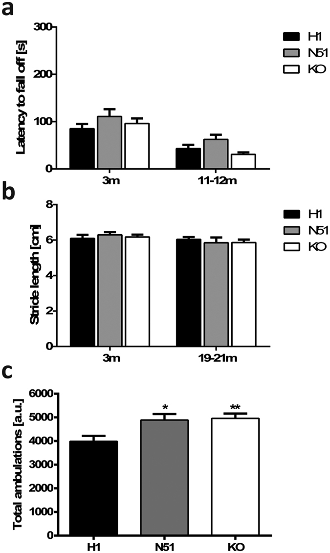
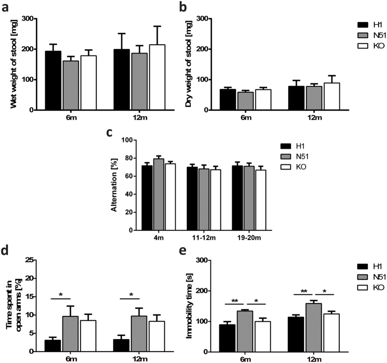
The microtubule-associated protein tau is implicated in various neurodegenerative diseases including Alzheimer's disease, progressive supranuclear palsy and corticobasal degeneration, which are characterized by intracellular accumulation of hyperphosphorylated tau. Mutations in the tau gene MAPT cause frontotemporal dementia with parkinsonism linked to chromosome 17 (FTDP-17). In the human central nervous system, six tau isoforms are expressed, and imbalances in tau isoform ratios are associated with pathology. To date, few animal models of tauopathy allow for the potential influence of these protein isoforms, relying instead on cDNA-based transgene expression. Using the P1-derived artificial chromosome (PAC) technology, we created mouse lines expressing all six tau isoforms from the human MAPT locus, harbouring either the wild-type sequence or the disease-associated N296H mutation on an endogenous Mapt-/- background. Animals expressing N296H mutant tau recapitulated early key features of tauopathic disease, including a tau isoform imbalance and tau hyperphosphorylation in the absence of somatodendritic tau inclusions. Furthermore, N296H animals displayed behavioural anomalies such as hyperactivity, increased time in the open arms of the elevated plus maze and increased immobility during the tail suspension test. The mouse models described provide an excellent model to study the function of wild-type or mutant tau in a highly physiological setting.


Conflict of interest statement

The authors declare no competing financial interests.

Figures

Figure 1
Figure 1. Generation of PAC-based tau transgenic mouse line expressing wild-type or N296H mutant human MAPT.
(a) Exon structure of the human MAPT genomic locus. p - promoter, s - Saitohin (STH) gene, indel - 238 bp insertion relative to H2 haplotype, black exons - constitutive expression, blue exons - alternatively spliced, white exons - not present in human CNS tau protein, green exons and exon 10 - microtubule binding domain. (b) Exon PCR for assessment of MAPT transgene integrity. All 16 exons were amplified as well as the Saitohin gene (s) nested between exons 9 and 10 and the 238 bp insertion (indel) that characterizes the H1 haplotype. (c) Localization of H1 wild-type and N51 mutant transgenes with chromosome paints by fluorescence in situ hybridization. A single integration transgene site (red) was confirmed for both H1 (chromosome 6, green) and N51 (chromosome 9, green) line. (d) MAPT transgene mRNA expression assessed by RNA in situ hybridization of H1 and N51 transgenic line compared to Mapt KO control. Hc: hippocampus; Ctx: cortex. (e) Tau transgene protein expression in whole brain extract of 3–5 month old H1 and N51 animals compared to Mapt KO nontransgenic littermates and C57BL6 animals with endogenous tau expression. (f) Quantification of expression levels normalized to GAPDH loading control. One-way ANOVA followed by Bonferroni post-hoc correction. Results represent mean signal ± SEM. ****p < 0.0001 (compared to H1), ####p < 0.0001 (compared to H1 and N51), N = 3 samples per group.
Figure 2
Figure 2. N296H-MAPT transgenic mice display tau hyperphosphorylation and increased expression of 4R tau isoforms.
(a) Phosphorylation of tau at the disease-relevant T181 residue in old (18–22 months) tau transgenic whole hemispheres. Blots were probed with the phospho-T181 antibody AT270, stripped and reprobed with the pan-tau antibody A0024. (b) Signal quantification revealed a significant upregulation of T181 phosphorylation (N = 6 animals per genotype, one-tailed Student’s t-test). (c) Dephosphorylation of whole brain lysates revealed the presence of all six human tau isoforms in H1 and N51 animals (+dephosphorylated sample, −untreated sample) using a total tau antibody. 20 ug of H1 samples and 10 ug of N51 samples were loaded for better visualization. (d) Quantification of separated tau isoforms revealed a significant upregulation of 4R to total tau ratio in N51 compared to H1 animals (N = 3 animals per genotype, Student’s t test). (e) Detection of tau isoforms in dephosphorylated brain samples with the 4R-specific tau antibody RD4. (f) Quantification of 4R tau showed a significant increase of 4R tau isoforms in N51 mutant animals using the RD4 antibody (N = 4 animals per genotype, two-way ANOVA followed by Bonferroni post-hoc analysis). All results represent mean ± SEM. *p < 0.05, **p < 0.01.
Figure 3
Figure 3. N296H-MAPT and MAPT-H1 transgenic mice do not display somatodendritic accumulation of tau.
Coronal sections of 3–5 and 18–21 month old animals were stained with human-specific HT7 antibody to assess the formation of somatodendritic tau inclusions at a young (3–5 months) and old (18–21 months) age. No age-related changes in staining or evidence of tau inclusion formation were found in cortex (a) or hippocampus (b) of wild-type (H1) and N296H mutant (N51) animals in comparison to their non-transgenic tau knockout (KO) littermates. (c) Coronal sections of 18–21 month old animals were stained with the phospho-tau antibody AT8. Both H1 wild-type and N51 mutant tau expressing lines showed no accumulation of AT8 phospho-tau compared to the knockout negative control or the P301S mutant positive control. Positive control sections (pos ctrl, obtained from P301S transgenic animal) showing heavy HT7 and AT8 staining confirm the detection of somatodendritic tau inclusions. Scale bars = 100 μm.
Figure 4
Figure 4. N296H-MAPT transgenic mice display a hyperactivity phenotype.
(a) Accelerating rotarod performance at 3 m and 11–12 m. Animals were tested on a rotarod accelerating from 4–40 rpm over a 5 min period with three trials per day and the average latency to fall off was recorded. No significant differences between genotypes were observed at either tested time points in pairwise post-hoc comparisons despite a main effect of genotype. (b) Stride length (average distance between middle toe of a step and heel of the next step) at 3 m and 19–21 m of age. No differences in stride length between genotypes were observed in young (3 months) or old (19–21 months) mice. (c) Locomotor activity in an unfamiliar environment. Tau KO and N51 mutant tau expressing animals showed increased locomotor activity in 5 month old animals compared to H1 wild-type expressing animals. Results for all tests represent mean ± SEM for (a,b) N = 10–12, (c) N = 24–31, (d) N = 5–6 animals per genotype. *p < 0.05, **p < 0.01, ***p < 0.001. One-way (c) or two-way ANOVA (a,b,d) followed by Bonferroni post-hoc analysis.
Figure 5
Figure 5. N296H-MAPT transgenic mice display non-motor phenotypes.
(a,b) Assessment of an overt gastrointestinal phenotype revealed no differences between genotypes. (a) Wet weight, (b) dry weight of fecal boli collected over a one hour period from 6 and 12 month old animals. At both 6 months and 12 months of age, gastrointestinal function of transgenic animals was assessed by collecting fecal boli. (c) Spontaneous alternation in the T-maze, assessed at 4 months, 11–12 months and 19–20 months of age. No differences between genotypes were observed either in 4 month, 11–12 months or 19–20 months of age. (d) Elevated plus maze, assessed at 6 months and 12 months of age. At both age points, N51 mutant tau animals and tau knockout animals spent more time in the open arms of the elevated plus maze compared to H1 animals. (e) Tail suspension test, assessed at 6 months and 12 months of age. At both age points, N51 mutant animals displayed significantly higher immobility time compared to H1 and KO animals. Results for all tests represent mean ± SEM; for (a,b) N = 9–10, (c) N = 24–31 (4 m) and N = 11–13 (11–12 m, 19–20 m), (d,e) N = 10–12 animals per genotype. * p < 0.05, ** p < 0.01. Two-way ANOVA followed by Bonferroni post-hoc analysis.

References

    1. Crawford F. et al.. No genetic association between polymorphisms in the Tau gene and Alzheimer’s disease in clinic or population based samples. Neurosci. Lett. 266, 193–196 (1999). - PubMed
    1. Myers A. J. et al.. The H1c haplotype at the MAPT locus is associated with Alzheimer’s disease. Hum. Mol. Genet. 14, 2399–2404 (2005). - PubMed
    1. Baker M. et al.. No association between TAU haplotype and Alzheimer’s disease in population or clinic based series or in familial disease. Neurosci. Lett. 285, 147–149 (2000). - PubMed
    1. Russ C. et al.. The microtubule associated protein Tau gene and Alzheimer’s disease–an association study and meta-analysis. Neurosci. Lett. 314, 92–96 (2001). - PubMed
    1. Abraham R. et al.. An association study of common variation at the MAPT locus with late-onset Alzheimer’s disease. Am. J. Med. Genet. Part B Neuropsychiatr. Genet. Off. Publ. Int. Soc. Psychiatr. Genet. 150B, 1152–1155 (2009). - PubMed

Publication types

LinkOut - more resources