. 2017 Feb 24:8:14393.
doi: 10.1038/ncomms14393.

Sponge-associated bacteria mineralize arsenic and barium on intracellular vesicles

Affiliations

Sponge-associated bacteria mineralize arsenic and barium on intracellular vesicles

Ray Keren et al. Nat Commun. .

Abstract

Arsenic and barium are ubiquitous environmental toxins that accumulate in higher trophic-level organisms. Whereas metazoans have detoxifying organs to cope with toxic metals, sponges lack organs but harbour a symbiotic microbiome performing various functions. Here we examine the potential roles of microorganisms in arsenic and barium cycles in the sponge Theonella swinhoei, known to accumulate high levels of these metals. We show that a single sponge symbiotic bacterium, Entotheonella sp., constitutes the arsenic- and barium-accumulating entity within the host. These bacteria mineralize both arsenic and barium on intracellular vesicles. Our results indicate that Entotheonella sp. may act as a detoxifying organ for its host.


Conflict of interest statement

The authors declare no competing financial interests.

Figures

Figure 1
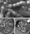
Figure 1. Entotheonella sp. is the arsenic and barium accumulating entity in the T. swinhoei holobiont.
(a,b) Compatible SEM micrographs of Entotheonella sp. in sponge tissue. (a) Secondary electron detection mode with arrows indicating some of the Entotheonella sp. filaments. Scale bar, 100 μm. (b) Backscattered electrons detection mode reveals Entotheonella sp. filaments as the most-electron-dense objects in the sponge. Scale bar, 50 μm. (c,d) Dry weight concentration (μg g−1) of barium and arsenic (respectively) in cell-enriched fractions (n=5 samples, each one from a different sponge). FBAC, unicellular bacterial cells; FEC, Entotheonella sp. with cyanobacteria; FENTO, Entotheonella sp. cells; FSC, sponge cells. Error bars show s.e.
Figure 2
Figure 2. Intracellular mineralization in Entotheonella sp.
(a) SEM micrograph (secondary electron mode) of Entotheonella sp. showing mineralized spherical structures inside their cells. Scale bar, 10 μm. (b,c) TEM micrographs of thin sections of Entotheonella sp. Black arrows in b mark nucleation of minerals from the vesicles' membrane. Black arrows in c mark the thickening of the mineral inwards from the membrane of the vesicle (white arrows). Scale bar, 500 nm.
Figure 3
Figure 3. Characterization of spheres in Entotheonella sp.
(a) Membrane staining with DiOC6 dye (excitation (ex), 482 nm; emission (em), 504 nm) shows that the vesicles (marked by arrows) of Entotheonella sp. are lipid based. Scale bar, 5 μm. (b) SEM micrograph (secondary electron) of cross-section of sphere in Entotheonella sp. The sphere wall is highly porous, with resemblance of channels (marked by arrows). Scale bar, 200 nm. (c) Average weight ratios (n=3) of detected elements determined by EDS. Carbon (C), nitrogen (N) and barium (Ba) are marked in bold.
Figure 4
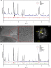
Figure 4. XRD and TEM analysis of spheres inside Entotheonella sp.
(a) Rietveld refinement plot of preheated freeze-dried Entotheonella sp. reveals the resulted fit for the observed (blue dots) and calculated (black line) diffraction patterns and the difference between them (red line). Black notches indicate the positions of the diffraction peaks of crystalline barite (BaSO4), the major phase in the sample. Diffraction peaks highlighted by arrows in the difference curve are compatible with an arsenate or phosphate phase. These minor phases are not clearly visible in the observed diffraction pattern as they overlap with the diffraction peaks of barite. (b) HRTEM micrograph of a portion of the freeze-dried Entotheonella sp. Insert shows higher magnification image revealing the lattice, and with its Fast Fourier Transform (FFT). (c) Rietveld refinement plot of a freeze-dried Entotheonella sp. after thermal annealing, revealing minor phases of sodium chloride (NaCl), calcium arsenate (Ca3(AsO4)2) and calcium sulfide phosphate (Ca10(PO4)6S). Observed (Obs.), calculated (Calc.) and difference curves are presented in the same schematics as in a.
Figure 5
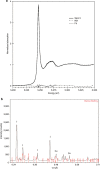
Figure 5. X-ray microprobe analysis of arsenic in Entotheonella sp. at 95 °K.
(a) X-ray fluorescence distribution map of As in filaments. Pixel size is 2 μm. Inset: As map of a single filament, pixel size is 0.8 μm, scale bar, 4 μm. (b) As K edge spectrum of Entotheonella sp. compared with As(V) and As(III) standards. Least-square linear combination fitting using 47% sodium arsenate, 39% pharmacolite and 14% As2O3 is shown as dotted line (residuals in dashed line, norm. sum-sq=7.6e−4). (c) X-ray fluorescence chemical map showing the distribution of As(III) and As(V) in filaments in a area. Yellow circle points to location of XANES analysis (b). Scale bar, 20 μm.
Figure 6
Figure 6. X-ray microprobe analysis of barium in Entotheonella sp. at 95 °K.
(a) Ba L3 edge XANES on a filament (spot3) along with LCF fit (dotted line) to 100% barite (residuals in dashed line, norm. sum-sq=3.54e−4). (b) Indexing of the XRD pattern recorded at 17 keV at that location confirms the presence of crystalline barite. I- peaks from Ice crystals; Cu- peaks from copper grid.
Figure 7
Figure 7. Membrane system of Entotheonella sp.
(a) Cryo-SEM micrograph of freeze-fractured Entotheonella sp. filament. Red arrows mark the filament sheath. Yellow arrows mark the outer membrane. White arrows mark the internal membrane harbouring the mineralized spheres. Scale bar, 1 μm. (b) TEM micrograph of thin sections of Entotheonella sp. showing vesicles in the space between the two membranes. Yellow arrow marks the outer membrane. White arrow marks the internal membrane. Black arrow marks a single membrane vesicle. Green arrow marks a double membrane vesicle. Scale bar, 500 nm.

References

    1. Oremland R. S. & Stolz J. F. The ecology of arsenic. Science 300, 939–944 (2003). - PubMed
    1. Dehairs F., Chesselet R. & Jedwab J. Discrete suspended particles of barite and the barium cycle in the open ocean. Earth Planet. Sci. Lett. 49, 528–550 (1980).
    1. Neff J. M. Ecotoxicology of arsenic in the marine environment. Environ. Toxicol. Chem. 16, 917–927 (1997).
    1. Menzie C. A., Southworth B., Stephenson G. & Feisthauer N. The importance of understanding the chemical form of a metal in the environment: the case of barium sulfate (barite). Hum. Ecol. Risk Assess. 14, 974–991 (2008).
    1. Slyemi D. & Bonnefoy V. How prokaryotes deal with arsenic. Environ. Microbiol. Rep. 4, 571–586 (2012). - PubMed

Publication types

MeSH terms

LinkOut - more resources