. 2017 Feb 24:7:43190.
doi: 10.1038/srep43190.

Epitopes of anti-RIFIN antibodies and characterization of rif-expressing Plasmodium falciparum parasites by RNA sequencing

Affiliations

Epitopes of anti-RIFIN antibodies and characterization of rif-expressing Plasmodium falciparum parasites by RNA sequencing

Jun-Hong Ch'ng et al. Sci Rep. .

Abstract

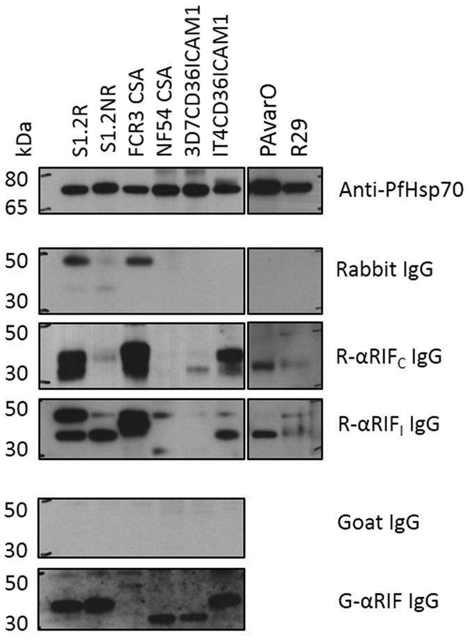
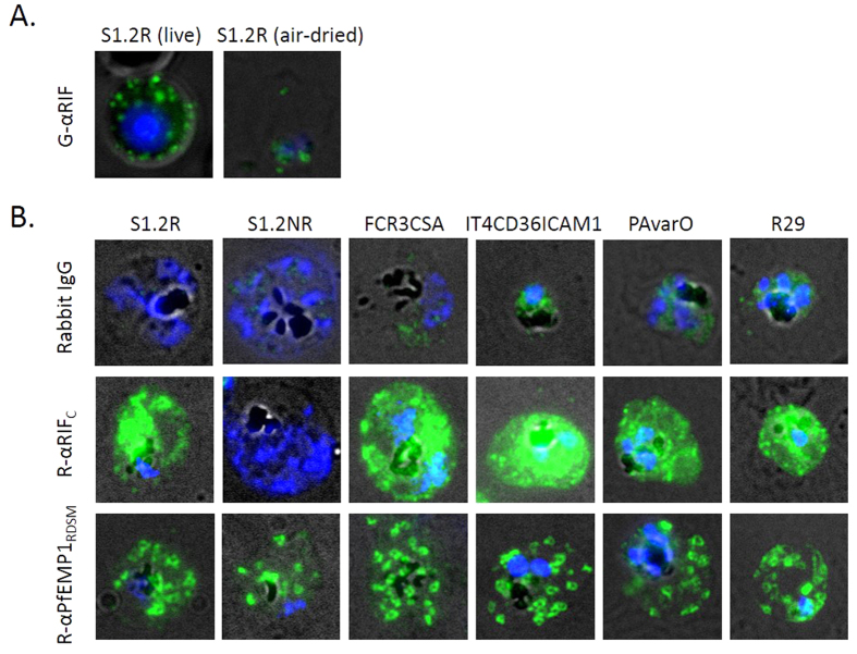
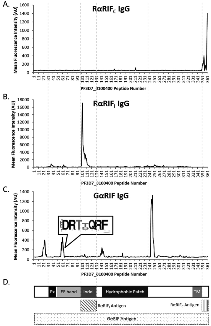
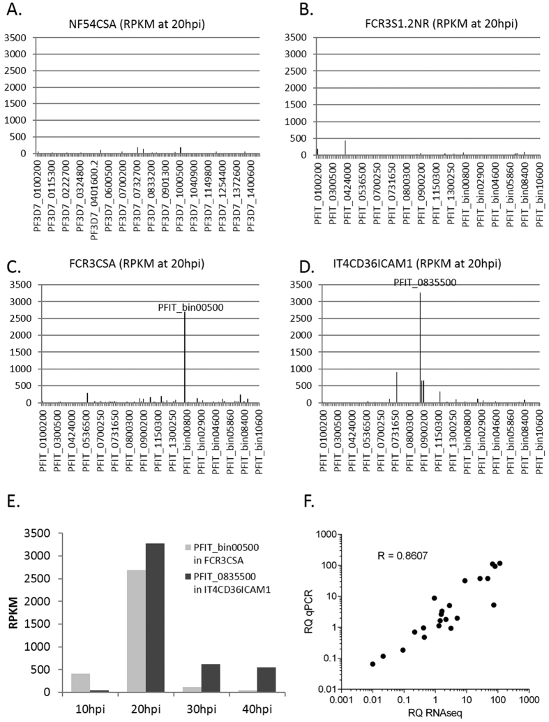
Variable surface antigens of Plasmodium falciparum have been a major research focus since they facilitate parasite sequestration and give rise to deadly malaria complications. Coupled with its potential use as a vaccine candidate, the recent suggestion that the repetitive interspersed families of polypeptides (RIFINs) mediate blood group A rosetting and influence blood group distribution has raised the research profile of these adhesins. Nevertheless, detailed investigations into the functions of this highly diverse multigene family remain hampered by the limited number of validated reagents. In this study, we assess the specificities of three promising polyclonal anti-RIFIN antibodies that were IgG-purified from sera of immunized animals. Their epitope regions were mapped using a 175,000-peptide microarray holding overlapping peptides of the P. falciparum variable surface antigens. Through immunoblotting and immunofluorescence imaging, we show that different antibodies give varying results in different applications/assays. Finally, we authenticate the antibody-based detection of RIFINs in two previously uncharacterized non-rosetting parasite lines by identifying the dominant rif transcripts using RNA sequencing.


Conflict of interest statement

MW is a co-founder and board member of Modus Therapeutics AB, a company developing drugs for severe malaria. All other authors declare no financial conflicts of interest.

Figures

Figure 1
Figure 1. Western blots of RIFINs in multiple parasite strains.
SDS-extracted parasite lysates of S1.2R, S1.2NR, FCR3CSA, NF54CSA, 3D7CD36ICAM1 and IT4CD36ICAM1 were run on SDS-PAGE (lanes 1-6 respectively), transferred to nitrocellulose membrane and blotted with rabbit anti-PfHsp70 (1:2000), non-immune rabbit IgG (10 μg/ml), RαRIFC (rabbit anti-A-RIFIN C-terminus) IgG (10 μg/ml), RαRIFI (rabbit anti-A-RIFIN indel) IgG (10 μg/ml), non-immune goat IgG (10 μg/ml) and GαRIF (goat anti-A-RIFIN full length) IgG (10 μg/ml). Corresponding HRP-conjugated secondary anti-rabbit and anti-goat IgG antibodies were used together with ECL reagent for detection. Description of the antigens used for immunizations of RαRIFC, RαRIFI and GαRIF are described in Supplementary Table S1 and diagrammatically illustrated in Fig. 3D.
Figure 2
Figure 2. Indirect immunofluorescence micrographs.
(A) GαRIF IgG labelling of RIFINs on a live S1.2R-infected erythrocyte and an air-dried iRBC. (B) Air-dried monolayers of erythrocytes infected with S1.2R, S1.2NR, FCR3CSA, IT4CD36ICAM1, PAvarO and R29 parasites stained with non-immune rabbit IgG (10 μg/ml), anti-RIFIN RαRIFC rabbit IgG (10 μg/ml) and Maurer’s cleft labelling by R-αPfEMP1RDSM IgG (8 μg/ml). Alexa488-conjugated secondary anti-rabbit or anti-goat antibodies were used (1:200) as well as Vectashield with DAPI (nuclear staining). Merged channels of transmission light (black and white), green (Alexa 488) and blue (DAPI) and representative cells are shown here.
Figure 3
Figure 3. Epitope region mapping of anti-RIFIN antibodies using an ultra-dense peptide array.
The reactivity for polyclonal IgG purified from (A) RαRIFC, (B) RαRIFI and (C) GαRIF are shown here against peptides from the RIFIN protein PF3D7_0100400. Y-axis shows mean median fluorescence intensity of duplicate spots and X-axis shows the peptide number from the N terminus to the C-terminus. (C) A conserved amino acid sequence that is recognized by GαRIF IgG is highlighted. (D) A diagram of RIFIN PF3D7_0100400 domains is provided together with the respective antigens used for immunizations: RαRIFC was immunized with the C-terminus, RαRIFI with the indel region and GαRIF with the full length of PF3D7_0100400. The abbreviation “Px” stands for PEXEL motif while “TM” refers to the transmembrane domain. Detailed intensity plots of the key epitope regions are highlighted in Supplementary Figure S2.
Figure 4
Figure 4. RNA sequencing counts of rif genes.
RNA from (A) NF54CSA, (B) S1.2NR, (C) FCR3CSA and (D) IT4CD36ICAM1 were extracted at four time points and sent for RNA sequencing. Results for the rif genes are presented here in RPKM at 20 hpi. (E) RPKM values at 10, 20, 30 and 40 hpi are provided to show the temporal expression of the two dominant rif genes (PFIT_bin00500 in FCR3CSA and PFIT_0835500 in IT4CD36ICAM1). (F) Quantitative real time PCR was performed to validate the RNA sequencing results and Pearson’s R shows level of correlation between the relative quantity (RQ) of qPCR fold expression and RNAseq RPKM values.

References

    1. Goel S. et al.. RIFINs are adhesins implicated in severe Plasmodium falciparum malaria. Nat Med 21, 314–317, doi: 10.1038/nm.3812 (2015). - DOI - PubMed
    1. Gardner M. J. et al.. Genome sequence of the human malaria parasite Plasmodium falciparum. Nature 419, 498–511, doi: 10.1038/nature01097 (2002). - DOI - PMC - PubMed
    1. Joannin N., Abhiman S., Sonnhammer E. L. & Wahlgren M. Sub-grouping and sub-functionalization of the RIFIN multi-copy protein family. BMC Genomics 9, 19, doi: 10.1186/1471-2164-9-19 (2008). - DOI - PMC - PubMed
    1. Cheng Q. et al.. stevor and rif are Plasmodium falciparum multicopy gene families which potentially encode variant antigens. Mol Biochem Parasitol 97, 161–176 (1998). - PubMed
    1. Helmby H., Cavelier L., Pettersson U. & Wahlgren M. Rosetting Plasmodium falciparum-infected erythrocytes express unique strain-specific antigens on their surface. Infect Immun 61, 284–288 (1993). - PMC - PubMed

Publication types