

Relevant human tissue resources and laboratory models for use in endometriosis research
- PMID: 28233896
- PMCID: PMC5485163
- DOI: 10.1111/aogs.13119
Relevant human tissue resources and laboratory models for use in endometriosis research
Abstract
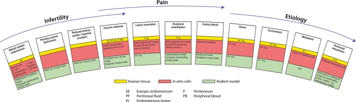
Endometriosis is characterized by the growth of endometrium-like tissue outside the uterus, most commonly on the pelvic peritoneum and ovaries. Although it may be asymptomatic in some women, in others it can cause debilitating pain, infertility or other symptoms including fatigue. Current research is directed both at understanding the complex etiology and pathophysiology of the disorder and at the development of new nonsurgical approaches to therapy that lack the unwanted side effects of current medical management. Tools for endometriosis research fall into two broad categories; patient-derived tissues, and fluids (and cells isolated from these sources) or models based on the use of cells or animals. In this review, we discuss the literature that has reported data from the use of these tools in endometriosis research and we highlight the strengths and weaknesses of each. Although many different models are reported in the literature, hypothesis-driven research will only be facilitated with careful experimental design and selection of the most appropriate human tissue from patients with and without endometriosis and combinations of physiologically relevant in vitro and in vivo laboratory models.
Keywords: Endometriosis; ex vivo; in vitro; in vivo; laboratory models.
© 2017 The Authors. Acta Obstetricia et Gynecologica Scandinavica published by John Wiley & Sons Ltd on behalf of Nordic Federation of Societies of Obstetrics and Gynecology (NFOG).
Figures
Comment in
-
When endometriosis is the barrier for fertility.Acta Obstet Gynecol Scand. 2017 Jun;96(6):621-622. doi: 10.1111/aogs.13155. Acta Obstet Gynecol Scand. 2017. PMID: 28556123 No abstract available.
References
-
- Giudice LC, Kao LC. Endometriosis. Lancet. 2004;364:1789–99. - PubMed
-
- Fassbender A, Rahmioglu N, Vitonis AF, Vigano P, Giudice LC, D'Hooghe TM, et al. World Endometriosis Research Foundation Endometriosis Phenome and Biobanking Harmonisation Project: IV. Tissue collection, processing, and storage in endometriosis research. Fertil Steril. 2014;102:1244–53. - PMC - PubMed
-
- Rahmioglu N, Fassbender A, Vitonis AF, Tworoger SS, Hummelshoj L, D'Hooghe TM, et al. World Endometriosis Research Foundation Endometriosis Phenome and Biobanking Harmonization Project: III. Fluid biospecimen collection, processing, and storage in endometriosis research. Fertil Steril. 2014;102:1233–43. - PMC - PubMed
Publication types
MeSH terms
Substances
Grants and funding
LinkOut - more resources
Full Text Sources
Other Literature Sources
Medical
