. 2017 Jun;65(6):864-882.
doi: 10.1002/glia.23131. Epub 2017 Feb 24.

Axon contact-driven Schwann cell dedifferentiation

Affiliations

Axon contact-driven Schwann cell dedifferentiation

Jennifer Soto et al. Glia. 2017 Jun.

Abstract

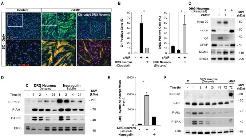
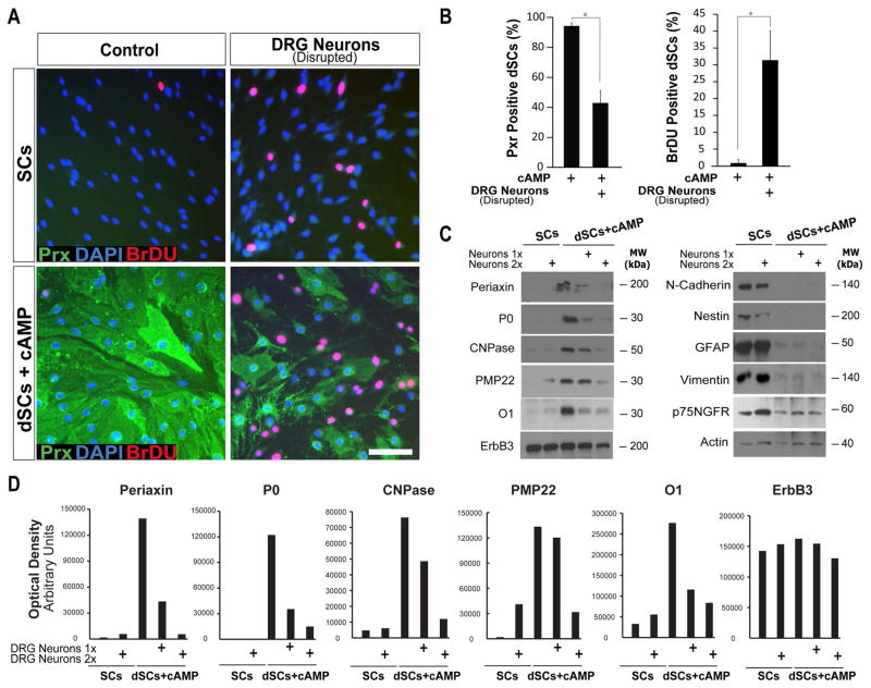
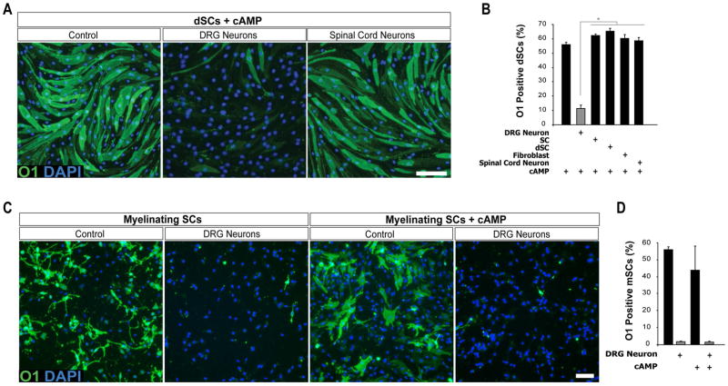
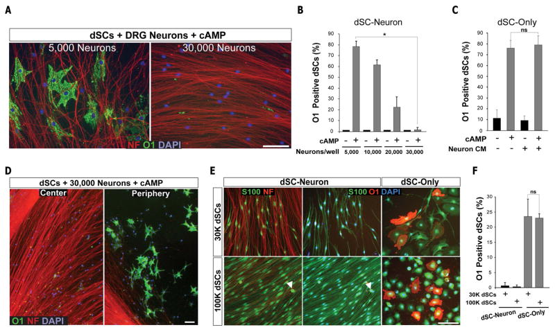
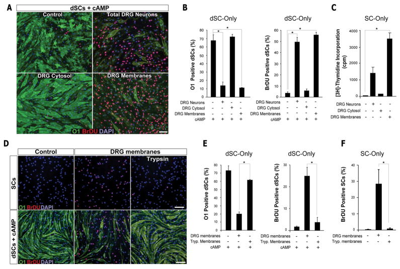
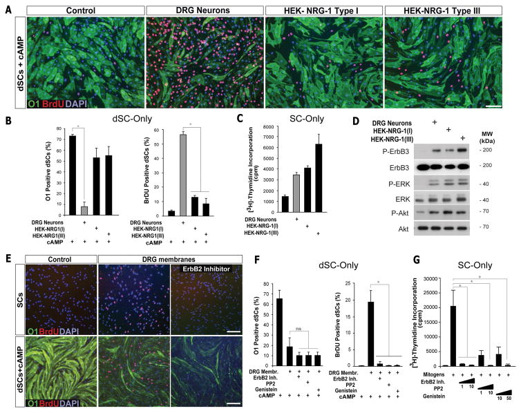
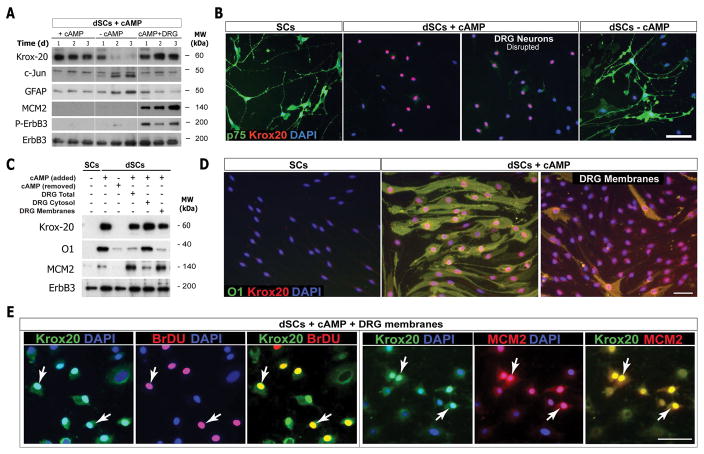
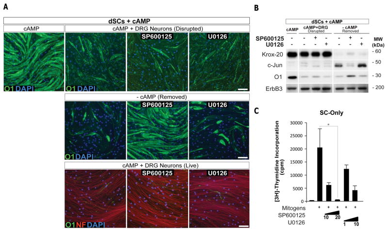
Mature Schwann cells (SCs) retain dedifferentiation potential throughout adulthood. Still, how dedifferentiation occurs remains uncertain. Results from a variety of cell-based assays using in vitro cultured cAMP-differentiated and myelinating SCs revealed the existence of a novel dedifferentiating activity expressed on the surface of dorsal root ganglion (DRG) axons. This activity had the capacity to prevent SC differentiation and elicit dedifferentiation through direct SC-axon contact. Evidence is provided showing that a rapid loss of myelinating SC markers concomitant to proliferation occurred even in the presence of elevated cAMP, a signal that is required to drive and maintain a differentiated state. The dedifferentiating activity was a membrane-bound protein found exclusively in DRG neurons, as judged by its subcellular partitioning, sensitivity to proteolytic degradation and cell-type specificity, and remained active even after disruption of cellular organization. It differed from the membrane-anchored neuregulin-1 isoforms that are responsible for axon contact-induced SC proliferation and exerted its action independently of mitogenic signaling emanating from receptor tyrosine kinases and mitogen-activated protein kinases such as ERK and JNK. Interestingly, dedifferentiation occurred without concomitant changes in the expression of Krox-20, a transcriptional enhancer of myelination, and c-Jun, an inhibitor of myelination. In sum, our data indicated the existence of cell surface axon-derived signals that override pro-differentiating cues, drive dedifferentiation and allow SCs to proliferate in response to axonal mitogens. This axonal signal may negatively regulate myelination at the onset or reversal of the differentiated state. GLIA 2017;65:851-863.

Keywords: cAMP; cell signaling; cell-cell interactions; differentiation; dorsal root ganglion neurons; in vitro systems; myelination; proliferation.


Figures

FIGURE 1
FIGURE 1
Differentiation failure of axon-related SCs. SCs (undifferentiated) were plated onto DRG axons (SC-Neuron) or laminin-coated dishes (SC-only) and allowed to proliferate for 3 days prior to stimulation with CPT-cAMP (250 μM) in the presence of BrDU labeling reagent. The medium of SC-only cultures was additionally supplemented with neuregulin (10 nM) and 10% FBS (referred to as ‘mitogens’) to mimic pro-mitogenic cues present in DRG neurons. Three days after CPT-cAMP administration, the cultures were evaluated for the following markers: (1) O1 (green) to reveal the state of SC differentiation; (2) BrDU (red, insets) to reveal the location of proliferating SCs; and (3) neurofilament (NF, red, upper panels) to reveal the axonal substrate. Nuclei were revealed with DAPI in this and all subsequent images (blue). The arrowheads indicate selected BrDU positive axon-related SCs. A quantification of O1 positive (differentiated) and BrDU positive (proliferating) SCs based on image data is shown in panels B and C, respectively. In this and all subsequent figures, scale bars, 50 μM; *, p <0.05.
FIGURE 2
FIGURE 2
DRG neurons express a component that prevents cAMP-induced SC differentiation. A–C. Blockage of SC differentiation by treatment with a preparation of disrupted DRG neurons. Isolated SCs were left untreated (control) or treated with CPT-cAMP in the absence or presence of a mitogenically active DRG neuron homogenate (indicated as ‘disrupted DRG neurons’ in this and subsequent figures). Cells were analyzed for O1 expression (green) and BrDU incorporation (red) 3 days after treatment initiation. Representative images and quantification of results are provided in A and B, respectively. The lower panels in A are a close up view of the sections denoted by the white frames (upper panels). In C, cultures subjected to identical experimental conditions were pre-labelled with O1 antibodies prior to Western blot analysis of the indicated markers of differentiation (Krox-20, c-Jun, O1 and GFAP) and proliferation (MCM2). D–F. Mitogenic and differentiating activity of the disrupted DRG neurons. SCs (undifferentiated) were left untreated (Control, C) or treated for the indicated time points with disrupted DRG neurons, soluble neuregulin (10 nM, positive control for proliferation) or 250 μM CPT-cAMP (positive control for SC differentiation), as indicated. The phosphorylation of ErbB3 (Tyr-1289), Akt (Thr-473), and ERK1/2 (Tyr-204) (D) are shown along with the incorporation of [3H]-thymidine (E) to confirm the mitogenic activity of the disrupted neurons. The expression Krox-20 and c-Jun (Western blots) is shown to confirm the differentiating activity of CPT-cAMP (F). Disrupted DRG neurons promoted ErbB activation (D and F) and proliferation (E) without changing the basal levels of Krox-20 and c-Jun (C, right lanes, and F).
FIGURE 3
FIGURE 3
Contact with live DRG axons drives SC dedifferentiation by counteracting the effect of cAMP. Undifferentiated SCs (SCs) or SCs induced to differentiate with CPT-cAMP (dSCs) were trypsin-dissociated and re-plated as a single cell suspension onto cultures of DRG neurons (SC-neuron) or laminin-coated dishes (SC-only). Experiments were carried out in the absence or presence of CPT-cAMP (A–B) or other cAMP-stimulating agents (D–E) and analyzed by immunofluorescence microscopy (A–B and D–E) and Western blot (C) using the indicated antibodies. In A–B, cultures were analyzed 40 h after co-culture initiation to reveal early changes in cell morphology and differentiation. In D–E, cultures were analyzed 72 h post-plating. Note that differentiated SCs lost the expression of O1 and re-acquired an elongated shape only when plated on DRG neurons. A quantification of O1 positive cells (B and E) is provided. Arrowheads in A (middle panel) point out to a representative S100 positive mitotic figure, indicative of cell division. Note that reduced levels of O1 and P0 (along with increased levels of MCM2) occur without concurrent changes in O4 and ErbB3 in differentiated SCs co-cultured with DRG neurons (C, right lanes). O4 was highly expressed in cAMP-differentiated SCs, as expected based on its well-known sensitivity to cAMP stimulation.
FIGURE 4
FIGURE 4
cAMP-differentiated SCs undergo proliferation upon contact with live DRG axons. Single cell suspensions of undifferentiated (SCs) and cAMP-differentiated cells (dSCs) were plated and stimulated as described in Figure 3 (legend). Proliferation was determined 3 days after co-culture initiation by means of BrDU incorporation assays (A), [3H]-thymidine incorporation assays (C–D) and quantification of the number of S100 positive cell division profiles (B). dSCs lost the expression of O1 (A, lower panels) and resumed proliferation when placed in co-culture with DRG neurons (A and C). dSCs growing in an axon-free environment failed to proliferate despite stimulation with soluble mitogens (D). Undifferentiated SCs were used as controls in all experiments.
FIGURE 5
FIGURE 5
Stimulation with live or disrupted DRG neurons induces SC dedifferentiation. cAMP-differentiated SCs (A, dSCs) or undifferentiated SCs (B, SCs) were stimulated with live DRG neurons (re-plated as a single cell suspension onto the SC cultures) or a preparation of disrupted neurons in medium containing CPT-cAMP and BrDU. Detection of O1, BrDU and neurofilament (NF) was done 3 days after treatment. An enlarged section (a) is provided in A (bottom panels) to show the axonal engagement of proliferating (BrDU positive) SCs. The low levels of O1 expression in some of these cells (arrowhead) was interpreted as indication of dedifferentiation. In C, SCs and dSCs were treated for the indicated time points with disrupted neurons and the levels of MCM2 expression and ErbB3 phosphorylation (P-ErbB3) were determined by Western blot. Note that MCM2 expression was increased to similar levels regardless of the state of differentiation and CPT-cAMP stimulation. In D–E, dSCs were incubated for 3 days in medium containing (middle panel) or lacking (left panel) disrupted DRG neurons and CPT-cAMP. Cultures were analyzed by immunofluorescence microscopy (D, O1/BrDU co-staining) or Western blot (E, O1/ErbB3). As expected, removal of the cAMP stimulus (-cAMP) induces O1 loss (D–E) in the absence of concomitant proliferation (D).
FIGURE 6
FIGURE 6
An activity of DRG neurons reduces the expression of myelin markers without increasing the expression of immature SC markers in the presence of CPT-cAMP. Undifferentiated and cAMP-differentiated SCs were treated with disrupted DRG neurons for 3 days, as indicated, prior to analysis by immunofluorescence microscopy (A–B) and Western blot (C). Representative images of cultures double-immunostained with BrDU (red) and periaxin (Prx, green) antibodies (A) are provided along with quantitative data (B). Western blot analysis was performed in parallel samples (C) using several markers known to discriminate between differentiated (left panels) and immature (right panels) SCs. Two concentrations (10 and 20 μg) of disrupted neurons were assayed (referred to as 1x and 2x in the figure). Densitometric data (optical density) for the expression of myelin markers is presented in panel D to illustrate the relative, dose-dependent changes in each individual marker.
FIGURE 7
FIGURE 7
An activity specifically expressed in DRG neurons reduces O1 expression in cAMP-differentiated and myelinating SCs. In A–B, cultures of cAMP-differentiated SCs (dSCs) were stimulated with an equal protein concentration (20 μg) of disrupted cells obtained from: purified DRG neurons, purified spinal cord neurons, SCs, dSCs, and Thy-1.1 positive fibroblasts. Stimulation was carried out for 3 days in the presence of CPT-cAMP. Cultures were immunostained with O1 antibodies and analyzed by fluorescence microscopy. Images from representative cultures (A) and quantification of O1 expression (B) is provided. In C–D, heavily myelinated SC-neuron cultures were subjected to enzymatic dissociation and separation of myelinating SCs by magnetic-activated cell sorting. Populations enriched in O1 positive SCs were plated in 24-well dishes and treated for 2 days with disrupted DRG neurons (20 μg) in the absence (left panels) or presence (right panels) of CPT-cAMP. Analysis and quantification of O1 expression was performed as described in A–B.
FIGURE 8
FIGURE 8
Contact between the SC membrane and the DRG axolemma is required for SC dedifferentiation. A–B. Axon density dependency of O1 loss in axon-related SCs. Differentiated SCs (dSCs) were seeded onto DRG cultures consisting of increasing neuronal densities, either in the absence or presence of CPT-cAMP. Note that a compact axonal web induced more extensive SC dedifferentiation (i.e. reduction of O1 expression) than an open axonal web. C. Lack of a dedifferentiating activity in the conditioned medium from high density neuronal cultures. dSCs were treated with freshly prepared neuronal medium or the conditioned medium from 30,000 DRG neurons (Neuron CM) in the absence or presence of CPT-cAMP. D. Maintenance of a differentiated O1 positive state in axon-deprived SCs. dSCs-neuron cultures were established and analyzed as described in A with the exception that an area devoid of axons located in the periphery of the culture well was included in the analysis. Representative pictures of the periphery (axons present only in the bottom left corner of the image) and the center (within the thick axonal outgrowth) of the culture are shown to denote that dSCs located in axon-free areas preserved high levels of O1 expression. E–F. Lack of effect of increased SC density on O1 loss in axon-related SCs. dSCs were plated at low (30,000 cells) and high (100,000 cells) density onto live DRG neurons (SC-neuron) or a laminin substrate (dSC-only). Note that dSCs lose O1 expression, acquire an elongated shape and proliferate (arrowhead in E, lower panel) upon contact with axons. O1 expression was determined 3 days after treatment initiation in all experiments.
FIGURE 9
FIGURE 9
The dedifferentiating activity of DRG axons is a membrane-bound protein. A–C. Recovery of a dedifferentiating activity in a membrane-enriched fraction from DRG neurons. Homogenized DRG neurons were subjected to differential centrifugation to obtain membrane-enriched (DRG membranes) and soluble (cytosol) fractions which were then used to stimulate non-differentiated (SCs, C) and differentiated (dSCs, A–B) SCs, respectively. D–F. Sensitivity of the neuronal dedifferentiating activity to trypsin digestion. SCs and dSCs were stimulated with control (no trypsin) or trypsin-digested (trypsin) DRG membranes in the absence or presence of CPT-cAMP and BrDU, as indicated. Representative images (A and D) and quantitative analysis (B and E) of O1/BrDU co-staining in cultures fixed 3 days after treatment initiation are provided. The mitogenic activity of all preparations and the effectiveness of trypsinization were confirmed via [3H]-thymidine incorporation assays (C and F).
FIGURE 10
FIGURE 10
The axonal dedifferentiating activity is different from the mitogenic activity. A–D. Lack of a dedifferentiating activity in membranes enriched in neuregulin-1. cAMP-differentiated SCs (dSCs) were left untreated (control) or stimulated with an equal protein concentration (20 μg) of membranes isolated from DRG neurons (DRG membranes) or HEK293 overexpressing neuregulin-1 (NRG-1) Type I and III. The incorporation of [3H]-thymidine (C) and the activation of ErbB signaling (D, Western blots) in undifferentiated SCs was used to confirm the mitogenic activity of all membrane preparations. E–G. Effect of tyrosine kinase antagonists on proliferation and dedifferentiation. SCs and dSCs were stimulated with DRG membranes in the absence or presence of CPT-cAMP provided alone or together with the indicated pharmacological inhibitors. These inhibitors were chosen based on their demonstrated specificity for ErbB receptors (EGFR/ErbB2 inhibitor), Src (PP2) and tyrosine kinases in general (genistein). Note that inhibitors effectively blocked S-phase entry, as determined by the incorporation of BrDU in SCs (E and G) and dSCs (E–F), without preventing O1 loss in response to DRG membrane stimulation (E–F). The dose-dependent anti-mitogenic action of the inhibitors was determined by [3H]-thymidine incorporation assays in undifferentiated SCs. Inhibitors were used at 10 μM unless indicated in the figure.
FIGURE 11
FIGURE 11
An activity of DRG neurons induces SC dedifferentiation without reducing the expression of Krox-20 or increasing the expression of c-Jun, GFAP or p75NGFR. Differentiated SCs (dSCs) were stimulated with disrupted DRG neurons (A–B) or DRG membranes collected by centrifugation (C–E) in the presence of CPT-cAMP for 3 days. A condition in which CPT-cAMP was removed from the culture medium (- cAMP) was also included in the analysis to re-establish p75NGFR, GFAP and c-Jun expression (A–B) and concomitantly reduce that of Krox-20 (A–C). The cultures were analyzed by immunofluorescence microscopy and Western blot using the markers indicated in each panel. Undifferentiated cells (SCs) were used as controls in panels B–D. In E, Krox-20 immunolabeling (green) was performed together with BrDU (red, left panels) and MCM2 (red, right panels). A subset of Krox-20 positive SCs labelled positive for nuclear BrDU and MCM2 (arrows) indicative of cell cycle re-entry.
FIGURE 12
FIGURE 12
Axon-induced dedifferentiation is independent of JNK and ERK activation. Differentiated SCs (dSCs) were treated with disrupted (A, upper panels, and B) or live DRG neurons (A, lower panels) in the absence or presence of 10 μM SP600125 or U0126, as indicated. Experiments were carried out in the presence of CPT-cAMP with the exception of a condition in which CPT-cAMP was removed to initiate JNK-dependent dedifferentiation (-cAMP). The cultures were analyzed by immunofluorescence microscopy (A) and Western blot (B) 3 days after stimulation or co-culture initiation. In C, the incorporation of [3H]-thymidine was determined in undifferentiated SCs stimulated with mitogens (neuregulin/FBS) to confirm the anti-mitogenic activity of the inhibitors used.

Similar articles

Cited by

References

    1. Andersen ND, Srinivas S, Pinero G, Monje PV. A rapid and versatile method for the isolation, purification and cryogenic storage of Schwann cells from adult rodent nerves. Sci Rep. 2016;6:31781. - PMC - PubMed
    1. Arthur-Farraj PJ, Latouche M, Wilton DK, Quintes S, Chabrol E, Banerjee A, Woodhoo A, Jenkins B, Rahman M, Turmaine M, et al. c-Jun reprograms Schwann cells of injured nerves to generate a repair cell essential for regeneration. Neuron. 2012;75:633–47. - PMC - PubMed
    1. Bacallao K, Monje PV. Requirement of cAMP Signaling for Schwann Cell Differentiation Restricts the Onset of Myelination. PLoS One. 2015;10:e0116948. - PMC - PubMed
    1. Chen ZL, Yu WM, Strickland S. Peripheral regeneration. Annu Rev Neurosci. 2007;30:209–33. - PubMed
    1. Doddrell RD, Dun XP, Moate RM, Jessen KR, Mirsky R, Parkinson DB. Regulation of Schwann cell differentiation and proliferation by the Pax-3 transcription factor. Glia. 2012;60:1269–78. - PMC - PubMed

Publication types

MeSH terms