

Sexual epigenetic dimorphism in the human placenta: implications for susceptibility during the prenatal period
- PMID: 28234023
- PMCID: PMC5331919
- DOI: 10.2217/epi-2016-0132
Sexual epigenetic dimorphism in the human placenta: implications for susceptibility during the prenatal period
Abstract
Aim: Sex-based differences in response to adverse prenatal environments and infant outcomes have been observed, yet the underlying mechanisms for this are unclear. The placental epigenome may be a driver of these differences.
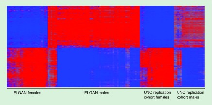
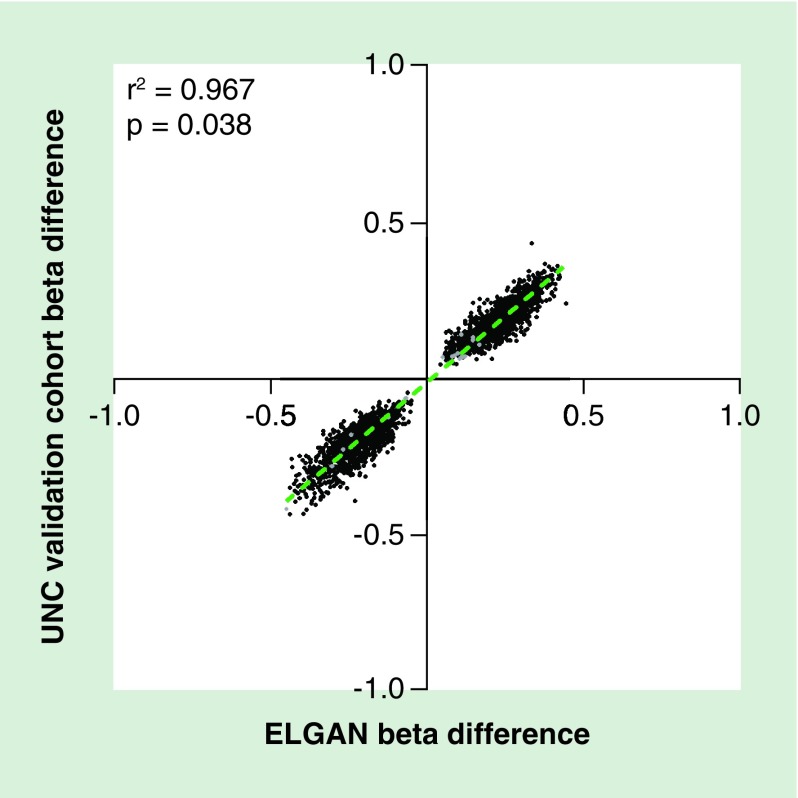
Methods: Placental DNA methylation was assessed at more than 480,000 CpG sites from male and female infants enrolled in the extremely low gestational age newborns cohort (ELGAN) and validated in a separate US-based cohort. The impact of gestational age on placental DNA methylation was further examined using the New Hampshire Birth Cohort Study for a total of n = 467 placentas.
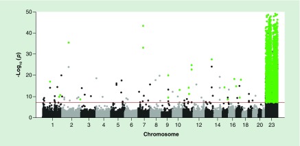
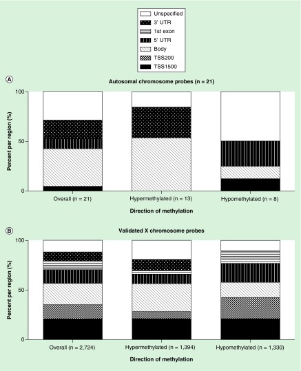
Results: A total of n = 2745 CpG sites, representing n = 587 genes, were identified as differentially methylated (p < 1 × 10⁻⁷). The majority (n = 582 or 99%) of these were conserved among the New Hampshire Birth Cohort. The identified genes encode proteins related to immune function, growth/transcription factor signaling and transport across cell membranes.
Conclusion: These data highlight sex-dependent epigenetic patterning in the placenta and provide insight into differences in infant outcomes and responses to the perinatal environment.
Keywords: CpG DNA methylation; epigenetics; placenta; sexual dimorphism.
Conflict of interest statement
This research was supported by grants from the NIH including the Environmental Influences on Child Health Outcomes (ECHO) award (1U2COD023375, UG33OD023348 and 1UG3OD023275), the National Institute of Environmental Health Sciences (P42-ES007126, T32-ES007018, P42-ES007373, P01-ES022832) and the National Institute of Neurological Disorders and Stroke (5U01NS040069 and 2R01NS040069). Further support was provided by the Wake Forest School of Medicine Innovation Pilot Grant, the Harold M and Mary Earnhardt Eagle Endowed Fund for Pediatric and Neonatal Research, and the EPA (RD83544201). The authors have no other relevant affiliations or financial involvement with any organization or entity with a financial interest in or financial conflict with the subject matter or materials discussed in the manuscript apart from those disclosed.
No writing assistance was utilized in the production of this manuscript.
Figures
References
-
- Godfrey KM. The role of the placenta in fetal programming-a review. Placenta. 2002;23(Suppl. A):S20–S27. - PubMed
-
- Kippler M, Wagatsuma Y, Rahman A, et al. Environmental exposure to arsenic and cadmium during pregnancy and fetal size: a longitudinal study in rural Bangladesh. Reprod. Toxicol. 2012;34(4):504–511. - PubMed
Publication types
MeSH terms
Grants and funding
LinkOut - more resources
Full Text Sources
Other Literature Sources