. 2017 May;24(5):819-831.
doi: 10.1038/cdd.2017.14. Epub 2017 Feb 24.

AMPK improves gut epithelial differentiation and barrier function via regulating Cdx2 expression

Affiliations

AMPK improves gut epithelial differentiation and barrier function via regulating Cdx2 expression

Xiaofei Sun et al. Cell Death Differ. 2017 May.

Abstract

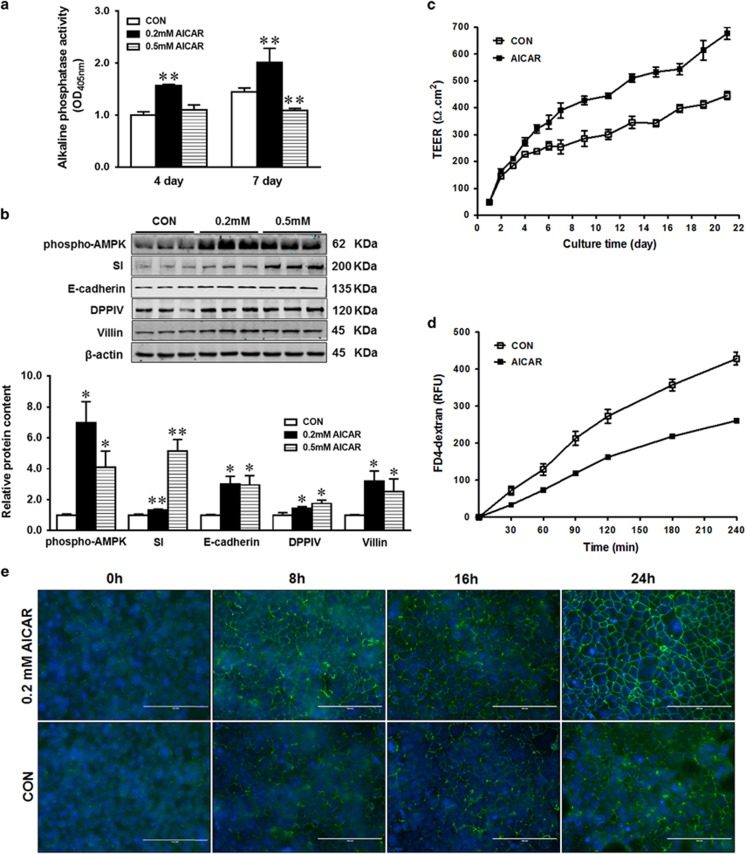
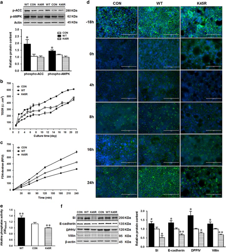
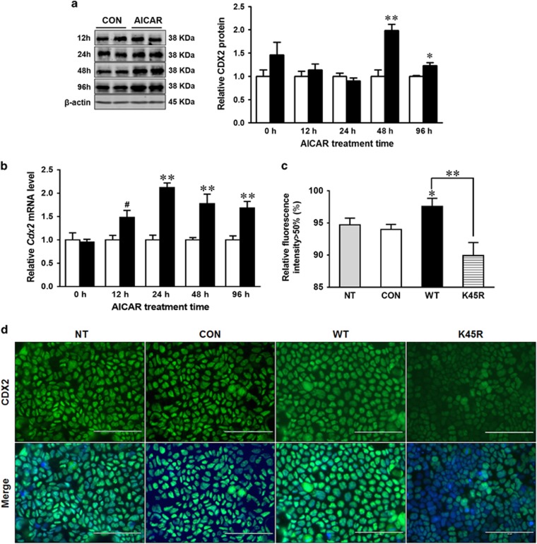
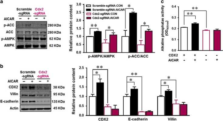
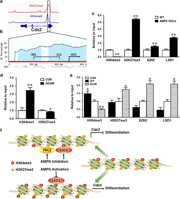
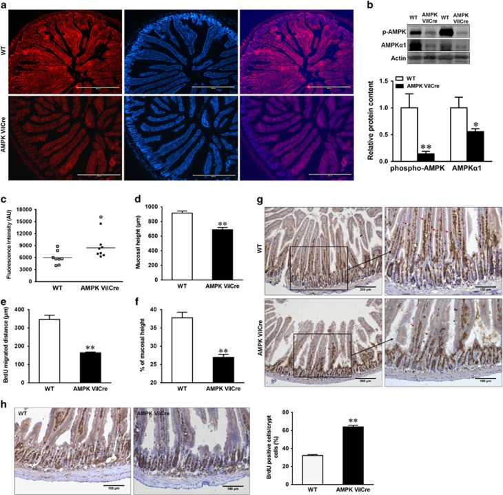
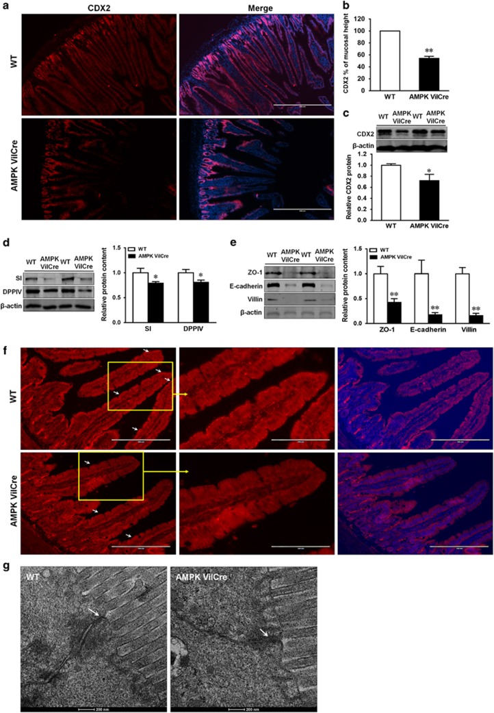
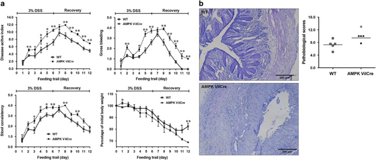
Impairment in gut epithelial integrity and barrier function is associated with many diseases. The homeostasis of intestinal barrier is based on a delicate regulation of epithelial proliferation and differentiation. AMP-activated protein kinase (AMPK) is a master regulator of energy metabolism, and cellular metabolites are intrinsically involved in epigenetic modifications governing cell differentiation. We aimed to evaluate the regulatory role of AMPK on intestinal epithelial development and barrier function. In this study, AMPK activator (AICAR) improved the barrier function of Caco-2 cells as indicated by increased transepithelial electrical resistance and reduced paracellular FITC-dextran permeability; consistently, AICAR enhanced epithelial differentiation and tight junction formation. Transfection of Caco-2 cells with AMPK WT plasmid, which enhances AMPK activity, improved epithelial barrier function and epithelial differentiation, while K45R (AMPK dominant negative mutant) impaired; these changes were correlated with the expression of caudal type homeobox 2 (CDX2), the key transcription factor committing cells to intestinal epithelial lineage. CDX2 deficiency abolished intestinal differentiation promoted by AMPK activation. Mechanistically, AMPK inactivation was associated with polycomb repressive complex 2 regulated enrichment of H3K27me3, the inhibitory histone modification, and lysine-specific histone demethylase-1-mediated reduction of H3K4me3, a permissive histone modification. Those histone modifications provide a mechanistic link between AMPK and CDX2 expression. Consistently, epithelial AMPK knockout in vivo reduced CDX2 expression, impaired intestinal barrier function, integrity and ultrastructure of tight junction, and epithelial cell migration, promoted intestinal proliferation and exaggerated dextran sulfate sodium-induced colitis. In summary, AMPK enhances intestinal barrier function and epithelial differentiation via promoting CDX2 expression, which is partially mediated by altered histone modifications in the Cdx2 promoter.


Conflict of interest statement

The authors declare no conflict of interest.

Figures

Figure 1
Figure 1
AICAR treatment enhances differentiation of Caco-2 cells. (a) Alkaline phosphatase activity in Caco-2 cells treated with 0, 0.2 or 0.5 mM AICAR. (b) Enhanced protein content of phospho-AMPK, SI (sucrase-isomaltase), E-cadherin, DPPIV (dipeptidyl peptidase-4) and villin in Caco-2 cells treated with 0.2 or 0.5 mM AICAR. (c) Transepithelial electrical resistance over time. (d) FITC-dextran paracellular intestinal epithelial permeability at 21 days post incubation. (e) Immunofluorescence staining of tight junction protein ZO-1 pre- and post- calcium switch assay. Caco-2 cells were grown to confluence with or without 0.2 mM AICAR and subjected to a calcium switch assay. Cells were fixed at various time points (0, 8, 16 and 24 h) after restoration of Ca2+ containing medium. Data are representative of three separate experiments. Scale bar is 100 μm. Mean±S.E.M., n=4, *P<0.05 versus CON; **P<0.01 versus CON
Figure 2
Figure 2
AMPK promotes intestinal epithelial differentiation. Caco-2 cells were transfected with EGFP (CON), AMPKα WT (WT) or AMPKα K45R (K45R) plasmid. (a) Phospho-ACC and phospho-AMPK activation. (b) Transepithelial electrical resistance over time. (c) FITC-dextran paracellular intestinal epithelial permeability at 21 days post incubation. (d) Immunofluorescent staining of tight junction protein ZO-1 pre- and post- calcium switch assay. Caco-2 cells were grown to confluence and subjected to a calcium switch assay. Cells were fixed 18 h before calcium switch (−18 h) and various time points (0, 4, 8, 16 and 24 h) after restoration of Ca2+ containing medium. Scale bar is 100 μm. (e) Alkaline phosphatase activity. (f) Protein contents of SI (sucrase-isomaltase), E-cadherin, DPPIV (dipeptidyl peptidase-4) and villin. Data are representative of three separate experiments. Mean±S.E.M., n=4, #P<0.1 versus CON *P<0.05 versus CON; **P<0.01 versus CON
Figure 3
Figure 3
AMPK enhances CDX2 expression in intestinal epithelial cells. Caco-2 cells were treated with 0 or 0.2 mM AICAR for 96 h when cells were collected for analysis. (a) Protein content of CDX2. (b) mRNA expression of Cdx2. (c and d) Immunofluorescent staining of CDX2 in non-transfected (NT) Caco-2 cells, or Caco-2 cells transfected with EGFP (CON), or AMPKα WT (WT) or AMPKα K45R (K45R) plasmid. Scale bar is 100 μm. Data are representative of three separate experiments. Mean±S.E.M., n=4, #P<0.1 versus CON; *P<0.05 versus CON; **P<0.01 versus CON
Figure 4
Figure 4
CDX2 knockout abolishes the positive effects of AICAR on intestinal differentiation. Caco-2 cells were transfected with scrambled CRISPR/Cas9 plasmid (scramble-sgRNA) or CDX2 CRISPR/Cas9 plasmid (Cdx2 sgRNA) to delete Cdx2 or not. Transfected cells with GFP expression (plasmid carries GFP gene) were isolated using cell sorting, then treated with 0 or 0.2 mM AICAR for 4 days. (a) Protein contents of phospho-ACC, ACC, phospho-AMPK and AMPK. (b) Protein contents of CDX2, villin, E-cadherin and beta-actin. (c) Alkaline phosphatase activity. Data are representative of three separate experiments. Mean±S.E.M., n=3, *P<0.05 versus Scramble-sgRNA; **P<0.01 versus Scramble-sgRNA
Figure 5
Figure 5
AMPK regulates Cdx2 transcription through inducing histone modifications. (a) Data analysis for epigenetic modifications in the Cdx2 promoter based on NCBI ChIP-Seq profile of H3K4me3 and H3K27me3. (b) CpG sites profile and genomic structure in the Cdx2 promoter. Blue regions show CpG islands. Red lines show CpG dinucleotides. −391 bp to −121 bp represents primer amplification region. (c) H3K4me3 and H3K27me3 modifications as well as binding status of EZH2 and LSD1 in the Cdx2 promoter of AMPK VilCre KO and WT mice using ChIP-PCR. (d) H3K4me3 and H3K27me3 modifications in the Cdx2 promoter of Caco-2 cells treated with or without 0.2 mM AICAR and measured by ChIP-PCR. (e) H3K4me3 and H3K27me3 modifications as well as binding status of EZH2 and LSD1 in the Cdx2 promoter of Caco-2 cells transfected with EGFP (CON), AMPKα WT (WT) or AMPKα K45R (K45R) plasmid. (f) A proposed model for the effects of AMPK on intestinal differentiation via inducing histone modifications by regulating methylase and demethylase in the Cdx2 promoter. Data are representative of three separate experiments. Mean±S.E.M.; n=3, #P<0.1 versus CON; *P<0.05 versus CON; **P<0.05 versus CON
Figure 6
Figure 6
AMPK enhances intestinal barrier function and migration, and alters proliferation in vivo. Jejunum tissues of AMPK VilCre KO and WT male mice (2.5 months of age) were used. (a) Immunofluorescent staining of phospho-AMPK. Scale bars represent 400 μm. (b) Protein contents of phospho-AMPK and AMPKα1. (c) In vivo paracellular intestinal epithelial permeability of FITC-dextran. (d) Mucosal height. (e) Enterocyte migration. (f) Enterocyte migration relative to mucosal height. (g) 24 h post a single BrdU injection for intestinal migration staining. Arrows indicate the farthest BrdU-positive enterocytes. Scale bars represent 200 μm on the left and 100 μm on the right. (h) 2 h post a single BrdU injection for intestinal proliferation analysis. Scale bars represent 100 μm. Data are representative of three separate experiments. Mean±S.E.M.; n=8, *P<0.05 versus WT; **P<0.01 versus WT
Figure 7
Figure 7
AMPK knockout decreases CDX2 content and intestinal differentiation in vivo. Jejunum tissues of AMPK VilCre KO and WT male mice (2.5 months of age) were used. (a) Immunofluorescent staining of CDX2. Scale bar is 400 μm. (b) CDX2 staining relative to mucosal height. (c) Protein content of CDX2. (d) Protein content of SI (sucrase-isomaltase) and DPPIV (dipeptidyl peptidase-4). (e) Protein content of ZO-1, E-cadherin and villin. (f) Immunofluorescent staining of ZO-1 in jejunum tissues. Arrows indicate ZO-1 at the border of villus. Scale bar is 200 μm. (g) Transmission electron microscopy (TEM) ultrastructure of tight junction. Arrows indicate tight junctions. Scale bar is 200 nm. Mean±S.E.M.; n=8, *P<0.05 versus WT; **P<0.01 versus WT
Figure 8
Figure 8
AMPK deletion exacerbates DSS-induced colitis. AMPK VilCre KO and WT male mice (3 months of age) were induced colitis by 3% DSS for 6 days, followed by 6 days of recovery. (a) Disease activity index including body weight, gross bleeding and stool consistency were monitored and scored daily. (b) Pathological scores in DSS-induced distal colon. Scale bars are 200 μm. Mean±S.E.M.; n=5, *P<0.05 versus WT; **P<0.01 versus WT

References

    1. van der Flier LG, Clevers H. Stem cells, self-renewal, and differentiation in the intestinal epithelium. Annu Rev Physiol 2009; 71: 241–260. - PubMed
    1. Turner JR. Intestinal mucosal barrier function in health and disease. Nat Rev Immunol 2009; 9: 799–809. - PubMed
    1. Hardie DG. AMP-activated/SNF1 protein kinases: conserved guardians of cellular energy. Nat Rev Mol Cell Biol 2007; 8: 774–785. - PubMed
    1. Viollet B, Horman S, Leclerc J, Lantier L, Foretz M, Billaud M et al. AMPK inhibition in health and disease. Crit Rev Biochem Mol Biol 2010; 45: 276–295. - PMC - PubMed
    1. Meddings J. The significance of the gut barrier in disease. Gut 2008; 57: 438–440. - PubMed

Publication types

MeSH terms