Review
. 2018 Feb 14;118(3):889-918.
doi: 10.1021/acs.chemrev.6b00737. Epub 2017 Feb 24.

Ubiquitin-like Protein Conjugation: Structures, Chemistry, and Mechanism

Affiliations
Review

Ubiquitin-like Protein Conjugation: Structures, Chemistry, and Mechanism

Laurent Cappadocia et al. Chem Rev. .

Abstract

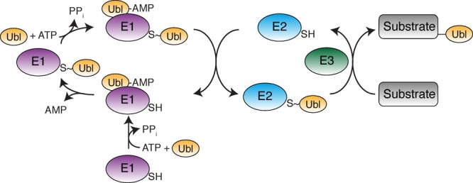
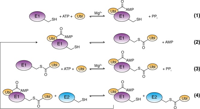
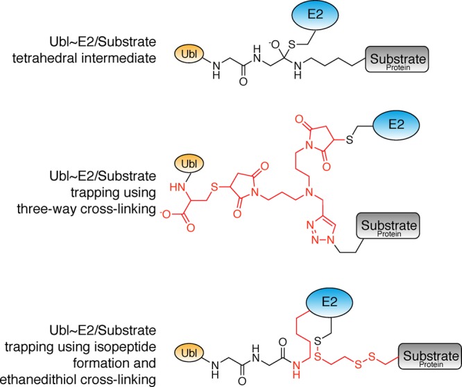
Ubiquitin-like proteins (Ubl's) are conjugated to target proteins or lipids to regulate their activity, stability, subcellular localization, or macromolecular interactions. Similar to ubiquitin, conjugation is achieved through a cascade of activities that are catalyzed by E1 activating enzymes, E2 conjugating enzymes, and E3 ligases. In this review, we will summarize structural and mechanistic details of enzymes and protein cofactors that participate in Ubl conjugation cascades. Precisely, we will focus on conjugation machinery in the SUMO, NEDD8, ATG8, ATG12, URM1, UFM1, FAT10, and ISG15 pathways while referring to the ubiquitin pathway to highlight common or contrasting themes. We will also review various strategies used to trap intermediates during Ubl activation and conjugation.


Conflict of interest statement

The authors declare no competing financial interest.

Figures

Figure 1
Figure 1
Ubl conjugation cascade where E1, E2, E3, and Ubl designate an E1 conjugating enzyme, an E2 conjugation enzyme, an E3 ligase, and a ubiquitin-like protein, respectively.
Figure 2
Figure 2
Structure of select Ubl’s. (A) Structure of ubiquitin (PDB 1UBQ). (B) Structure of ISG15 (PDB 1Z2M). The first β-grasp domain is colored gray. (C) Structure of ATG8 (GATE-16; PDB 1EO6). The N-terminal extension that contains two α-helices is colored gray. (D) Structure of SUMO (SUMO1; PDB 1A5R). The flexible N-terminal extension is colored gray. Root-mean-square deviations (RMSDs) are calculated between ubiquitin Cα and the Cα of ISG15, ATG8, or SUMO1 that are colored in yellow.
Figure 3
Figure 3
Ubl binding motifs. (A) Structure of SUMO/RANBP2 (PDB 1Z5S). (B) Structure of ATG8/ATG19 (PDB 2ZPN). (C) Structure of UFM1/UBA5 (PDB 5HKH). (D) Structure of ATG12/ATG3 (PDB 4NAW). The Ubl’s are in cartoon representation colored yellow, while the Ubl binding motifs are in cartoon representation colored gray. Selected residues of the Ubl binding motifs are presented in stick representation. Backbone interactions that mediate β-strand complementation for SUMO/RANBP2 and ATG8/ATG19 are highlighted at the top.
Figure 4
Figure 4
Canonical E1 chemical reactions. (1) The E1 binds a Ubl and ATP and catalyzes adenylation of the Ubl. (2) The E1 catalytic cysteine attacks the Ubl∼AMP resulting in formation of an E1∼Ubl thioester. (3) The E1 adenylates a second Ubl. (4) The Ubl is transferred to an E2 through a transthioesterification reaction.
Figure 5
Figure 5
Domain organization of canonical E1s. (A) Primary structure of canonical E1s for NEDD8, SUMO, and ubiquitin. (B) Structure of human NEDD8∼E1NAE1/UBA3/E2UBC12/ATP (PDB 2NVU). Adenylation domains are shown in a Gaussian surface representation, while other domains are in cartoon representation. The sulfur atom of the active site cysteine is in sphere representation colored yellow. Positions for ATP, the crossover loop, and the active site cysteine are highlighted by arrows.
Figure 6
Figure 6
Adenylation domains of E1 and E1-like proteins. (A) Structure of E. coli MoaD/MoeB/ATP (PDB 1JWA). (B) Structure of human E1SAE1/UBA2/SUMO/ATP (PDB 1Y8R). In both cases, Ubl and adenylation domains are depicted in cartoon and Gaussian surface representations. The ATP and last two residues of the Ubl’s are in stick representation. (C) Close-up view of interactions between ATP, the SUMO C-terminus, and the E1 adenylation pocket.
Figure 7
Figure 7
Thioester formation in the SUMO E1. (A) Structure of human E1SAE1/UBA2/SUMO–AMSN (PDB 3KYC). (B) Structure of human E1SAE1/UBA2/SUMO–AVSN (PDB 3KYD). For simplicity, only the CYS domain and SUMO–AMSN or SUMO–AVSN is presented. The sulfur atom of the active site cysteine is in sphere representation colored in yellow. A color gradient is applied on the CYS domain to highlight the different orientations observed in the two structures. As an additional landmark, Lys336, a lysine residue in the CYS domain, is presented in sphere representation. AMSN and AVSN are nonhydrolyzable AMP mimics with AVSN covalently linked to the E1 catalytic cysteine.
Figure 8
Figure 8
E1–E2 interaction for canonical E1 proteins. (A) Structure of human E1NAE1/UBA3/E2UBC12/NEDD8/ATP (PDB 2NVU) showing the bipartite binding of the E2UBC12 to E1. In this structure, the catalytic cysteine residues of E1 and E2 are separated by ∼20 Å. (B) Structure of S. pombe E1UBA1/E2UBC4/ubiquitin/ATP (PDB 4II2) showing juxtaposition of E1 and E2 active sites. Adenylation domains are shown in Gaussian surface representation. Other domains are in cartoon representation. The sulfur atoms of the active site cysteine residues of E1 and E2 are in sphere representation colored yellow.
Figure 9
Figure 9
E1–E2 interaction for ATG7. (A) Structure of S. cerevisiae E1ATG7–E2ATG3 (PDB 4GSL). (B) Structure of S. cerevisiae E1ATG7–E2ATG10 (PDB 4GSK). In both cases, E2 binding occurs between the NTD and CTD domains of E1ATG7. Dashed lines represent regions missing elements in the crystal structure. Sulfur atoms of the catalytic cysteine residues of E1ATG7 and E2ATG3 are in sphere representation colored yellow. The catalytic cysteine of E2ATG10 is not visible in the structure. The position of ATG8 or ATG12 on the adenylation domain is indicated by a yellow oval.
Figure 10
Figure 10
Canonical and noncanonical E2s. (A) A structure of E2UBC9 (PDB 1Z5S) was chosen as a representative example of canonical E2s. (B) Structure of E2ATG10 (PDB 3VX7), a noncanonical E2. (C) Structure of E2UFC1 (PDB 3EVX), a second noncanonical E2 that differs from both E2UBC9 and E2ATG10. Proteins are in cartoon representation colored cyan except for divergent structural elements that are colored white. The sulfur atoms of the catalytic cysteine residues are in sphere representation to highlight the shift in position of this residue in E2ATG10 as compared to E2UBC9 and E2UFC1.
Figure 11
Figure 11
E2 chemical reactions (A) Scheme illustrating how the Ubl moiety of a E2∼Ubl thioester can be transferred to the primary amine group of a lysine residue of a protein substrate (top), to the primary amine group of PE (middle), or to a HECT or RBR E3 for subsequent transfer to the lysine residue of a protein substrate (bottom). Transfer to PE is performed by E2ATG3. Ubl transfer to E3s of the HECT or RBR families are limited to E2UBCH8, and this only allows the ISGylation of a limited number of protein substrates. (B) Mechanism for the aminolysis reaction. In this case, the primary amines are presented in their deprotonated states.
Figure 12
Figure 12
E2 active site. (A) Overall view and (B) close-up view of E2UBC9/SUMO–RANGAP1 (PDB 1Z5S) illustrating how the RANGAP1 substrate and SUMO are positioned in the E2UBC9 active site. This state represents a product complex after conjugation where SUMO, colored yellow, has been transferred to a lysine of RANGAP1 colored gray. SUMOD designates a SUMO protein in donor (D) configuration. E2UBC9 is in cartoon representation colored cyan. Certain residues of the E2 active site are in stick representation. The consensus sequence for substrate recognition by E2UBC9 is indicated on top. (C) Close-up of E2UBC9/RANGAP1 (PDB 1KPS) representing RANGAP binding prior to catalysis in the absence of SUMO.
Figure 13
Figure 13
E2∼Ubl complexes. (A) Structure of E2UBC9 in complex with SUMO in a closed conformation (PDB 1Z5S). (B) Structure of E2UBC9 in complex with SUMO that binds E2UBC9 on the E2 backside (PDB 2PE6). Proteins are in cartoon representation with catalytic cysteine residues in sphere representation. SUMOD and SUMOB represent SUMO proteins in donor (D) and backside (B) configurations, respectively.
Figure 14
Figure 14
Representative RING E3/E2 interaction. (A) Overall view and (B) close-up view of human E3TRIM25/E2UBCH5A (PDB 5FER). Both proteins are in cartoon representation. A white-to-green gradient running from the N- to C-terminus has been applied to E3TRIM25. Two zinc ions are depicted as gray spheres. Residues contributing to the E3TRIM25/E2UBCH5A interaction are in stick representation.
Figure 15
Figure 15
E3 stabilization of a closed E2∼Ubl conformation. (A) Structure of human NEDD8∼E2UBC12/E3RBX1 (PDB 4P5O). The position of the catalytic cysteine (a serine residue in the structure) is indicated by a yellow sphere. (B) Structure of yeast SUMO∼E2UBC9/E3SIZ1 (PDB 5JNE). (C) Structure of human SUMO∼E2UBC9/E3RANBP2 (PDB 1Z5S). (D) Structure of human SUMO∼E2UBC9/E3ZNF451 (PDB 5D2M). Zinc atoms are in gray sphere representations. SUMOD and SUMOB represent SUMO proteins in donor (D) and backside (B) configurations, respectively. NEDD8D designates a NEDD8 protein in donor (D) configuration.
Figure 16
Figure 16
E3/E2∼Ubl/substrate complexes. (A) Structure of human co-E3DCN1/E3RBX1/E2UBC12∼NEDD8/CUL1 (PDB 4P5O). The target residue 720 (an arginine in the structure) is in stick representation. NEDD8D designates a NEDD8 protein in donor (D) configuration. (B) Structure of yeast SUMO–E3SIZ1/SUMO∼ E2UBC9–PCNA (PDB 5JNE). The target residue 164 (a cysteine in the structure) and its linkage to the E2 catalytic cysteine via ethanedithiol are presented in stick representation. SUMOD and SUMOB represent SUMO proteins in donor (D) and backside (B) configurations, respectively. (C) Structure of human SUMO–RANGAP1/E2UBC9/E3RANBP2 (PDB 1Z5S). The isopeptide linkage between the target residue 524 and the C-terminal glycine of SUMO is in stick representation.
Figure 17
Figure 17
E3/E2 complex in the ATG8 system. Structure of E3ATG12–ATG5/E2ATG3 (PDB 4NAW). Proteins are in cartoon representation with the isopeptide linkage between ATG12 and ATG5 in stick representation.
Figure 18
Figure 18
Chemical structures representing methods for trapping the E1 thioesterification step. Non-native linkages are colored red.
Figure 19
Figure 19
Chemical structures representing methods for trapping E1/E2 complexes. Non-native linkages are colored red.
Figure 20
Figure 20
E2∼Ubl mimics. Non-native linkages or amino acid residues are colored red.
Figure 21
Figure 21
Chemical structures representing methods for trapping Ubl∼E2/substrate complexes. Non-native linkages or residues are colored red.

Similar articles

Cited by

References

    1. Lorenz S.; Cantor A. J.; Rape M.; Kuriyan J. Macromolecular Juggling by Ubiquitylation Enzymes. BMC Biol. 2013, 11, 65.10.1186/1741-7007-11-65. - DOI - PMC - PubMed
    1. Metzger M. B.; Pruneda J. N.; Klevit R. E.; Weissman A. M. RING-Type E3 Ligases: Master Manipulators of E2 Ubiquitin-Conjugating Enzymes and Ubiquitination. Biochim. Biophys. Acta, Mol. Cell Res. 2014, 1843, 47–60. 10.1016/j.bbamcr.2013.05.026. - DOI - PMC - PubMed
    1. Spratt D. E.; Walden H.; Shaw G. S. RBR E3 Ubiquitin Ligases: New Structures, New Insights, New Questions. Biochem. J. 2014, 458, 421–437. 10.1042/BJ20140006. - DOI - PMC - PubMed
    1. Berndsen C. E.; Wolberger C. New Insights into Ubiquitin E3 Ligase Mechanism. Nat. Struct. Mol. Biol. 2014, 21, 301–307. 10.1038/nsmb.2780. - DOI - PubMed
    1. Streich F. C. Jr.; Lima C. D. Structural and Functional Insights to Ubiquitin-Like Protein Conjugation. Annu. Rev. Biophys. 2014, 43, 357–379. 10.1146/annurev-biophys-051013-022958. - DOI - PMC - PubMed

Publication types

MeSH terms