. 2017 Feb 24;12(2):e0172736.
doi: 10.1371/journal.pone.0172736. eCollection 2017.

Expression patterns of Slit and Robo family members in adult mouse spinal cord and peripheral nervous system

Affiliations

Expression patterns of Slit and Robo family members in adult mouse spinal cord and peripheral nervous system

Lauren Carr et al. PLoS One. .

Abstract

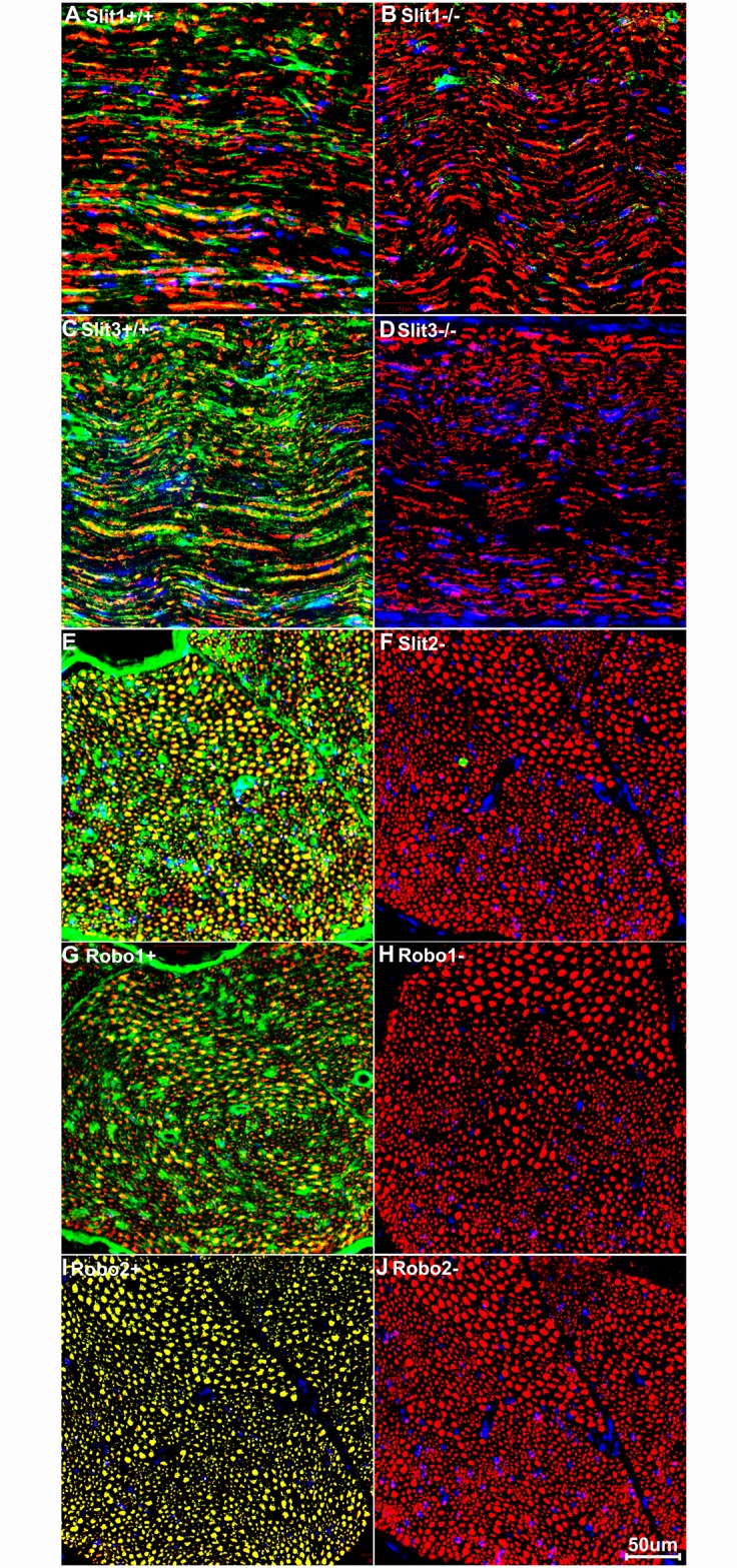
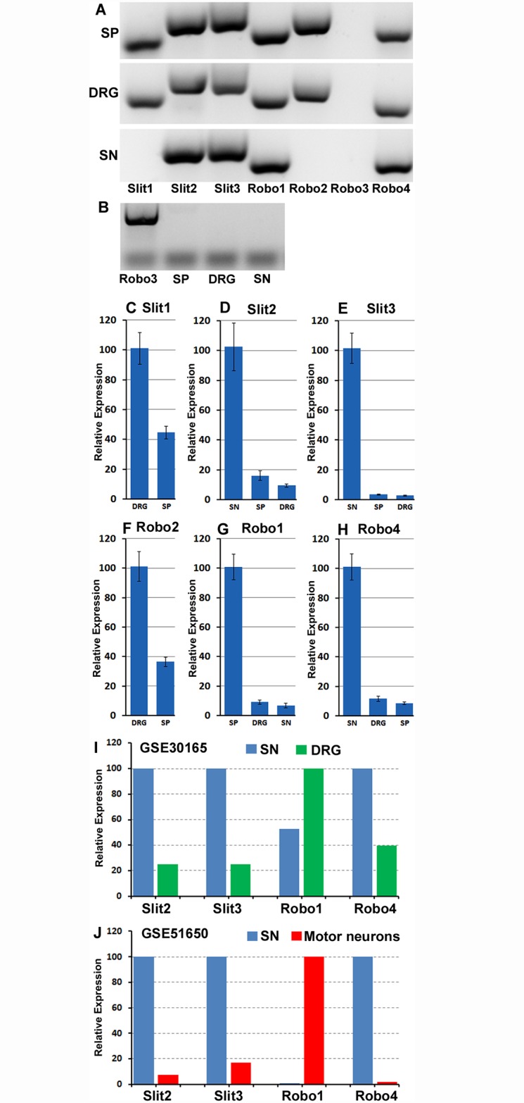
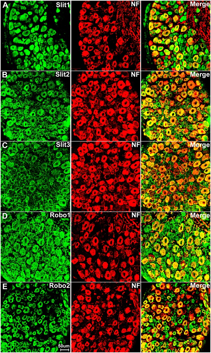
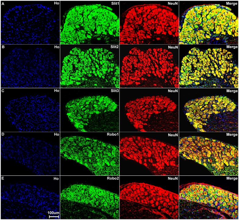
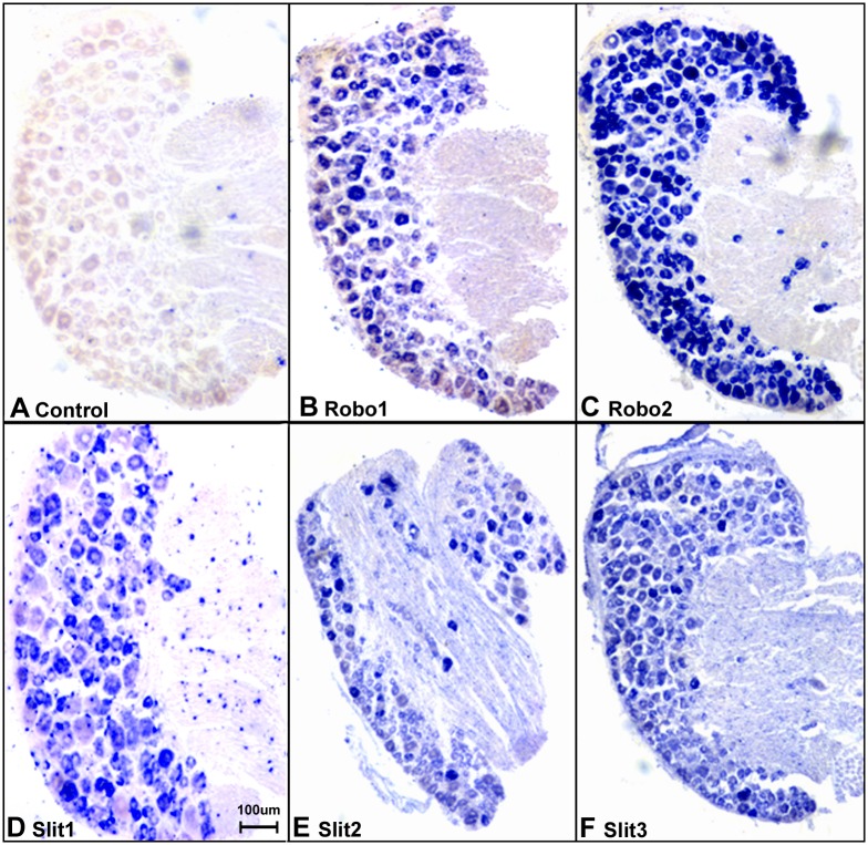
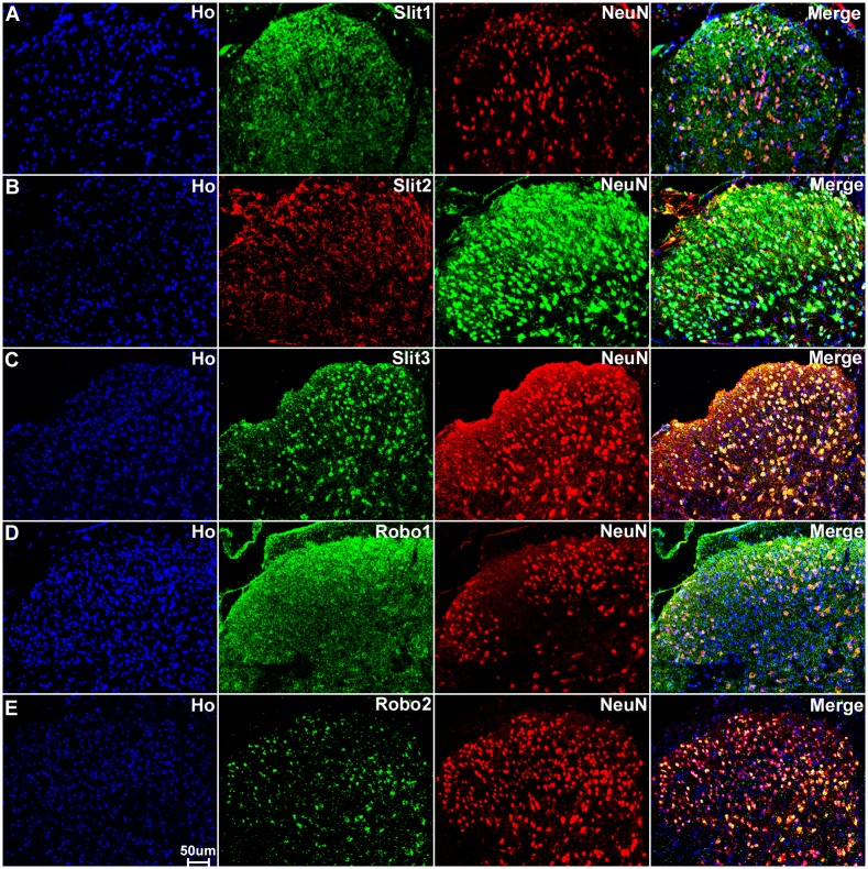
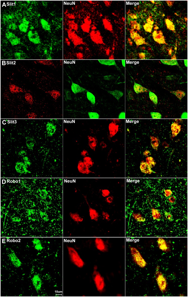
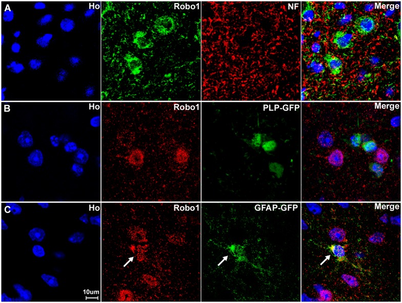
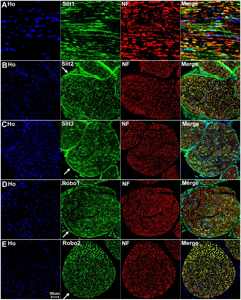
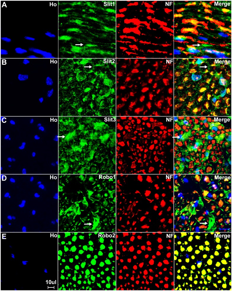
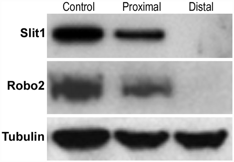
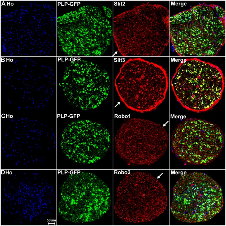
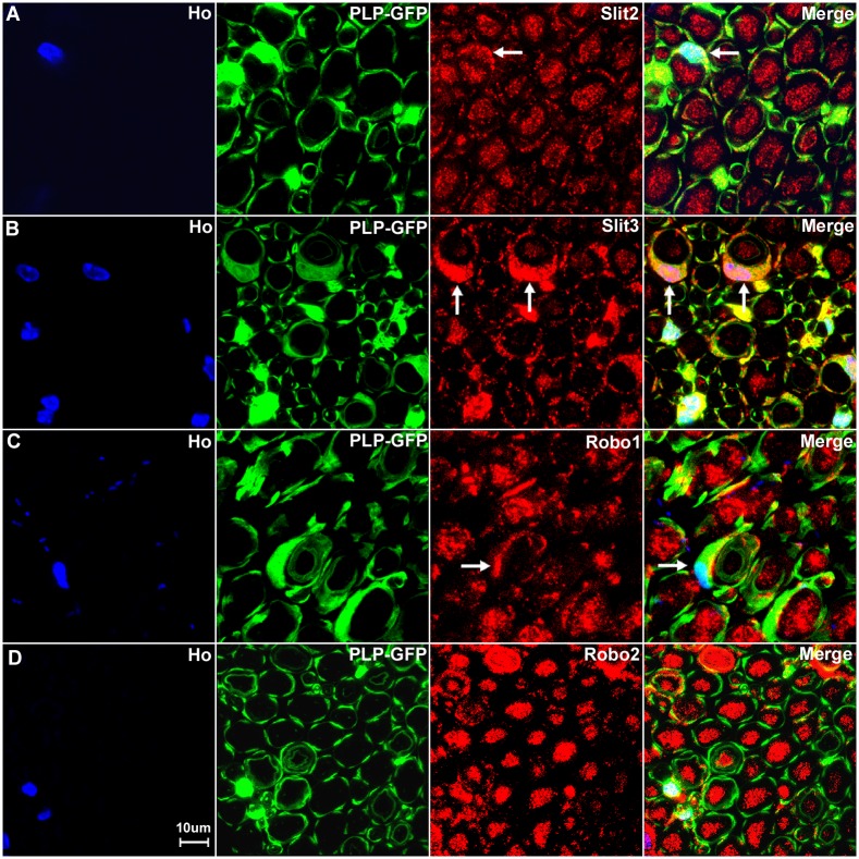
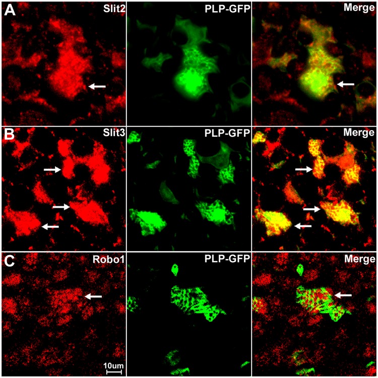
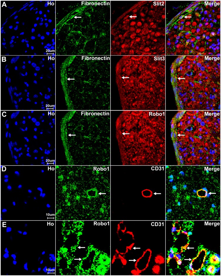
The secreted glycoproteins, Slit1-3, are classic axon guidance molecules that act as repulsive cues through their well characterised receptors Robo1-2 to allow precise axon pathfinding and neuronal migration. The expression patterns of Slit1-3 and Robo1-2 have been most characterized in the rodent developing nervous system and the adult brain, but little is known about their expression patterns in the adult rodent peripheral nervous system. Here, we report a detailed expression analysis of Slit1-3 and Robo1-2 in the adult mouse sciatic nerve as well as their expression in the nerve cell bodies within the ventral spinal cord (motor neurons) and dorsal root ganglion (sensory neurons). Our results show that, in the adult mouse peripheral nervous system, Slit1-3 and Robo1-2 are expressed in the cell bodies and axons of both motor and sensory neurons. While Slit1 and Robo2 are only expressed in peripheral axons and their cell bodies, Slit2, Slit3 and Robo1 are also expressed in satellite cells of the dorsal root ganglion, Schwann cells and fibroblasts of peripheral nerves. In addition to these expression patterns, we also demonstrate the expression of Robo1 in blood vessels of the peripheral nerves. Our work gives important new data on the expression patterns of Slit and Robo family members within the peripheral nervous system that may relate both to nerve homeostasis and the reaction of the peripheral nerves to injury.


Conflict of interest statement

Competing Interests: The authors have declared that no competing interests exist.

Figures

Fig 1
Fig 1. Validation of Slit1-3 and Robo1-2 antibodies.
Staining with Slit1-3 and Robo1-2 antibodies is shown in green; staining with neurofilament heavy chain antibody is shown in in red. The yellow signal in panels A, C, E, G and I shows co-localization of the Slit1-3 and Robo1-2 signal with neurofilament staining. (A-B) Slit1 staining on transverse sections of sciatic nerve from Slit1 control (+/+, A) and Slit1 null (-/-, B) mice. (C-D) Slit3 staining on sciatic nerve samples from Slit3+/+ (C) and Slit3-/- (D) mice. (E-J) Slit2, Robo1 and Robo2 staining on sciatic nerve samples from C57BL/6 mice. Slit2, Robo1 and Robo2 primary antibodies were used in E, G and I but were omitted in F, H and J. Staining with Hoechst dye is also shown (blue) to identify cell nuclei within the tissue.
Fig 2
Fig 2. Slit1-3 and Robo1-4 mRNA expression in adult mouse (C57BL/6) spinal cord, DRG and sciatic nerve.
(A) RT-PCR shows Slit1-3 and Robo1-4 mRNA expression in spinal cord (SP), DRG and sciatic nerve (SN). Slit1-3, Robo1-2 and Robo4 mRNA are expressed in spinal cord and DRG. In sciatic nerve, only Slit2, Slit3, Robo1 and Robo4 mRNA are detectable. Robo3 mRNA is not expressed in the adult mouse spinal cord and the peripheral nervous system. (B) Validation of Robo3 primers with a mouse Robo3 cDNA plasmid (Robo3). (C-H) qRT-PCR compares Slit1-3, Robo1-2 and Robo4 relative expression levels between spinal cord, DRG and sciatic nerve samples. The highest mRNA level has been set as 100% for each individual graph. Error bars show one standard deviation of the mean from 3 repeat experiments. Slit1 and Robo2 have their highest relative mRNA level in the DRG. Slit2, Slit3 and Robo4 have their highest mRNA level in the sciatic nerve. Robo1 has its highest mRNA level in spinal cord. (I-J), comparison of Slit2, Slit3, Robo1 and Robo4 relative levels from two microarray data sets (GSE30165 and GSE51650). As for Panels C-H, the highest mRNA level has been set as 100% for each individual graph. This analysis also shows that Slit2, Slit3 and Robo4 appear to have their highest mRNA level in the sciatic nerve. Robo1 appears to have its highest mRNA level in the motor neurons of the spinal cord.
Fig 3
Fig 3. Double staining of Slit1-3 and Robo1-2 with neurofilament heavy chain in DRG.
The neurofilament heavy chain antibody (NF) labels large diameter sensory neurons within the DRG. Merged images show that Slit1 (A), Slit2 (B), Slit3 (C), Robo1 (D) and Robo2 (E) are seemingly all expressed by large diameter sensory neurons. The yellow colour in merged images shows the co-localization of Slit1-3 and Robo1-2 signal with neurofilament heavy chain staining. Slit1-3 and Robo1-2 also appear to be expressed in small diameter cells in addition to their expression in large diameter sensory neurons. Slit1 (A) and Robo2 (E) show clear neuronal cell body staining. In contrast, Slit2 (B), Slit3 (C) and Robo1 (D) also show positive staining in the gaps between the cell bodies of sensory neurons in addition to their expression in the neuronal cell bodies.
Fig 4
Fig 4. Sensory neurons in the DRG express Slit1-3 and Robo1-2.
Double staining of Slit1-3 and Robo1-2 with NeuN in the DRG shows all sensory neurons in the DRG express Slit1-3 and Robo1-2. The neuronal marker NeuN labels both large and small diameter sensory neurons. Merged images show that Slit1 (A), Slit2 (B), Slit3 (C), Robo1 (D) and Robo2 (E) are all expressed by both large and small diameter sensory neurons. The yellow colour in merged images shows the co-localization of Slit1-3 and Robo1-2 signal with NeuN staining. Double staining of NeuN with Slit1-3 and Robo1-2 antibodies thus confirms that all the sensory neurons in the DRG express Slit1-3 and Robo1-2. Staining with Hoechst dye (Ho) is also shown (blue) to identify cell nuclei within the tissue.
Fig 5
Fig 5. In situ hybridization confirms the expression of Slit1-3 and Robo1-2 in sensory neurons of the DRG.
(A) Sense mRNA probe control, no blue signal has been developed in all the control samples using Slit1-3 and Robo1-2 sense probes for in situ hybridization (Robo1 sense mRNA probe control was shown in A, images for Robo2 and Slit1-3 sense mRNA probe control not shown). A positive signal for Robo1 (B), Robo2 (C), Slit1 (D), Slit2 (E) and Slit3 (F) were observed in sensory neuronal cell bodies. Slit2 (E) and Slit3 (F) signal also could be observed in the nerve in addition to their strong signal in the neuronal cell bodies of the DRG. The in situ hybridization results thus help to further confirm the specificity of the Slit1-3 and Robo1-2 antibodies used for immunohistochemistry.
Fig 6
Fig 6. Satellite cells in the DRG express Slit2, Slit3 and Robo1.
DRG tissues are from PLP-GFP transgenic mice which express cytoplasmic GFP within the satellite cells. Signals from Slit2-3 and Robo1 staining shows co-localization with GFP-positive satellite cells surrounding the neuronal cell bodies. Lower magnification images show Slit2 (A), Slit3 (C) and Robo1 (E) staining in the whole DRG sections. Higher magnification images show Slit2 (B), Slit3 (D) and Robo1 (F) signals co-localize with the GFP signal in satellite cells. Staining with Hoechst dye (Ho) is also shown (blue) to identify cell nuclei within the tissue.
Fig 7
Fig 7. Slit1-3 and Robo1-2 expression in the dorsal region of adult mouse spinal cord.
Slit1-3 and Robo1-2 double staining with NeuN to show Slit1-3 and Robo1-2 expression in the dorsal region of adult mouse spinal cord. Spinal cord sections are from the L4/L5 region of adult C57BL/6 mice. Double staining with NeuN showed that Slit1 (A) and Robo1 (D) are not only expressed in dorsal neuronal cell bodies but are also expressed in other cells in the grey matter of the dorsal spinal cord. Slit2 (B) expression is low in dorsal spinal cord and seemingly restricted to neuronal cell bodies. Slit3 (C) and Robo2 (E) are also mainly expressed in the neuronal cell bodies in the dorsal spinal cord. Staining with Hoechst dye (Ho) is also shown (blue) to identify cell nuclei within the tissue.
Fig 8
Fig 8. Slit1-3 and Robo1-2 expression in the ventral region of adult mouse spinal cord.
Images show Slit1-3 and Robo1-2 expression in the neuronal cell bodies localizing in the ventral horn of the spinal cord. (A) Slit1, (B) Slit2, (C) Slit3, (D) Robo1 and (E) Robo2. Slit1-3 and Robo2 are mainly expressed in the neuronal cell bodies in the ventral horn of the spinal cord. Robo1 is highly expressed in the ventral horn neuronal cell bodies of the spinal cord but Robo1 also appears to show expression in other cells in the grey matter of the spinal cord (D). Staining with Hoechst dye (Ho) is also shown (blue) to identify cell nuclei within the tissue.
Fig 9
Fig 9. Astrocytes of the spinal cord express Robo1.
(A) Double staining for Robo1 with neurofilament heavy chain antibody shows that very few axons within the spinal cord are Robo1 positive. (B) The Robo1 signal does not co-localize with the GFP signal in the spinal cord of PLP-GFP mice, which labels oligodendrocytes with cytoplasmic GFP. (C) The Robo1 signal co-localizes with the GFP signal in the spinal cord of GFAP-GFP mice, which labels astrocytes with GFP (indicated by white arrow). The staining confirms that Robo1 positive signal from outside of the neuronal cell bodies in the grey matter is from astrocytes. Staining with Hoechst dye (Ho) is also shown (blue) to identify cell nuclei within the tissue.
Fig 10
Fig 10. Expression of Slit1-3 and Robo1-2 in the adult mouse sciatic nerve.
Lower magnification images show the expression of Slit1-3 and Robo1-2 in the adult mouse sciatic nerve by double staining of Slit1-3 and Robo1-2 antibody with the axon marker neurofilament heavy chain (NF). (A) Double staining of Slit1 with neurofilament heavy chain on a longitudinal sciatic nerve section shows that Slit1 is largely expressed in axons. Slit1 shows a better staining on sciatic nerve longitudinal sections compared to transverse sections. (B-D) Double staining of Slit2, Slit3 and Robo1 with neurofilament heavy chain on sciatic nerve transverse sections shows that Slit2, Slit3 and Robo1 are all expressed in axons. Slit2 (B), Slit3 (C) and Robo1 (D) also show positive staining outside the nerve fibres. In addition, the staining also shows that Slit2, Slit3 and Robo1 are expressed by cells in the nerve epineurium (indicated by white arrows). (E) Double staining of Robo2 with neurofilament heavy chain on sciatic nerve transverse section shows that the Robo2 signal completely co-localizes with the signal of the neurofilament heavy chain staining. Note that Robo2 is not expressed by the cells in the epineurium (indicated by white arrow in E). Staining with Hoechst dye (Ho) is also shown (blue) to identify cell nuclei within the tissue.
Fig 11
Fig 11. Higher magnification images from Fig 10 show Slit1-3 and Robo1-2 expression in the sciatic nerve.
(A) Longitudinal sciatic nerve section staining with Slit1 antibody shows that Slit1 is largely expressed in axons, Slit1 staining could also be observed outside of axons (indicated by white arrow in A). The Slit1 positive staining outside the axons might be due to the secretion of Slit1 from axons. (B-D) Transverse sciatic nerve sections show Slit2 (B), Slit3 (C) and Robo1 (D) expression in axons and also positive staining in other cells other than axons (indicated by white arrows). (E) Robo2 staining shows complete co-localization of Robo2 with neurofilament heavy chain indicating that Robo2 is exclusively expressed in the axons of the peripheral nerves. Staining with Hoechst dye (Ho) is also shown (blue) to identify cell nuclei within the tissue.
Fig 12
Fig 12. Slit1 and Robo2 protein are not expressed in the distal sciatic nerve stump after injury.
Western blot shows the disappearance of Slit1 and Robo2 protein in the distal nerve stump following a sciatic nerve transection injury. Slit1 and Robo2 protein are present in the intact sciatic nerve (Control) and also within the proximal nerve stump 4 days after sciatic nerve transection injury. However, Slit1 and Robo2 protein completely disappeared in the distal nerve stump 4 days following a sciatic nerve transection injury. The disappearance of Slit1 and Robo2 protein in the distal nerve stump is not due to down-regulation because Slit1 and Robo2 mRNAs are undetectable in the intact adult sciatic nerve (see Fig 2A).
Fig 13
Fig 13. Slit2-3 and Robo1-2 staining on the sciatic nerve transverse sections from PLP-GFP mice.
Lower magnification images show the staining of Slit2, Slit3, Robo1 and Robo2 on sciatic nerve transverse sections from PLP-GFP mice. The PLP-GFP mice express cytoplasmic GFP in both myelinating and non-myelinating Schwann cells. Slit2, Slit3 and Robo1 staining in the sciatic nerve transverse sections from PLP-GFP mice shows Slit2 (A), Slit3 (B) and Robo1 (C) co-localization with the GFP signal. In contrast, Robo2 (D) doesn’t show any co-localization with the GFP signal (D). Slit2, Slit3 and Robo1 are expressed by the cells in the epineurium (indicated by white arrows) but not Robo2 (D). Staining with Hoechst dye (Ho) is also shown (blue) to identify cell nuclei within the tissue.
Fig 14
Fig 14. Slit2, Slit3 and Robo1 expression in the cell bodies of myelinating Schwann cells.
Higher magnification images from Fig 13 show the expression of Slit2 (A), Slit3 (B) and Robo1 (C) in the cell bodies of myelinating Schwann cells (indicated by white arrows). Slit3 is highly expressed in the cell bodies of myelinating Schwann cells (B). In contrast, Robo2 staining does not show any co-localization with the GFP signal in further confirmation that Robo2 is expressed only in axons of the peripheral nerve (D). Staining with Hoechst dye (Ho) is also shown (blue) to identify cell nuclei within the tissue.
Fig 15
Fig 15. Slit2, Slit3 and Robo1 expression in the cell bodies of non-myelinating Schwann cells.
Higher magnification images from Fig 13 show the expression of Slit2, Slit3 and Robo1 in non-myelinating Schwann cells. Non-myelinating Schwann cells in the PLP-GFP mouse nerve could be easily distinguished by the morphology of Remak bundles via the GFP signal (A-C). Slit2 (A), Slit3 (B) and Robo1 (C) expression in non-myelinating Schwann cells was observed (indicated by white arrows). Slit2 and Robo1 staining appeared stronger in the small diameter axons of the Remak bundle rather than the non-myelinating Schwann cells. Slit3 showed strong expression in non-myelinating Schwann cells.
Fig 16
Fig 16. Slit2, Slit3 and Robo1 expression in fibroblasts and endothelial cells.
Fibroblasts of the external nerve epineurium express Slit2, Slit3 and Robo1. Endothelial cells of the blood vessels in the sciatic nerve express Robo1. (A-C) Double staining of Slit2, Slit3 and Robo1 with fibronectin (a fibroblast marker) shows that epineurial fibroblasts also show expression of Slit2 (A), Slit3 (B) and Robo1 (C), indicated by white arrows. (D-E) Double staining of Slit2, Slit3 and Robo1 with CD31 (an endothelial cell marker) showing that blood vessels of the sciatic nerve express Robo1 (indicated by white arrows). (D) A CD31 positive blood vessel inside the sciatic nerve expresses Robo1. (E) Blood vessels outside the epineurium of the sciatic nerve also express Robo1. Staining with Hoechst dye (Ho) is also shown (blue) to identify cell nuclei within the tissue.

References

    1. Kidd T, Bland KS, Goodman CS Slit is the midline repellent for the robo receptor in Drosophila. Cell 1999; 96: 785–794. - PubMed
    1. Wang KH, Brose K, Arnott D, Kidd T, Goodman CS, Henzel W, et al. Biochemical purification of a mammalian slit protein as a positive regulator of sensory axon elongation and branching. Cell 1999; 96: 771–784. - PubMed
    1. Yuan W, Zhou L, Chen JH, Wu JY, Rao Y, Ornitz DM. The mouse SLIT family: secreted ligands for ROBO expressed in patterns that suggest a role in morphogenesis and axon guidance. Dev Biol. 1999; 212: 290–306. 10.1006/dbio.1999.9371 - DOI - PubMed
    1. Brose K, Tessier-Lavigne M. Slit proteins: key regulators of axon guidance, axonal branching, and cell migration. Curr Opin Neurobiol. 2000; 10: 95–102. - PubMed
    1. Marillat V, Cases O, Nguyen-Ba-Charvet KT, Tessier-Lavigne M, Sotelo C, Tessier-Lavigne M, et al. Spatiotemporal expression patterns of slit and robo genes in the rat brain. J Comp Neurol. 2002; 442: 130–155. - PubMed

MeSH terms