. 2017 Feb 24;12(2):e0172014.
doi: 10.1371/journal.pone.0172014. eCollection 2017.

Early wound healing and refractive response of different pocket configurations following presbyopic inlay implantation

Affiliations

Early wound healing and refractive response of different pocket configurations following presbyopic inlay implantation

Aris Konstantopoulos et al. PLoS One. .

Abstract

Background: Presbyopic inlays have mostly been implanted under a corneal flap. Implantation in a pocket has advantages including less postoperative dry eye and neurotrophic effect, and better biomechanical corneal stability. This study investigated the effect of different pocket and flocket dimensions on corneal stability and refractive power after Raindrop™ implantation, and the associated wound healing response.

Methodology: Ten New Zealand White rabbits had bilateral pocket Raindrop™ implantation. Eyes were allocated to 4 groups: pockets with 4mm, 6mm, and 8mm diameters, and 8mm flocket. They were examined pre-operatively, at day 1, weeks 1, 2, 3 and 4 post-surgery with anterior segment optical coherence tomography, corneal topography and in-vivo confocal microscopy. After euthanasia (week 4), CD11b, heat shock protein (HSP) 47 and fibronectin corneal immunohistochemistry was performed.

Results: Corneal thickness (mean±SD) increased from 360.0±16.2μm pre-operatively to 383.9±32.5, 409.4±79.3, 393.6±35.2, 396.4±50.7 and 405±20.3μm on day 1, weeks 1,2,3 and 4 respectively (p<0.008, all time-points). Corneal refractive power increased by 11.1±5.5, 7.5±2.5, 7.5±3.1, 7.0±3.6 and 6.3±2.9D (p<0.001). Corneal astigmatism increased from 1.1±0.3D to 2.3±1.6, 1.7±0.7, 1.8±1.0, 1.6±0.9 and 1.6±0.9D respectively (p = 0.033). CT, refractive power change and astigmatism were not different between groups. The 8mm pocket and 8mm flocket groups had the least stromal keratocyte reflectivity. CD11b, fibronectin or HSP47 weren't detected.

Conclusions: Anatomical and refractive stability was achieved by 1 week; the outcomes were not affected by pocket or flocket configuration. No scarring or inflammation was identified. The 8mm pocket and flocket showed the least keratocyte activation, suggesting they might be the preferred configuration.


Conflict of interest statement

Competing Interests: The authors have declared that no competing interests exist.

Figures

Fig 1
Fig 1. Schematic diagrams showing the design and dimensions of the femtosecond laser pockets and flocket.
Figures A, B and C illustrate the 4 mm, 6 mm and 8 mm pockets respectively, figure D the 8 mm flocket. The blue line represents the access incision.
Fig 2
Fig 2. Implantation of the Raindrop inlay.
A. Fluorescein stained inlay on the introducer. B. Introduction of the inlay through the access incision. C. Advancement of the inlay in the flocket. D. Release of the inlay from the introducer.
Fig 3
Fig 3. Corneal thickness, refractive power change and astigmatism following Raindrop inlay implantation.
A. The optically transparent inlay (arrow) is non-reflective, resulting in the appearance of an empty stromal space. B. A significant increase in the mean corneal thickness of all eyes was observed by the first postoperative day compared to before surgery. Mean corneal thickness remained increased at all time points compared to before surgery and did not change significantly after day 1. C. The mean increase in refractive power of all eyes was significant as early as on the first postoperative day and remained significant at all time points; no significant change was observed between day 1 and week 1, weeks 1 and 2, weeks 2 and 3, and weeks 3 and 4. D. The mean astigmatism of all eyes was significantly increased at day 1 following surgery, but not at week 1 or later. (error bars: ± 1SD)
Fig 4
Fig 4
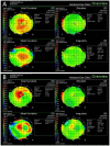
Anterior corneal surface topography before (A) and 4 weeks (B) after Raindrop implantation in a 6mm pocket. A focal increase in corneal curvature and irregularity that correspond to the position of the inlay were observed after implantation. A small increase in anterior elevation was also present.
Fig 5
Fig 5
In vivo confocal microscopy images at the inlay plane (A) and inlay edge (B). Highly reflective particles were observed in all eyes and, although they were more apparent in the early stages post-implantation, they decreased with time. (W1 = week 1, W2 = week 2, W3 = week 3, W4 = week 4)
Fig 6
Fig 6
Representative in vivo confocal microscopy micrographs for different time points at the areas anterior to the inlay edge (A), anterior to the inlay plane (B), and adjacent to the inlay (C). Highly reflective keratocytes were seen in these areas surrounding the inlays. (W1 = week 1, W2 = week 2, W3 = week 3, W4 = week 4)
Fig 7
Fig 7. Stromal keratocyte mean reflectivity for the different pocket/flocket groups.
The 4mm pocket group had significantly higher stromal keratocyte reflectivity than the other groups throughout the study period, whereas the 8mm pocket and flocket groups had lower reflectivity. (W1 = week 1, W2 = week 2, W3 = week 3, W4 = week 4)
Fig 8
Fig 8. Hematoxylin and eosin stained corneal sections and expression of CD11b, fibronectin and HSP47 four weeks post-implantation.
Hematoxylin and eosin stained sections of the 4mm pocket (A1), 6mm pocket (B1), 8mm pocket (C1) and 8mm flocket (D1) groups showed no inflammatory cells or fibrotic capsule formation around the inlay. No CD11b, fibronectin or HSP 47 positive cells were detected in the 4mm pocket (A2-4), 6mm pocket (B2-4J), 8mm pocket (C2-4) and 8mm flocket (D2-4) groups, respectively. Scale bar 100 μm. (arrow points to the inlay) Row 1 represents hematoxylin and eosin, row 2 CD11b, row 3 fibronectin and row 4 HSP47 corneal sections.
Fig 9
Fig 9
Representative Transmission Electron Microscopy micrographs of the collagen fibers in the 6mm pocket (A) and flocket groups (B). The collagen fibers of the corneal stroma surrounding the inlay were intact and regular with no disruption or distortion in all groups. Scale bar 5 μm. (asterisk indicates inlay)

References

    1. Ostrin LA, Glasser A. Accommodation measurements in a prepresbyopic and presbyopic population. J Cataract Refract Surg 2004; 30:1435–44. 10.1016/j.jcrs.2003.12.045 - DOI - PubMed
    1. Holden BA, Fricke TR, Ho SM, Wong R, Schlenther G, Cronjé S, et al. Global vision impairment due to uncorrected presbyopia. Arch Ophthalmol 2008; 126:1731–9. 10.1001/archopht.126.12.1731 - DOI - PubMed
    1. Konstantopoulos A, Mehta JS. Surgical compensation of presbyopia with corneal inlays. Expert Rev Med Devices. 2015;12:341–52. 10.1586/17434440.2015.1007124 - DOI - PubMed
    1. Ito M, Shimizu K, Amano R, Handa T. Assessment of visual performance in pseudophakic monovision. J Cataract Refract Surg 2009;35:710–4. 10.1016/j.jcrs.2008.12.019 - DOI - PubMed
    1. Finkelman YM, Ng JQ, Barrett GD. Patient satisfaction and visual function after pseudophakic monovision. J Cataract Refract Surg 2009;35:998–1002. 10.1016/j.jcrs.2009.01.035 - DOI - PubMed