

The natural killer cell response to West Nile virus in young and old individuals with or without a prior history of infection
- PMID: 28235099
- PMCID: PMC5325267
- DOI: 10.1371/journal.pone.0172625
The natural killer cell response to West Nile virus in young and old individuals with or without a prior history of infection
Abstract
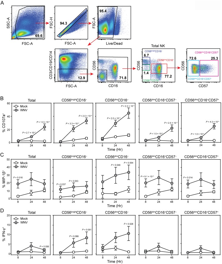
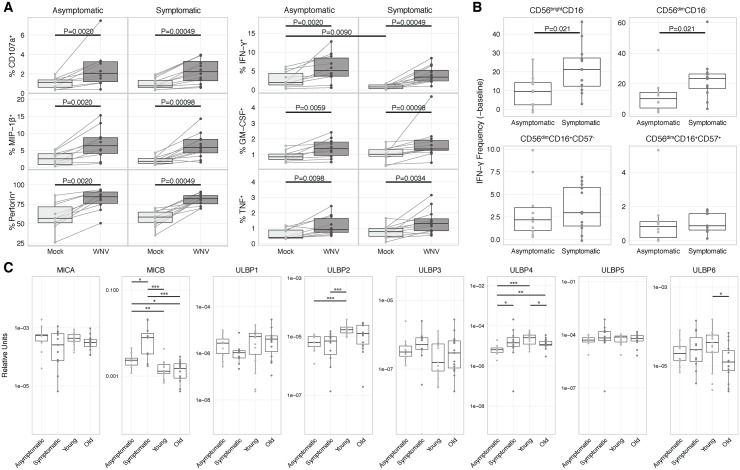
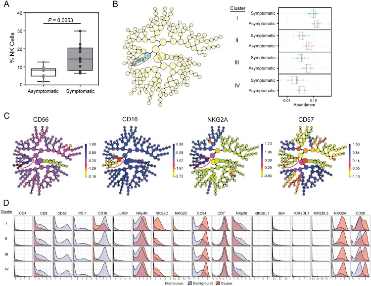
West Nile virus (WNV) typically leads to asymptomatic infection but can cause severe neuroinvasive disease or death, particularly in the elderly. Innate NK cells play a critical role in antiviral defenses, yet their role in human WNV infection is poorly defined. Here we demonstrate that NK cells mount a robust, polyfunctional response to WNV characterized by cytolytic activity, cytokine and chemokine secretion. This is associated with downregulation of activating NK cell receptors and upregulation of NK cell activating ligands for NKG2D. The NK cell response did not differ between young and old WNV-naïve subjects, but a history of symptomatic infection is associated with more IFN-γ producing NK cell subsets and a significant decline in a specific NK cell subset. This NK repertoire skewing could either contribute to or follow heightened immune pathogenesis from WNV infection, and suggests that NK cells could play an important role in WNV infection in humans.
Conflict of interest statement
Figures


Similar articles
-
Quantitative analysis of human NK cell reactivity using latex beads coated with defined amounts of antibodies.Eur J Immunol. 2020 May;50(5):656-665. doi: 10.1002/eji.201948344. Epub 2020 Feb 19. Eur J Immunol. 2020. PMID: 32027754
-
Anti-West Nile virus activity of in vitro expanded human primary natural killer cells.BMC Immunol. 2010 Jan 20;11:3. doi: 10.1186/1471-2172-11-3. BMC Immunol. 2010. PMID: 20089143 Free PMC article.
-
Balance between activating NKG2D, DNAM-1, NKp44 and NKp46 and inhibitory CD94/NKG2A receptors determine natural killer degranulation towards rheumatoid arthritis synovial fibroblasts.Immunology. 2014 Aug;142(4):581-93. doi: 10.1111/imm.12271. Immunology. 2014. PMID: 24673109 Free PMC article. Clinical Trial.
-
The Natural Cytotoxicity Receptors in Health and Disease.Front Immunol. 2019 May 7;10:909. doi: 10.3389/fimmu.2019.00909. eCollection 2019. Front Immunol. 2019. PMID: 31134055 Free PMC article. Review.
-
Role of natural killer and Gamma-delta T cells in West Nile virus infection.Viruses. 2013 Sep 20;5(9):2298-310. doi: 10.3390/v5092298. Viruses. 2013. PMID: 24061543 Free PMC article. Review.
Cited by
-
Natural killer cells and unconventional T cells in COVID-19.Curr Opin Virol. 2021 Aug;49:176-182. doi: 10.1016/j.coviro.2021.06.005. Epub 2021 Jun 19. Curr Opin Virol. 2021. PMID: 34217135 Free PMC article. Review.
-
Early cellular and molecular signatures correlate with severity of West Nile virus infection.iScience. 2023 Nov 2;26(12):108387. doi: 10.1016/j.isci.2023.108387. eCollection 2023 Dec 15. iScience. 2023. PMID: 38047068 Free PMC article.
-
Natural killer cells in antiviral immunity.Nat Rev Immunol. 2022 Feb;22(2):112-123. doi: 10.1038/s41577-021-00558-3. Epub 2021 Jun 11. Nat Rev Immunol. 2022. PMID: 34117484 Free PMC article. Review.
-
Natural killer cell responses to emerging viruses of zoonotic origin.Curr Opin Virol. 2020 Oct;44:97-111. doi: 10.1016/j.coviro.2020.07.003. Epub 2020 Aug 9. Curr Opin Virol. 2020. PMID: 32784125 Free PMC article. Review.
-
Identification of enhanced IFN-γ signaling in polyarticular juvenile idiopathic arthritis with mass cytometry.JCI Insight. 2018 Aug 9;3(15):e121544. doi: 10.1172/jci.insight.121544. eCollection 2018 Aug 9. JCI Insight. 2018. PMID: 30089725 Free PMC article.
References
-
- Centers for Disease Control and Prevention CDC. West Nile virus, Final Cumulative Maps & Data for 1999–2014 2015. http://www.cdc.gov/westnile/statsMaps/cumMapsData.html.
Publication types
MeSH terms
Substances
Grants and funding
LinkOut - more resources
Full Text Sources
Other Literature Sources
Medical
