

Mitophagy in neurodegeneration and aging
- PMID: 28235551
- PMCID: PMC5565781
- DOI: 10.1016/j.neuint.2017.02.007
Mitophagy in neurodegeneration and aging
Abstract
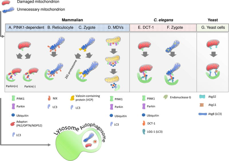
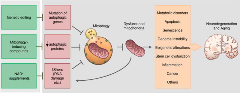
Mitochondrial dysfunction contributes to normal aging and a wide spectrum of age-related diseases, including neurodegenerative disorders such as Parkinson's disease and Alzheimer's disease. It is important to maintain a healthy mitochondrial population which is tightly regulated by proteolysis and mitophagy. Mitophagy is a specialized form of autophagy that regulates the turnover of damaged and dysfunctional mitochondria, organelles that function in producing energy for the cell in the form of ATP and regulating energy homeostasis. Mechanistic studies on mitophagy across species highlight a sophisticated and integrated cellular network that regulates the degradation of mitochondria. Strategies directed at maintaining a healthy mitophagy level in aged individuals might have beneficial effects. In this review, we provide an updated mechanistic overview of mitophagy pathways and discuss the role of reduced mitophagy in neurodegeneration. We also highlight potential translational applications of mitophagy-inducing compounds, such as NAD+ precursors and urolithins.
Published by Elsevier Ltd.
Figures
Similar articles
-
Mitophagy in Alzheimer's Disease and Other Age-Related Neurodegenerative Diseases.Cells. 2020 Jan 8;9(1):150. doi: 10.3390/cells9010150. Cells. 2020. PMID: 31936292 Free PMC article. Review.
-
The Role of Mitophagy in Various Neurological Diseases as a Therapeutic Approach.Cell Mol Neurobiol. 2023 Jul;43(5):1849-1865. doi: 10.1007/s10571-022-01302-8. Epub 2022 Nov 3. Cell Mol Neurobiol. 2023. PMID: 36326951 Free PMC article. Review.
-
Impaired Mitophagy in Neurons and Glial Cells during Aging and Age-Related Disorders.Int J Mol Sci. 2021 Sep 23;22(19):10251. doi: 10.3390/ijms221910251. Int J Mol Sci. 2021. PMID: 34638589 Free PMC article. Review.
-
Mitochondrial clearance: mechanisms and roles in cellular fitness.FEBS Lett. 2021 Apr;595(8):1239-1263. doi: 10.1002/1873-3468.14060. Epub 2021 Mar 8. FEBS Lett. 2021. PMID: 33615465 Review.
-
Mitochondrial autophagy in neural function, neurodegenerative disease, neuron cell death, and aging.Neurobiol Dis. 2011 Jul;43(1):46-51. doi: 10.1016/j.nbd.2010.09.009. Epub 2010 Sep 29. Neurobiol Dis. 2011. PMID: 20887789 Free PMC article. Review.
Cited by
-
Delineating the Role of Mitophagy Inducers for Alzheimer Disease Patients.Aging Dis. 2021 Jun 1;12(3):852-867. doi: 10.14336/AD.2020.0913. eCollection 2021 Jun. Aging Dis. 2021. PMID: 34094647 Free PMC article. Review.
-
Involvement of Mitochondrial Dynamics and Mitophagy in Sevoflurane-Induced Cell Toxicity.Oxid Med Cell Longev. 2021 Feb 26;2021:6685468. doi: 10.1155/2021/6685468. eCollection 2021. Oxid Med Cell Longev. 2021. Retraction in: Oxid Med Cell Longev. 2023 Aug 2;2023:9760436. doi: 10.1155/2023/9760436. PMID: 33728028 Free PMC article. Retracted. Review.
-
Mitochondrial fission and mitophagy are independent mechanisms regulating ischemia/reperfusion injury in primary neurons.Cell Death Dis. 2021 May 12;12(5):475. doi: 10.1038/s41419-021-03752-2. Cell Death Dis. 2021. PMID: 33980811 Free PMC article.
-
Beyond autophagy: LC3-associated phagocytosis and endocytosis.Sci Adv. 2022 Oct 28;8(43):eabn1702. doi: 10.1126/sciadv.abn1702. Epub 2022 Oct 26. Sci Adv. 2022. PMID: 36288309 Free PMC article. Review.
-
Dietary Mitophagy Enhancer: A Strategy for Healthy Brain Aging?Antioxidants (Basel). 2020 Sep 29;9(10):932. doi: 10.3390/antiox9100932. Antioxidants (Basel). 2020. PMID: 33003315 Free PMC article. Review.
References
-
- Rubinsztein DC, Mariño G, Kroemer G. Autophagy and aging. Cell. 2011;146(5):682–695. - PubMed
-
- Madeo F, Tavernarakis N, Kroemer G. Can autophagy promote longevity? Nature cell biology. 2010;12(9):842–846. - PubMed
-
- Hara T, et al. Suppression of basal autophagy in neural cells causes neurodegenerative disease in mice. Nature. 2006;441(7095):885–889. - PubMed
-
- Komatsu M, et al. Loss of autophagy in the central nervous system causes neurodegeneration in mice. Nature. 2006;441(7095):880–884. - PubMed
Publication types
MeSH terms
Grants and funding
LinkOut - more resources
Full Text Sources
Other Literature Sources
Medical
