Case Reports
. 2017 Jun:102:38-48.
doi: 10.1016/j.nbd.2017.02.006. Epub 2017 Feb 22.

SCN3A deficiency associated with increased seizure susceptibility

Affiliations
Case Reports

SCN3A deficiency associated with increased seizure susceptibility

Tyra Lamar et al. Neurobiol Dis. 2017 Jun.

Abstract

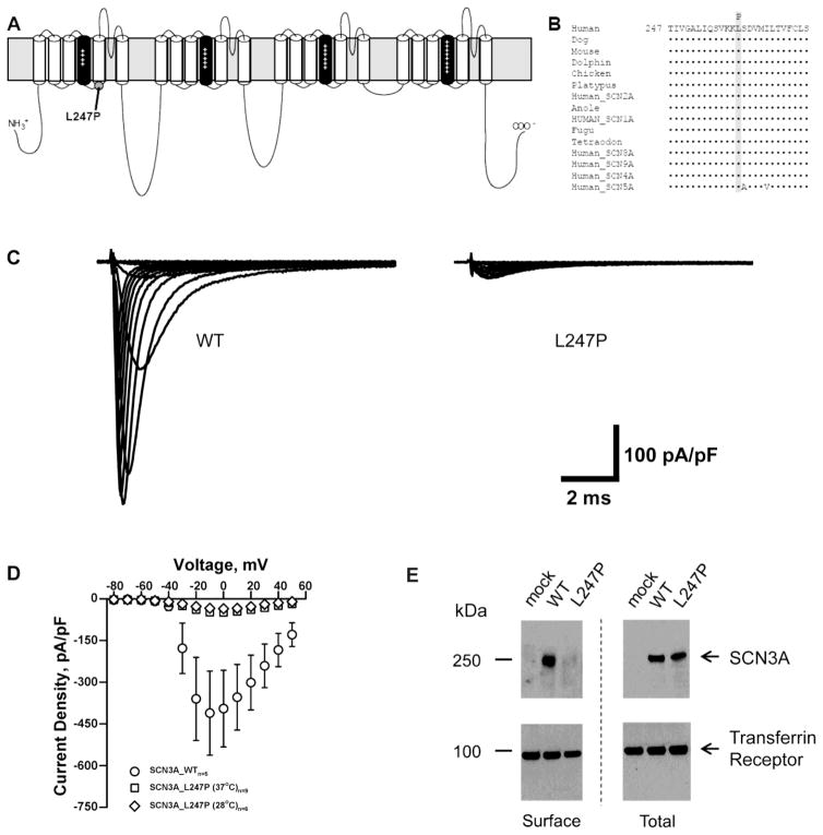
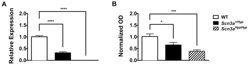
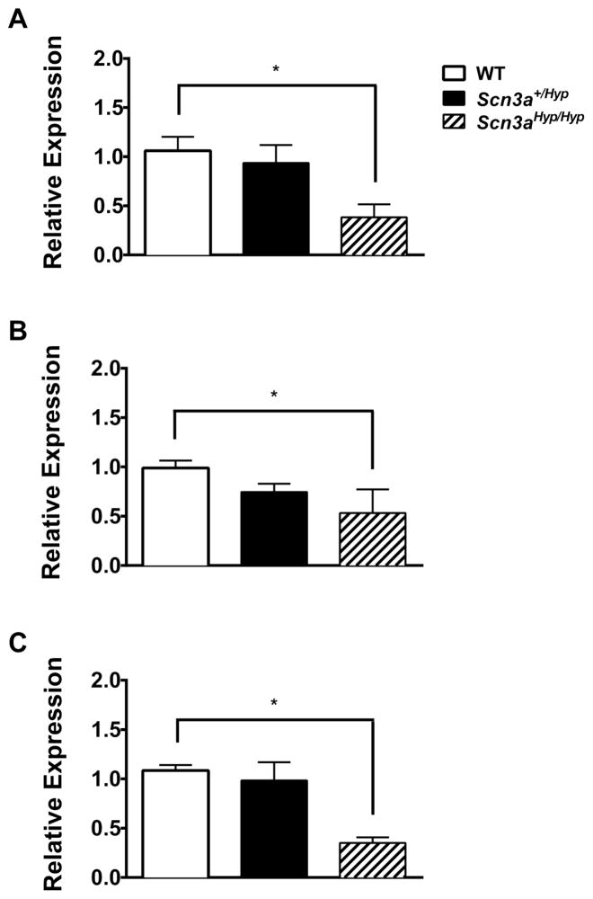
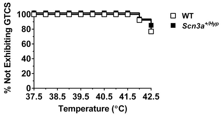
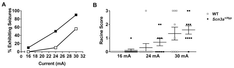
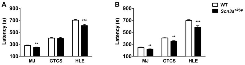
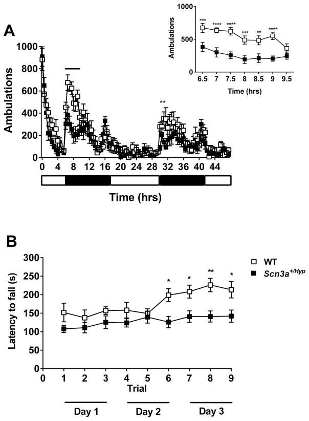
Mutations in voltage-gated sodium channels expressed highly in the brain (SCN1A, SCN2A, SCN3A, and SCN8A) are responsible for an increasing number of epilepsy syndromes. In particular, mutations in the SCN3A gene, encoding the pore-forming Nav1.3 α subunit, have been identified in patients with focal epilepsy. Biophysical characterization of epilepsy-associated SCN3A variants suggests that both gain- and loss-of-function SCN3A mutations may lead to increased seizure susceptibility. In this report, we identified a novel SCN3A variant (L247P) by whole exome sequencing of a child with focal epilepsy, developmental delay, and autonomic nervous system dysfunction. Voltage clamp analysis showed no detectable sodium current in a heterologous expression system expressing the SCN3A-L247P variant. Furthermore, cell surface biotinylation demonstrated a reduction in the amount of SCN3A-L247P at the cell surface, suggesting the SCN3A-L247P variant is a trafficking-deficient mutant. To further explore the possible clinical consequences of reduced SCN3A activity, we investigated the effect of a hypomorphic Scn3a allele (Scn3aHyp) on seizure susceptibility and behavior using a gene trap mouse line. Heterozygous Scn3a mutant mice (Scn3a+/Hyp) did not exhibit spontaneous seizures nor were they susceptible to hyperthermia-induced seizures. However, they displayed increased susceptibility to electroconvulsive (6Hz) and chemiconvulsive (flurothyl and kainic acid) induced seizures. Scn3a+/Hyp mice also exhibited deficits in locomotor activity and motor learning. Taken together, these results provide evidence that loss-of-function of SCN3A caused by reduced protein expression or deficient trafficking to the plasma membrane may contribute to increased seizure susceptibility.

Keywords: Focal epilepsy; Na(v)1.3; SCN3A; Seizure susceptibility; Voltage-gated sodium channel.


Figures

Fig. 1
Fig. 1
SCN3A-L247P is a trafficking deficient mutant. (A) Predicted transmembrane topology of Nav1.3 showing the location of the L247P variant characterized in this study. The S4 segments are shown in black with plus (+) signs and the grey rectangle behind the depiction represents the plasma membrane. (B) Multiple alignment of human homologs and species orthologs of Nav1.3 (Clustal Omega; (Sievers et al., 2011)). The variant amino acid L247P (shaded) is contained within a region of strong evolutionary conservation. (C) Average sodium current traces for SCN3A-WT and SCN3A-L247P co-expressed with β1 and β2 in a heterologous expression system (tsA201 cells). (D) Average current density-voltage relationships are shown for whole-cell currents tsA201 cells transiently co-expressing β1 and β2 with SCN3A-WT or SCN3A-L247P incubated at 37°C or 28°C for 24 hours prior to recording. (E) Total and surface protein were detected with anti-pan VGSC α subunit antibody or anti-transferrin receptor antibody as indicated. The WT SCN3A protein traffics normally to the cell surface, whereas trafficking of SCN3A L247P to the cell surface is significantly impaired.
Fig. 2
Fig. 2
Scn3a expression is reduced in Scn3a+/Hyp mice. (A) Scn3a mRNA expression is reduced by approximately 70% (p < 0.0001) and nearly 100% (p < 0.0001) in Scn3a+/Hyp and Scn3aHyp/Hyp mice respectively. mRNA expression, quantified from real-time PCR analysis, was normalized to β-actin. Expression values for each genotype are relative to WT levels. n = 4 (WT), 5 (Scn3a+/Hyp), 3 (Scn3aHyp/Hyp). (B) Scn3a protein expression, quantified from Western blot analysis, is reduced by approximately 35% (p < 0.05) and 60% (p < 0.001) in membrane-enriched whole brain homogenate from Scn3a+/Hyp and Scn3aHyp/Hyp mice, respectively. Optical density (OD) expression values are relative to WT and normalized to α-tubulin; n = 5 (WT), 6 (Scn3a+/Hyp), 4 (Scn3aHyp/Hyp). Protein results represent the means of triplicate values for each genotype. *p < 0.05, *** < 0.001, ****p < 0.0001; One-way ANOVA. Error bars indicate SEM.
Fig. 3
Fig. 3
Expression of Scn1a, Scn2a, and Scn8a in Scn3a+/Hyp mice. (A) Scn1a, (B) Scn2a), and (C) Scn8a mRNA expression, quantified from real-time PCR analysis, is significantly reduced in whole brain samples from P1 Scn3aHyp/Hyp mice. n = 4 (WT), 5 (Scn3a+/Hyp), 3 (Scn3aHyp/Hyp). Expression values are relative to WT and normalized to β-actin. *p < 0.05, One-way ANOVA. Error bars indicate SEM.
Fig. 4
Fig. 4
Scn3a+/Hyp mice do not exhibit increased susceptibility to hyperthermia-induced seizures. Seizure generation in response to increasing body temperature was comparable between Scn3a+/Hyp mutants (n = 12) and WT littermates (n = 13). Log-Rank Survival Test.
Fig. 5
Fig. 5
Scn3a+/Hyp mice exhibit increased susceptibility to 6 Hz psychomotor seizures. (A) The CC50 is significantly lower for Scn3a+/Hyp mice compared with WT littermates; n = 10/genotype. (B) The difference in the average Racine score between Scn3a+/Hyp mutants and WT littermates was not statistically significant for any current. Mann-Whitney Rank Sum test. Error bars indicate SEM.
Fig. 6
Fig. 6
Scn3a+/Hyp mice exhibit increased susceptibility to flurothyl-induced seizures. (A) Male Scn3a+/Hyp mutants exhibit significantly shorter latencies to the first MJ and HLE when compared to WT littermates. (B) Female Scn3a+/Hyp female mice have significantly shorter latencies to all three seizure components when compared to WT; male n = 33 (WT), 29 (Scn3a+/Hyp ); female n = 32 (WT), 29 (Scn3a+/Hyp). **p < 0.01, ***p < 0.001, Two-tailed Student’s t-test. Error bars indicate SEM.
Fig. 7
Fig. 7
Scn3a+/Hyp mice exhibit increased susceptibility to KA-induced seizures. (A) Following the administration of KA, Scn3a+/Hyp mutants (n = 8) exhibited significantly more severe seizure phenotypes when compared to WT littermates (n = 5). *p < 0.05; Two-way repeated measures ANOVA. (B) Average latency to the first electrographic seizure was shorter in Scn3a+/Hyp mutants. *p < 0.05, Two-tailed Student’s t-test. (C) A representative EEG recording during a KA-induced seizure in a mutant mouse. Seizure onset and termination are indicated within brackets. EEG montage: Cortical EEG 1, Cortical EEG 2, Cortical EEG 3, and Cortical EEG 4 – cortical electrodes. EMG – muscle electrodes. All cortical electrodes and EMG referenced to a second EMG electrode. Error bars indicate SEM.
Fig. 8
Fig. 8
Scn3a+/Hyp mice exhibit deficits in motor behavior. (A) Scn3a+/Hyp mice (n = 8) traveled significantly less than WT (n = 8) during the first dark phase (hours 6.5–9.5) and the second dark phase (hour 31.5–32.0). Inset shows locomotor activity during hours 6.5–9.5. **p < 0.01, ***p < 0.001, ****p < 0.0001; Two-way repeated measures ANOVA (Genotype × Time). White and black bars below the X-axis represent the light and dark periods, respectively. (B) Latency to fall from the rotarod was significantly shorter for Scn3a+/Hyp mutants (n = 11) when compared to WT littermates (n = 9) from trial 6 (Day 2) to the last trial (trial 9) on Day 3. *p < 0.05, **p < 0.01, Two-way repeated measures ANOVA (Genotype × Time). Error bars indicate SEM.

References

    1. Baasch AL, Huning I, Gilissen C, Klepper J, Veltman JA, Gillessen-Kaesbach G, Hoischen A, Lohmann K. Exome sequencing identifies a de novo SCN2A mutation in a patient with intractable seizures, severe intellectual disability, optic atrophy, muscular hypotonia, and brain abnormalities. Epilepsia. 2014;55:e25–9. - PubMed
    1. Barton ME, Klein BD, Wolf HH, White HS. Pharmacological characterization of the 6 Hz psychomotor seizure model of partial epilepsy. Epilepsy Res. 2001;47:217–27. - PubMed
    1. Bechi G, Rusconi R, Cestele S, Striano P, Franceschetti S, Mantegazza M. Rescuable folding defective NaV1.1 (SCN1A) mutants in epilepsy: Properties, occurrence, and novel rescuing strategy with peptides targeted to the endoplasmic reticulum. Neurobiol Dis. 2015;75:100–14. - PubMed
    1. Beckh S, Noda M, Lubbert H, Numa S. Differential regulation of three sodium channel messenger RNAs in the rat central nervous system during development. EMBO J. 1989;8:3611–6. - PMC - PubMed
    1. Black JA, Yokoyama S, Higashida H, Ransom BR, Waxman SG. Sodium channel mRNAs I, II and III in the CNS: cell-specific expression. Brain Res Mol Brain Res. 1994;22:275–89. - PubMed

Publication types

MeSH terms