. 2017 Apr 21;292(16):6438-6451.
doi: 10.1074/jbc.M117.779686. Epub 2017 Feb 24.

A trimeric structural fusion of an antagonistic tumor necrosis factor-α mutant enhances molecular stability and enables facile modification

Affiliations

A trimeric structural fusion of an antagonistic tumor necrosis factor-α mutant enhances molecular stability and enables facile modification

Masaki Inoue et al. J Biol Chem. .

Abstract

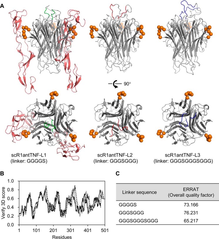
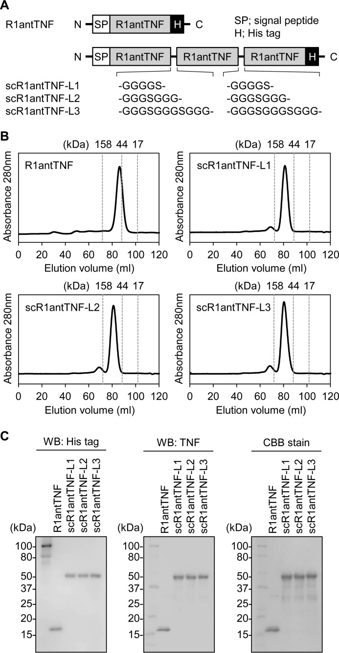
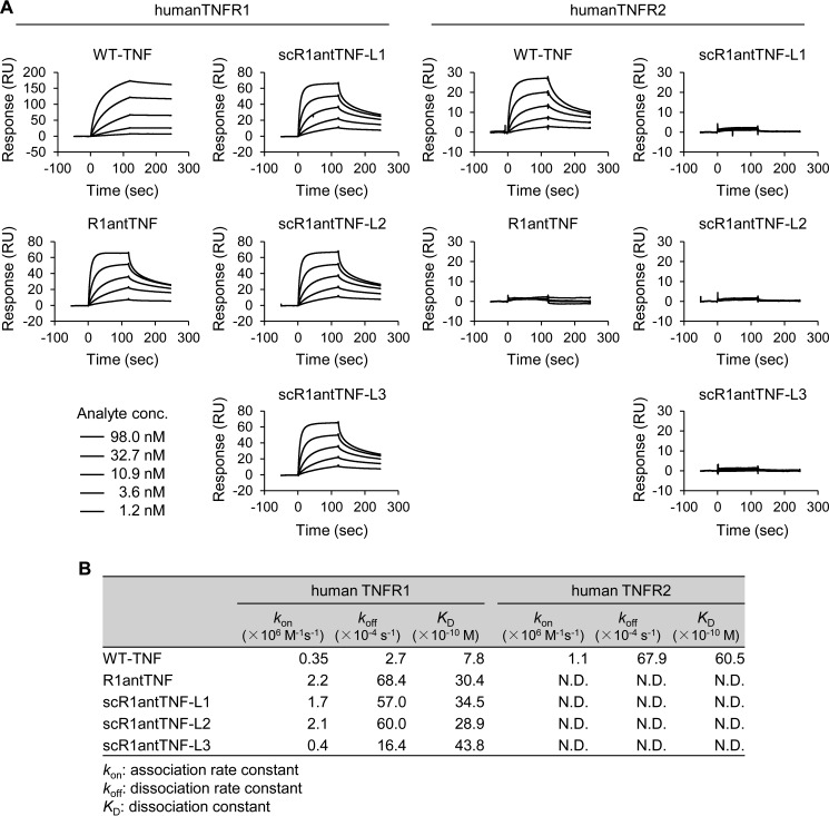
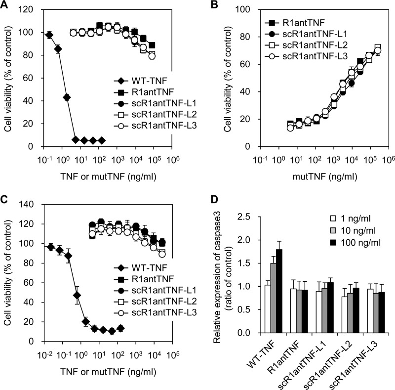
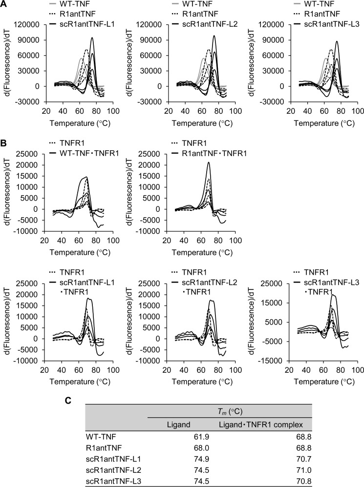
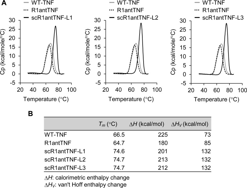
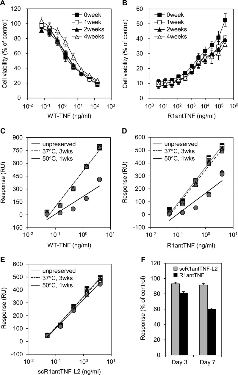
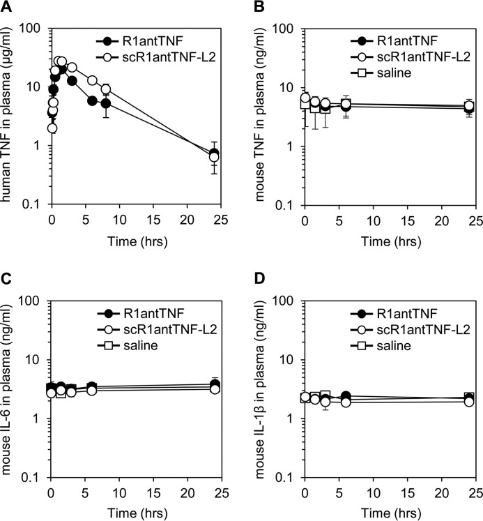
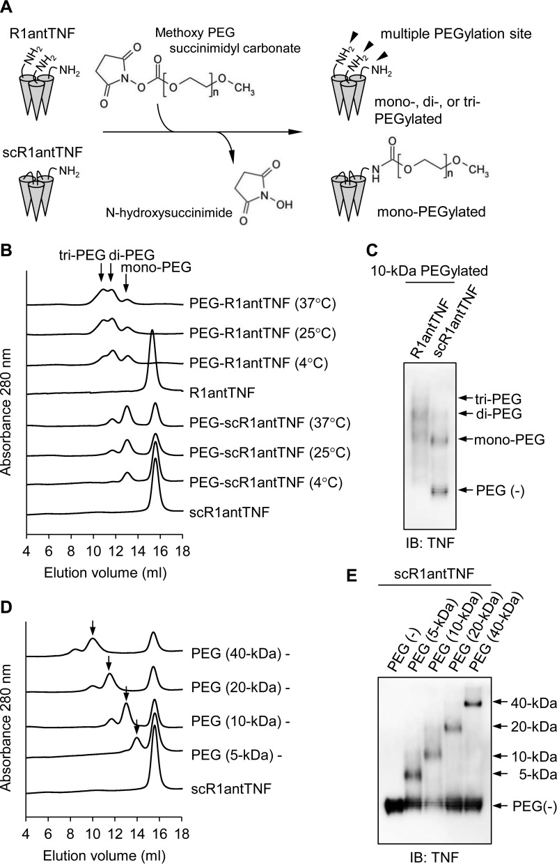
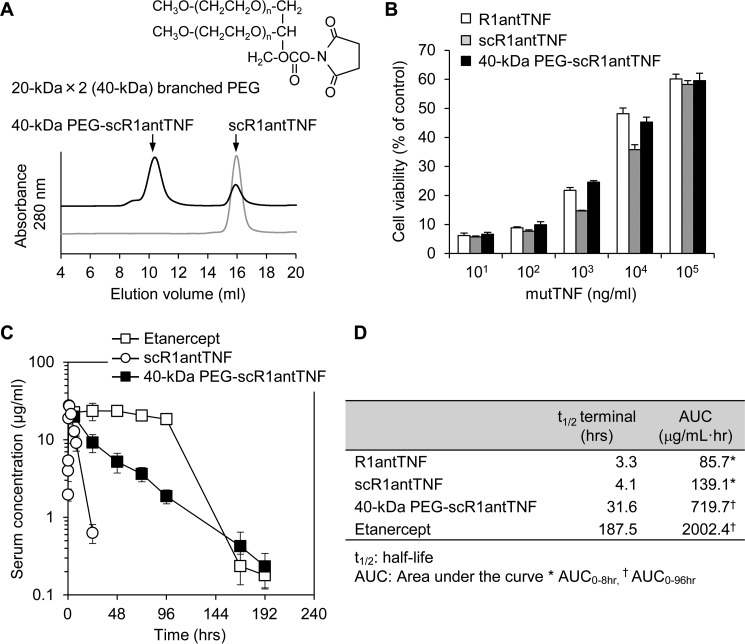
Tumor necrosis factor-α (TNF) exerts its biological effect through two types of receptors, p55 TNF receptor (TNFR1) and p75 TNF receptor (TNFR2). An inflammatory response is known to be induced mainly by TNFR1, whereas an anti-inflammatory reaction is thought to be mediated by TNFR2 in some autoimmune diseases. We have been investigating the use of an antagonistic TNF mutant (TNFR1-selective antagonistic TNF mutant (R1antTNF)) to reveal the pharmacological effect of TNFR1-selective inhibition as a new therapeutic modality. Here, we aimed to further improve and optimize the activity and behavior of this mutant protein both in vitro and in vivo Specifically, we examined a trimeric structural fusion of R1antTNF, formed via the introduction of short peptide linkers, as a strategy to enhance bioactivity and molecular stability. By comparative analysis with R1antTNF, the trimeric fusion, referred to as single-chain R1antTNF (scR1antTNF), was found to retain in vitro molecular properties of receptor selectivity and antagonistic activity but displayed a marked increase in thermal stability. The residence time of scR1antTNF in vivo was also significantly prolonged. Furthermore, molecular modification using polyethylene glycol (PEG) was easily controlled by limiting the number of reactive sites. Taken together, our findings show that scR1antTNF displays enhanced molecular stability while maintaining biological activity compared with R1antTNF.

Keywords: PEGylation; R1antTNF; TNF; autoimmune disease; biomaterials; drug design; molecular stability; protein chemical modification; protein design; protein engineering; single chain; thermal shift assay; tumor necrosis factor (TNF).


Conflict of interest statement

The authors declare that they have no conflicts of interest with the contents of this article

Figures

Figure 1.
Figure 1.
Structural model of scR1antTNFs. A, a structural model of scR1antTNF-L1 (GGGGS linker) with a TNFR1 extracellular domain was constructed using Modeller. The R1antTNF was composed of three monomers (gray schematic). The N- and C-terminal peptide linkers are shown as a green schematic. Monomeric TNFR1, which interacts with the two TNF monomers, is shown as a red schematic. For clarity, only two parts of the three monomers are shown. Amino acids mutated in R1antTNF that are thought to interact with TNFR1 are shown as orange spheres. The peptide linkers of scR1antTNF-L2 (GGGSGGG) and scR1antTNF-L3 (GGGSGGGSGGG) are shown as red and blue schematics, respectively. Verify 3D (B) and ERRAT (C) were used to check the quality of the protein models. The structural models of scR1antTNF with three different linker sequences, GGGGS (gray line), GGGSGGG (dotted line), and GGGSGGGSGGG (black line), were compared using Verify 3D.
Figure 2.
Figure 2.
Generation and characterization of scR1antTNFs. A, schematic primary structures of R1antTNF, scR1antTNF-L1, -L2, and -L3. Each scR1antTNF comprises three R1antTNF domains fused together by short peptide linkers as indicated. Each mutant form of the protein was fused to a signal peptide derived from a mouse IgG VH and His tag in N- and C-terminal regions, respectively. SEC purification (B) and Western blotting (WB) with anti-His tag antibody and anti-TNF antibody followed by Coomassie Brilliant Blue (CBB) stain (C). These results confirmed the expression of the recombinant proteins.
Figure 3.
Figure 3.
In vitro receptor binding ability of scR1antTNFs. A, binding ability of WT-TNF, R1antTNF, scR1andTNF-L1, -L2, and -L3 to human TNFR1 and human TNFR2 was analyzed by SPR. Each sensorgram was measured with serially diluted samples (1.2, 3.6, 10.9, 32.7, and 98.0 nm). B, kinetic parameters of WT-TNF, R1antTNF, and three scR1antTNFs for human TNFR1/R2 were calculated using BIAcore T200 evaluation software. Avidity values of TNF as trimer are shown. RU, response units.
Figure 4.
Figure 4.
In vitro bioactivity of the scR1antTNFs through TNFR1 and TNFR2. Agonistic (A) and antagonistic (B) activities of R1antTNF (closed squares), scR1antTNF-L1 (closed circles), scR1antTNF-L2 (open squares), and scR1antTNF-L3 (open circles) through TNFR1 were confirmed by LM cell assay. The activity of WT-TNF (closed diamonds) was used as a control. C, agonistic activity through TNFR2 was evaluated by cell death of hTNFR2/mFas preadipocytes. D, caspase-3 activation induced by serially diluted WT-TNF, R1antTNF, or scR1antTNFs (1, 10, and 100 ng/ml) using A673 cells was measured with Ac-DEVD-p-nitroanilide. All error bars represent S.D.; n = 3.
Figure 5.
Figure 5.
In vitro thermal stability of scR1antTNFs measured by thermal shift assay. A, thermal stability of WT-TNF (gray line), R1antTNF (dotted line), and scR1antTNFs (black line) was evaluated by TSA. The protein was serially diluted from 700 μg/ml by 2-fold dilution. The inflection point of the peak indicates a protein denaturation temperature (Tm). B, thermal stability of the complex of WT-TNF, R1antTNF, or scR1antTNFs with human TNFR1-Fc was evaluated using TSA of human TNFR1-Fc fusion protein and its complex as shown by the dotted line and black line, respectively. C, Tm values of WT-TNF, R1antTNF, scR1antTNFs, and each complex with TNFR1 are shown.
Figure 6.
Figure 6.
In vitro thermal stability of scR1antTNFs by differential scanning calorimetry. A, thermal stability of WT-TNF (gray line), R1antTNF (dotted line), and scR1antTNF (black line) was evaluated by DSC. The WT-TNF, R1antTNF, and scR1antTNFs were diluted to 900, 900, and 700 μg/ml, respectively. The inflection point of the peak indicates a protein denaturation temperature (Tm). B, values of Tm, ΔH, and ΔHV of WT-TNF, R1antTNF, and scR1antTNFs are shown. Cp, heat capacity.
Figure 7.
Figure 7.
Time-dependent alteration in bioactivity and binding ability of WT-TNF and TNF mutants. Agonistic activity of WT-TNF (A) and antagonistic activity of R1antTNF (B) was gradually reduced in a time-dependent manner by prolonged storage at 37 °C. Samples stored for 0, 1, 2, and 4 weeks are indicated by closed squares, open squares, closed triangles, and open triangles, respectively. WT-TNF (C), R1antTNF (D), and scR1antTNF-L2 (E) was incubated for 3 weeks at 37 °C or for 1 week at 50 °C. Each fitted line was calculated from the Rmax value of these proteins to human TNFR1 measured by SPR. Plots indicate serially diluted protein concentrations (0.05–5.00 ng/ml). Squares, triangles, and circles represent the concentration of protein at 4, 25, and 37 °C, respectively. Results were measured in triplicate. F, binding response alteration of TNF mutants stored at 50 °C for 3 and 7 days was evaluated. Error bars represent S.D.; n = 3. RU, response units.
Figure 8.
Figure 8.
In vivo plasma clearance and cytokine induction of scR1antTNF. A, plasma concentrations of R1antTNF and scR1antTNF-L2 after i.p. injection were measured by ELISA. Error bars represent S.D.; n = 5. Mouse TNF (B), mouse IL-6 (C), and mouse IL-1β (D) in plasma were detected by ELISA. R1antTNF, scR1antTNF-L2, and saline are indicated by closed circles, open circles, and open squares, respectively. Error bars represent S.D.; n = 5.
Figure 9.
Figure 9.
The difference of PEGylation efficiency between R1antTNF and scR1antTNF. A, the PEGylation reaction scheme of R1antTNF and scR1antTNF. R1antTNF, but not scR1antTNF, has multiple reactive sites as shown by arrowheads. Therefore, three types of PEGylated mutants were created. B, R1andTNF and scR1antTNF modified with 10-kDa PEG at different reaction temperatures (4, 25, and 37 °C) were analyzed by gel-filtration chromatography. The resultant chromatograms were overlaid. Peaks of mono-, di-, and tri-PEGylated protein are indicated by an arrow. C, each PEGylated mutant was also detected by native PAGE and Western blotting with an anti-human TNF antibody. Gel-filtration chromatography (D) and native PAGE followed by Western blotting (E) were performed to compare the PEGylated reaction efficacy due to differences of PEG size (5, 10, 20, and 40 kDa). Arrows indicate a peak of mono-PEGylated product. IB, immunoblotting.
Figure 10.
Figure 10.
Generation of 40-kDa branched PEGylated scR1antTNF and its in vitro activity and in vivo plasma clearance after a single dose. A, the scR1antTNF was incubated with 20-kDa × 2 (40-kDa) branched PEG to generate 40-kDa PEG-scR1antTNF. SEC analysis was performed to check the extent of reaction. B, the inhibition activity of TNF-induced cytotoxicity of 40-kDa PEG-scR1antTNF (black bars) was compared with R1antTNF (white bars) and scR1antTNF (gray bars) by LM cell assay. Error bars represent S.D.; n = 3. C, plasma concentrations of scR1antTNF (open circles) and 40-kDa PEG-scR1antTNF (closed squares) after a single i.p. injection were measured by ELISA. Etanercept (open squares), a human TNFR2-Fc fusion protein, was used as the positive control. Error bars represent S.D.; n = 5. D, the half-life and AUC of R1antTNF, scR1antTNF, and 40-kDa PEG-scR1antTNF were evaluated by moment analysis.

References

    1. Thalayasingam N., and Isaacs J. D. (2011) Anti-TNF therapy. Best Pract. Res. Clin. Rheumatol. 25, 549–567 - PubMed
    1. Bradley J. R. (2008) TNF-mediated inflammatory disease. J. Pathol. 214, 149–160 - PubMed
    1. Liang S., Dai J., Hou S., Su L., Zhang D., Guo H., Hu S., Wang H., Rao Z., Guo Y., and Lou Z. (2013) Structural basis for treating tumor necrosis factor α (TNFα)-associated diseases with the therapeutic antibody infliximab. J. Biol. Chem. 288, 13799–13807 - PMC - PubMed
    1. Gómez-Reino J. J., Carmona L., Valverde V. R., Mola E. M., Montero M. D., and BIOBADA SER Group (2003) Treatment of rheumatoid arthritis with tumor necrosis factor inhibitors may predispose to significant increase in tuberculosis risk: a multicenter active-surveillance report. Arthritis Rheum. 48, 2122–2127 - PubMed
    1. Lubel J. S., Testro A. G., and Angus P. W. (2007) Hepatitis B virus reactivation following immunosuppressive therapy: guidelines for prevention and management. Intern. Med. J. 37, 705–712 - PubMed

MeSH terms

Substances

LinkOut - more resources