. 2017 Apr 21;292(16):6600-6620.
doi: 10.1074/jbc.M116.742627. Epub 2017 Feb 24.

Nitric-oxide synthase trafficking inducer is a pleiotropic regulator of endothelial cell function and signaling

Affiliations

Nitric-oxide synthase trafficking inducer is a pleiotropic regulator of endothelial cell function and signaling

Shreeta Chakraborty et al. J Biol Chem. .

Abstract

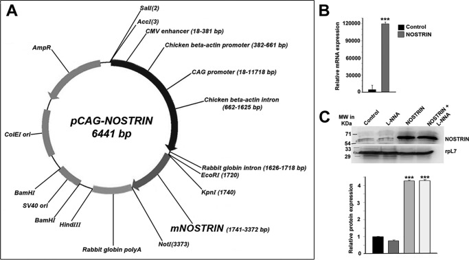
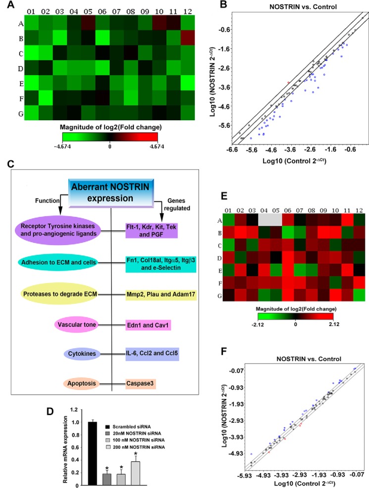
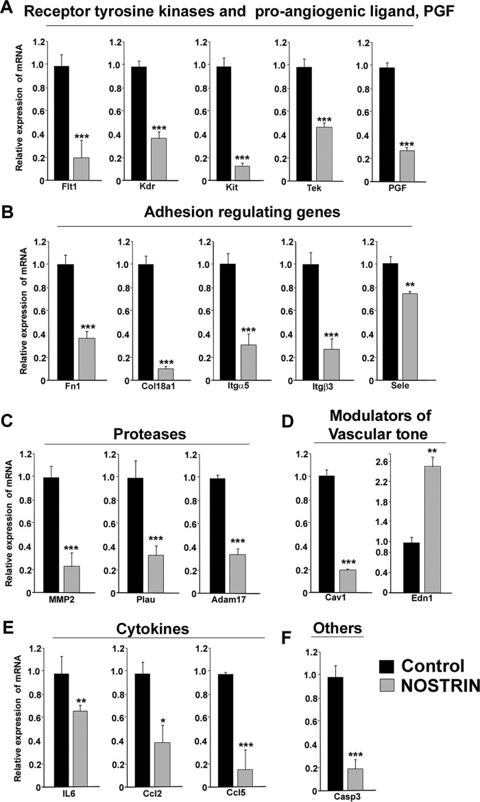
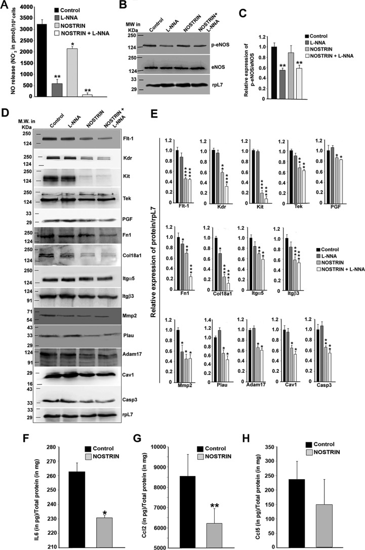
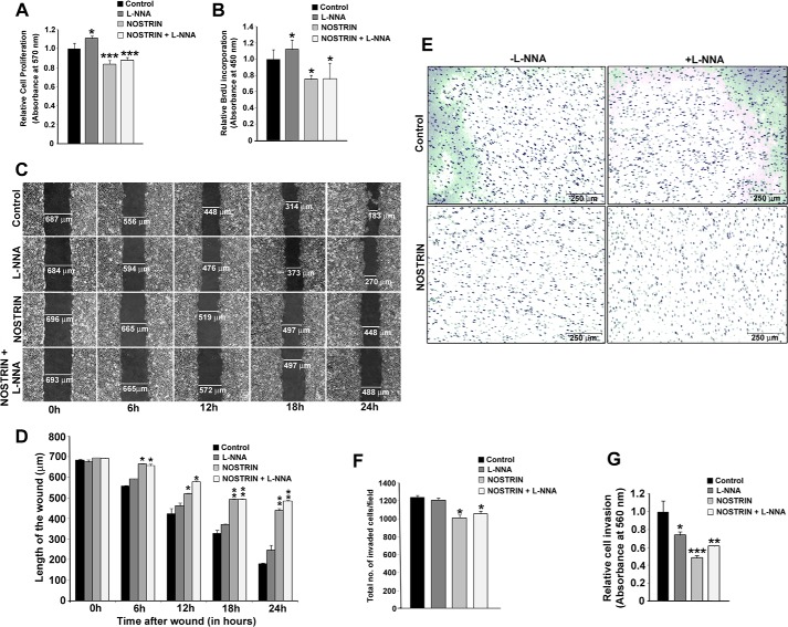
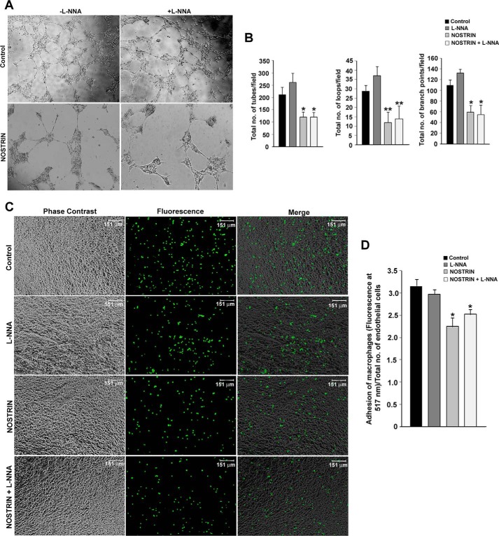
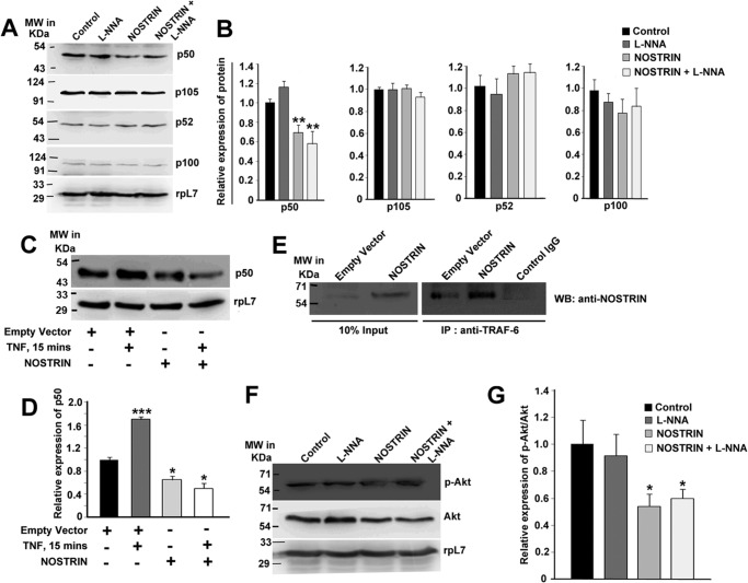
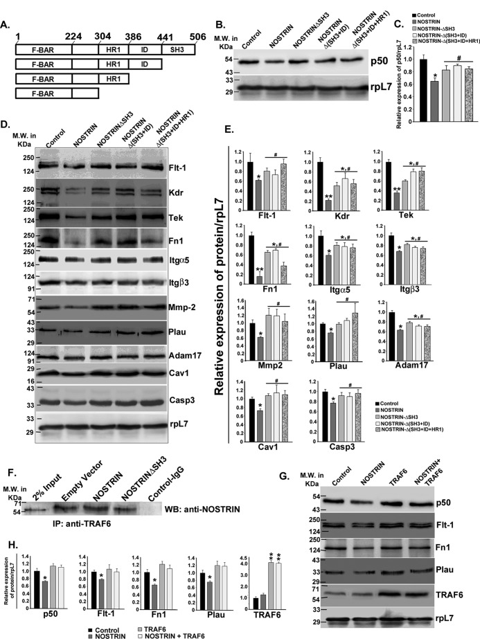
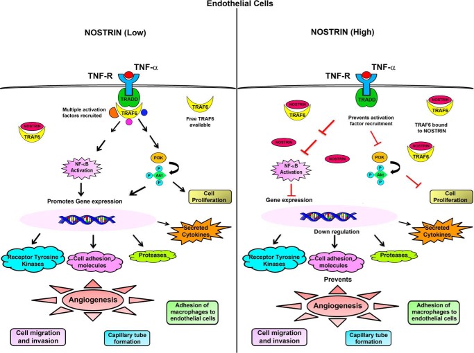
Endothelial nitric-oxide synthase (eNOS) and its bioactive product, nitric oxide (NO), mediate many endothelial cell functions, including angiogenesis and vascular permeability. For example, vascular endothelial growth factor (VEGF)-mediated angiogenesis is inhibited upon reduction of NO bioactivity both in vitro and in vivo Moreover, genetic disruption or pharmacological inhibition of eNOS attenuates angiogenesis during tissue repair, resulting in delayed wound closure. These observations emphasize that eNOS-derived NO can promote angiogenesis. Intriguingly, eNOS activity is regulated by nitric-oxide synthase trafficking inducer (NOSTRIN), which sequesters eNOS, thereby attenuating NO production. This has prompted significant interest in NOSTRIN's function in endothelial cells. We show here that NOSTRIN affects the functional transcriptome of endothelial cells by down-regulating several genes important for invasion and angiogenesis. Interestingly, the effects of NOSTRIN on endothelial gene expression were independent of eNOS activity. NOSTRIN also affected the expression of secreted cytokines involved in inflammatory responses, and ectopic NOSTRIN overexpression functionally restricted endothelial cell proliferation, invasion, adhesion, and VEGF-induced capillary tube formation. Furthermore, NOSTRIN interacted directly with TNF receptor-associated factor 6 (TRAF6), leading to the suppression of NFκB activity and inhibition of AKT activation via phosphorylation. Interestingly, TNF-α-induced NFκB pathway activation was reversed by NOSTRIN. We found that the SH3 domain of NOSTRIN is involved in the NOSTRIN-TRAF6 interaction and is required for NOSTRIN-induced down-regulation of endothelial cell proteins. These results have broad biological implications, as aberrant NOSTRIN expression leading to deactivation of the NFκB pathway, in turn triggering an anti-angiogenic cascade, might inhibit tumorigenesis and cancer progression.

Keywords: TNF receptor-associated factor (TRAF); angiogenesis; endothelial cell; invasion; nitric-oxide synthase.


Conflict of interest statement

The authors declare that they have no conflicts of interest with the contents of this article

Figures

Figure 1.
Figure 1.
Overexpression of Nostrin in MS1 endothelial cells. A, mammalian expression vector containing full-length Nostrin cDNA cloned from murine placenta under synthetic CAG promoter. B, overexpression of full-length Nostrin in MS1 endothelial cells by transfection of pCAG-NOSTRIN was quantified by real-time PCR, and a massive up-regulation in mRNA transcript was obtained. In each experiment requiring Nostrin overexpression, up-regulation of Nostrin was confirmed by real-time PCR. C, Western blotting analysis of NOSTRIN. The four treatment groups included were endothelial cells transfected with vector backbone (Control), vector backbone along with l-NNA treatment, Nostrin cDNA, and Nostrin cDNA followed by l-NNA treatment. The experiments were repeated using at least three independent biological replicates. rpL7 was used as a loading control. Quantification of protein bands is show in the bar graph (lower panel), normalized to the loading control using ImageJ software. Values are represented as mean ± S.E. of three independent experiments. ***, p < 0.005 (B and C).
Figure 2.
Figure 2.
PCR array analysis profiling the functional transcriptome of endothelial cells by ectopic overexpression or siRNA-mediated down-regulation of Nostrin. A, heat map represents the fold change in expression of 84 different genes in Nostrin-overexpressing endothelial cells as compared with control cells transfected with empty vector backbone (see also Fig. 1, A and B). Normalization was done using the housekeeping gene, which showed no change in NOSTRIN-overexpressed MS1 cells compared with control in three biological replicates using online software provided by SABiosciences. Up-regulated genes are shown in red, down-regulated in green, and those not regulated in black. B, scatter plot shows genes with similar expression levels (within the black lines) that are close to the line of regression. The red dots above the black lines represent up-regulated genes, whereas the blue dots below the lines represent down-regulated genes. The scatter plot demonstrates the log10 of normalized gene expression levels in the control group (x axis) versus that in NOSTRIN-overexpressing condition (y axis). Values are represented as mean ± S.E. of three independent experiments. C, function-based annotation of the 19 genes that are differentially regulated in all three experiments. Results about aberrant Nostrin expression are shown in a flowchart. D, real-time PCR showing siRNA-mediated down-regulation of Nostrin using a combination of two prevalidated Silencer Select siRNAs at three different dosages. A 20 nm dose of total siRNAs (at 10 nm each) was used for further experiments. E, heat map representing the change in expression of 84 genes by PCR array using mRNA from NOSTRIN down-regulated (20 nm concentration) endothelial cells compared with scrambled siRNA treatment. Colors depicted in individual wells are analyzed similarly as described above in A. F, scatter plot of Nostrin down-regulation shows that most of the genes displayed similar expression levels (within black lines), and -fold change values were less than 1 (not significant). The graphs and plots were generated using the mean values of three biological replicates. *, p < 0.05 (D).
Figure 3.
Figure 3.
Real-time PCR analysis of transcripts in endothelial cells affected by aberrant NOSTRIN expression. Quantitative real-time PCR analysis of endothelial function-associated genes shows significant differences in Nostrin-overexpressed versus control endothelial cells. The amount of a specific mRNA was normalized relative to the amount of rpL7 (ΔCt = Ctgene − CtrpL7). The -fold change of gene expression was measured by using 2−ΔΔCt, where ΔΔCt denotes the change in ΔCt values between samples and reference sample (here, vector backbone-transfected endothelial cells sample was used as the reference sample, ΔΔCt = ΔCtgene_NOSTRIN − ΔCtgene_control). Error bars represent S.E. from three different biological replicates. A, the receptor tyrosine kinases including Flt-1, Kdr, Kit, and Tek along with the pro-angiogeneic ligand Pgf, which can bind to Flt-1, were down-regulated significantly with overexpression of NOSTRIN. B, NOSTRIN overexpression led to significant down-regulation of transcripts of several genes involved in adhesion and invasion such as Fn1, Col18a1, Itgα5, Itgβ3, and Sele. C, expression of the three proteases Mmp-2, Plau, and Adam-17 was found to be diminished remarkably by NOSTRIN overexpression. D, Cav1, a well-known regulator of vascular tone, was down-regulated, whereas Edn-1, a vasoconstrictor, was up-regulated significantly by NOSTRIN overexpression. E, a significant decrease in mRNA levels of cytokines such as IL6, Ccl2, and Ccl5 was induced by NOSTRIN overexpression. F, Casp3, which induces apoptosis, was found to be down-regulated on NOSTRIN overexpression. *, p < 0.05; **, p < 0.01; and ***, p < 0.005.
Figure 4.
Figure 4.
NOSTRIN decreases NO levels in endothelial cells without affecting the phopho-eNOS levels, and the effect of NOSTRIN on endothelial cell function-associated proteins is eNOS-independent. A, NO production was measured in culture supernatants in the form of nitrites. Error bars represent S.E. from three different biological replicates. B, Western blotting analysis of eNOS and phospho-eNOS from endothelial cells from the four treatment groups. C, relative expression of phospho-eNOS with respect to eNOS was quantified by ImageJ software after normalization with rpL7 using three biological replicates from B. D, Western blotting analysis of protein levels of 14 of 19 genes, associated with various endothelial cell functions were reproducibly altered by NOSTRIN overexpression in three biological replicates. The positions of the molecular-weight markers for the spliced blots are shown on the left side of the image. E, quantification by ImageJ software of the proteins relative to rpL7 using three biological replicates from D. F–H, ELISA analysis of secreted cytokines from endothelial cells transfected with NOSTRIN and vector backbone (Control). Cytokines were quantified in pg relative to total protein estimated in mg. Each experiment was repeated three times with different biological samples. Error bars represent S.E. *, p < 0.05; **, p < 0.01; and ***, p < 0.005.
Figure 5.
Figure 5.
NOSTRIN curtails the angiogenic potential of endothelial cells by inhibiting cell proliferation, migration and invasion. A, MTT assay showing NOSTRIN-mediated repression of endothelial cell proliferation. The four treatment groups are the same as described in the legend for Fig. 4. Error bars represent S.E. from three different biological replicates. B, BrdU incorporation assay demonstrating inhibition of cell proliferation by NOSTRIN. Error bars represent S.E. from three different biological replicates. C, photomicrographic images from the scratch wound assay depicting wound closure at various time points in the four treatment groups. D, quantification by Leica LAS software of the length of the wound at various time intervals from a minimum of five measurements in each experiment using three different biological samples. At each time point, the length of the wound was compared with that of control. Starting from 6 h, the length of the wound was significantly bigger in the presence of NOSTRIN as compared with control. E, photomicrograph of invaded endothelial cells from the cell invasion assay. F, cell count per microscopic field from the four treatment groups. Error bars represent S.E. calculated using values of the count from three different biological replicates and five microscopic fields per replicate. G, colorimetric quantification of the stained, invaded cells at the lower surface of the membrane in the four treatment groups. Error bars represent S.E. from three biological replicates. *, p < 0.05; **, p < 0.01; and ***, p < 0.005.
Figure 6.
Figure 6.
NOSTRIN inhibits VEGF-induced tube formation and adhesion of macrophages to endothelial cells. A, photomicrograph of tube formation in the four treatment groups at 12 h following plating of cells on basement membrane matrix-coated plates. Experiments were repeated three times with different biological samples. B, quantification of capillary tube formation in four treatment groups using data from three replicate experiments and five random microscopic fields per experiment. Data were analyzed using Wimasis image analysis software. Error bars represent S.E. C, photomicrograph images of calcein-labeled macrophages adhered to endothelial cell monolayer from four treatment groups. D, quantification of fluorescence of the adhered macrophages normalized to total number of endothelial cells available per well. *, p < 0.05; and **, p < 0.01.
Figure 7.
Figure 7.
NOSTRIN suppresses NFκB- and AKT-signaling pathway and reverses TNFα-induced NFκB activation by directly interacting with TRAF6. A, Western blotting analysis of proteins from endothelial cells of the four treatment groups evaluating the levels of NFκB1 (p105/p50) and NFκB2 (p100/p52). B, densitometric quantification of proteins from the Western blotting analysis depicted in A normalized to loading control rpL7 using ImageJ software. Error bars represent S.E. from three biological replicates. C, immunoblot analysis of p50 from endothelial cells in the absence or presence of TNF-α (25 ng/ml) stimulation for 15 min (lanes 1 and 2) and of p50 from NOSTRIN-overexpressing endothelial cells in the absence or presence of TNF-α (25 ng/ml) stimulation for 15 min (lanes 3 and 4). D, quantification of p50 levels from C by densitometric analysis normalized to loading control rpL7 using ImageJ software. Error bars represent S.E. from three experiments using three different biological samples. E, immunoprecipitation with anti-TRAF6 antibody followed by immunoblotting (WB) using anti-NOSTRIN antibody in endothelial cells transfected with empty vector or NOSTRIN cDNA. The experiment was repeated three times to ensure reproducibility. F, Western blotting analysis of p-AKT and AKT from endothelial cells from the four treatment groups. RpL7 was used as a loading control. G, densitometric quantification of p-AKT protein level relative to total AKT protein from three replicates using ImageJ software. Error bars represent S.E. from three biological replicates. *, p < 0.05; **, p < 0.01; and ***, p < 0.005.
Figure 8.
Figure 8.
NOSTRIN-mediated effects on endothelial cell functional protein are reversed by deletion of SH3 domain of NOSTRIN, responsible for NOSTRIN-TRAF6 interaction. A, domain structure of full-length NOSTRIN and deletion mutants cloned and used for experiments. Amino acid numbers are marked at the borders of each domain. B, Western blotting analysis using proteins from endothelial cells to evaluate the transcriptionally active form of NFκB1 (p50) in the absence or presence of either full-length NOSTRIN or various deletion mutants of NOSTRIN. C, densitometric quantification of p50 from the Western blotting depicted in B, normalized to loading control rpL7 using ImageJ software. Error bars represent S.E. from three biological replicates. D, Western blotting analysis of endothelial cell proteins known to be affected by NOSTRIN in the absence or presence of either full-length or various deletion mutant of NOSTRIN. E, quantification of protein levels from those mentioned in D by densitometric analysis, normalized to loading control rpL7 using ImageJ software. Error bars represent S.E. from three experiments using three different biological samples. F, immunoprecipitation (IP) with anti-TRAF6 antibody followed by immunoblotting (WB) using anti-NOSTRIN antibody in endothelial cells transfected with empty vector, NOSTRIN, or NOSTRINΔSH3 mutant cDNA. The experiment was repeated three times to ensure reproducibility. G, Western blotting analysis of p50 and proteins representing three different functional aspect of endothelial cell affected by NOSTRIN in the four treatment groups shows that TRAF6 overexpression overrides the effect of NOSTRIN. H, densitometric quantification of proteins from Western blotting depicted in G, normalized to loading control rpL7 using ImageJ software. Error bars represent S.E. from three biological replicates. *, p < 0.05; and **, p < 0.01 compared with control; #, p < 0.05 compared with full-length NOSTRIN overexpression.
Figure 9.
Figure 9.
A schematic representation or summary of the alteration in endothelial cell signaling by aberrant NOSTRIN expression. Binding of TNF-α to its cognate receptor leads to the recruitment of several adaptor proteins and activation factors that in turn can activate the NFκB pathway and also promote phosphorylation of AKT. When NOSTRIN levels are low, free TRAF6 molecules are available to bind to TRADD and transduce the signal to downstream activation factors. Rapid activation of the NFκB pathway leads to expression of several pro-angiogenic genes along with production of pro-inflammatory cytokines that are direct targets of the transcription factor NFκB. On the other hand, when NOSTRIN levels are very high, NOSTRIN can bind directly to TRAF6 and thus inhibit its binding to TRADD on TNFα stimulation. This is followed by suppression of NFκB activity along with inhibition of AKT phosphorylation. As a consequence, gene expression is inhibited, and multiple functions of endothelial cells required for angiogenesis are severely compromised.

References

    1. Yoo S. Y., and Kwon S. M. (2013) Angiogenesis and its therapeutic opportunities. Mediators Inflamm. 2013, 127170. - PMC - PubMed
    1. Carmeliet P. (2003) Angiogenesis in health and disease. Nat. Med. 9, 653–660 - PubMed
    1. Chakraborty S., and Ain R. (2016) Endothelial cell biology: assessment of endothelial cell function, in Trends in Experimental Biology, pp. 1–21, Excel India Publishers, New Delhi, India
    1. Adams R. H., and Alitalo K. (2007) Molecular regulation of angiogenesis and lymph-angiogenesis. Nat. Rev. Mol. Cell Biol. 8, 464–478 - PubMed
    1. De Caterina R., Libby P., Peng H. B., Thannickal V. J., Rajavashisth T. B., Gimbrone M. A. Jr., Shin W. S., and Liao J. K. (1995) Nitric oxide decreases cytokine-induced endothelial activation. Nitric oxide selectively reduces endothelial expression of adhesion molecules and proinflammatory cytokines. J. Clin. Invest. 96, 60–68 - PMC - PubMed

Publication types

MeSH terms