

Corpus callosal atrophy and associations with cognitive impairment in Parkinson disease
- PMID: 28235816
- PMCID: PMC5373777
- DOI: 10.1212/WNL.0000000000003764
Corpus callosal atrophy and associations with cognitive impairment in Parkinson disease
Abstract
Objective: To investigate atrophy of the corpus callosum on MRI in Parkinson disease (PD) and its relationship to cognitive impairment.
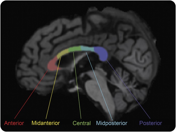
Methods: One hundred patients with PD and 24 healthy control participants underwent clinical and neuropsychological evaluations and structural MRI brain scans. Participants with PD were classified as cognitively normal (PD-NC; n = 28), having mild cognitive impairment (PD-MCI; n = 47), or having dementia (PDD; n = 25) by Movement Disorder Society criteria. Cognitive domain (attention/working memory, executive function, memory, language, visuospatial function) z scores were calculated. With the use of FreeSurfer image processing, volumes for total corpus callosum and its subsections (anterior, midanterior, central, midposterior, posterior) were computed and normalized by total intracranial volume. Callosal volumes were compared between participants with PD and controls and among PD cognitive groups, covarying for age, sex, and PD duration and with multiple comparison corrections. Regression analyses were performed to evaluate relationships between callosal volumes and performance in cognitive domains.
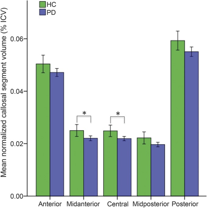
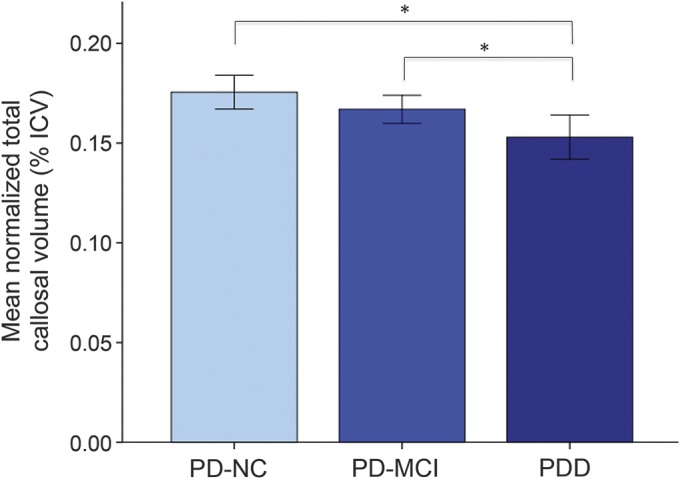
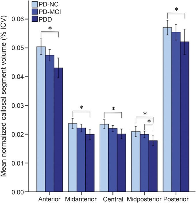
Results: Participants with PD had reduced corpus callosum volumes in midanterior and central regions compared to healthy controls. Participants with PDD demonstrated decreased callosal volumes involving multiple subsections spanning anterior to posterior compared to participants with PD-MCI and PD-NC. Regional callosal atrophy predicted cognitive domain performance such that central volumes were associated with the attention/working memory domain; midposterior volumes with executive function, language, and memory domains; and posterior volumes with memory and visuospatial domains.
Conclusions: Notable volume loss occurs in the corpus callosum in PD, with specific neuroanatomic distributions in PDD and relationships of regional atrophy to different cognitive domains. Callosal volume loss may contribute to clinical manifestations of PD cognitive impairment.
© 2017 American Academy of Neurology.
Figures
Similar articles
-
White matter abnormalities in the corpus callosum with cognitive impairment in Parkinson disease.Neurology. 2018 Dec 11;91(24):e2244-e2255. doi: 10.1212/WNL.0000000000006646. Epub 2018 Nov 14. Neurology. 2018. PMID: 30429273 Free PMC article.
-
Cortical thinning associated with mild cognitive impairment in Parkinson's disease.Mov Disord. 2014 Oct;29(12):1495-503. doi: 10.1002/mds.25982. Epub 2014 Aug 7. Mov Disord. 2014. PMID: 25100674
-
Corpus callosum in neurodegenerative diseases: findings in Parkinson's disease.Dement Geriatr Cogn Disord. 2005;20(6):345-51. doi: 10.1159/000088526. Epub 2005 Sep 26. Dement Geriatr Cogn Disord. 2005. PMID: 16192724
-
Corpus callosum atrophy is a possible indicator of region- and cell type-specific neuronal degeneration in Alzheimer disease: a magnetic resonance imaging analysis.Arch Neurol. 1998 Feb;55(2):193-8. doi: 10.1001/archneur.55.2.193. Arch Neurol. 1998. PMID: 9482361 Review.
-
Corpus callosum damage to account for cognitive, affective, and social-cognitive dysfunctions in multiple sclerosis: A model of callosal disconnection syndrome?Mult Scler. 2023 Feb;29(2):160-168. doi: 10.1177/13524585221091067. Epub 2022 Apr 27. Mult Scler. 2023. PMID: 35475386 Review.
Cited by
-
Primary CNS lymphoma of the corpus callosum: presentation and neurocognitive outcomes.J Neurooncol. 2022 May;158(1):99-109. doi: 10.1007/s11060-022-04014-7. Epub 2022 Apr 21. J Neurooncol. 2022. PMID: 35445956
-
Deep learning to differentiate parkinsonian disorders separately using single midsagittal MR imaging: a proof of concept study.Eur Radiol. 2019 Dec;29(12):6891-6899. doi: 10.1007/s00330-019-06327-0. Epub 2019 Jul 1. Eur Radiol. 2019. PMID: 31264017
-
Automatic deep learning multicontrast corpus callosum segmentation in multiple sclerosis.J Neuroimaging. 2022 May;32(3):459-470. doi: 10.1111/jon.12972. Epub 2022 Jan 26. J Neuroimaging. 2022. PMID: 35083815 Free PMC article.
-
Structural neuroimaging markers of normal pressure hydrocephalus versus Alzheimer's dementia and Parkinson's disease, and hydrocephalus versus atrophy in chronic TBI-a narrative review.Front Neurol. 2024 Mar 21;15:1347200. doi: 10.3389/fneur.2024.1347200. eCollection 2024. Front Neurol. 2024. PMID: 38576534 Free PMC article. Review.
-
Longitudinal Macro/Microstructural Alterations of Different Callosal Subsections in Parkinson's Disease Using Connectivity-Based Parcellation.Front Aging Neurosci. 2020 Nov 4;12:572086. doi: 10.3389/fnagi.2020.572086. eCollection 2020. Front Aging Neurosci. 2020. PMID: 33328954 Free PMC article.
References
-
- Levy G, Tang MX, Louis ED, et al. . The association of incident dementia with mortality in PD. Neurology 2002;59:1708–1713. - PubMed
-
- Hely MA, Reid WG, Adena MA, Halliday GM, Morris JG. The Sydney multicenter study of Parkinson's disease: the inevitability of dementia at 20 years. Movement Disord 2008;23:837–844. - PubMed
-
- Duncan GW, Firbank MJ, O'Brien JT, Burn DJ. Magnetic resonance imaging: a biomarker for cognitive impairment in Parkinson's disease? Movement Disord 2013;28:425–438. - PubMed
MeSH terms
Grants and funding
LinkOut - more resources
Full Text Sources
Other Literature Sources
Medical