. 2017 Jul 5;25(7):1606-1615.
doi: 10.1016/j.ymthe.2017.01.014. Epub 2017 Feb 22.

Targeted Intraceptor Nanoparticle for Neovascular Macular Degeneration: Preclinical Dose Optimization and Toxicology Assessment

Affiliations

Targeted Intraceptor Nanoparticle for Neovascular Macular Degeneration: Preclinical Dose Optimization and Toxicology Assessment

Xiaohui Zhang et al. Mol Ther. .

Abstract

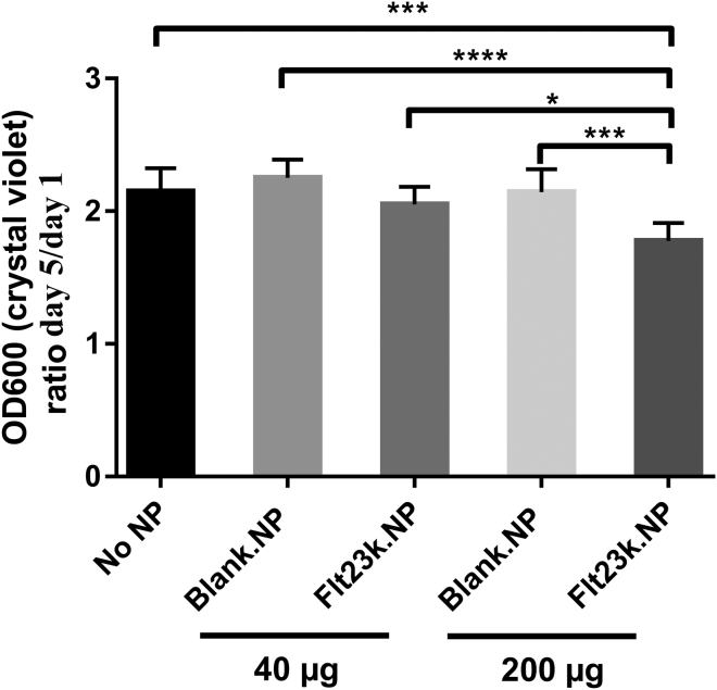
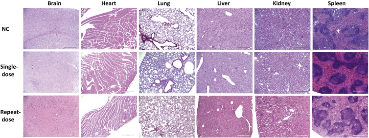
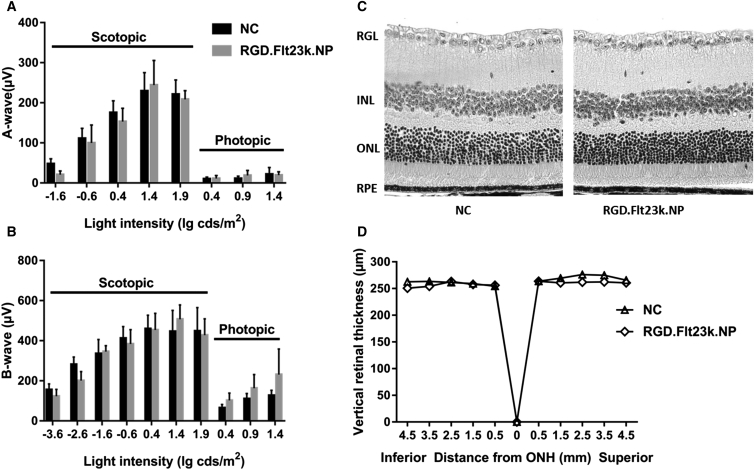
Neovascular age-related macular degeneration (AMD) is treated with anti-VEGF intravitreal injections, which can cause geographic atrophy, infection, and retinal fibrosis. To minimize these toxicities, we developed a nanoparticle delivery system for recombinant Flt23k intraceptor plasmid (RGD.Flt23k.NP) to suppress VEGF intracellularly within choroidal neovascular (CNV) lesions in a laser-induced CNV mouse model through intravenous administration. In the current study, we examined the efficacy and safety of RGD.Flt23k.NP in mice. The effect of various doses was determined using fluorescein angiography and optical coherence tomography to evaluate CNV leakage and volume. Efficacy was determined by the rate of inhibition of CNV volume at 2 weeks post-treatment. RGD.Flt23k.NP had peak efficacy at a dose range of 30-60 μg pFlt23k/mouse. Using the lower dose (30 μg pFlt23k/mouse), RGD.Flt23k.NP safety was determined both in single-dose groups and in repeat-dose (three times) groups by measuring body weight, organ weight, hemoglobin levels, complement C3 levels, and histological changes in vital organs. Neither toxicity nor inflammation from RGD.Flt23k.NP was detected. No side effect was detected on visual function. Thus, systemic RGD.Flt23k.NP may be an alternative to standard intravitreal anti-VEGF therapy for the treatment of neovascular AMD.

Keywords: RGD.Flt23k.NP efficacy; RGD.Flt23k.NP safety; age-related macular degeneration.


Figures

Figure 1
Figure 1
Flt23k Overexpression Reduced Endogenous VEGF-A Expression in HeLa Cells (A) As transfection control, pMaxGFP (0, 0.1, 0.5, 1.0, 2.5, and 5.0 μg) was transfected to HeLa cells with nucleofection. GFP expression increased with increasing plasmid concentration. (B) Western blot analysis demonstrates that Flt23k decreased endogenous VEGF expression, showing a dose-dependent trend.
Figure 2
Figure 2
Flt23k.NP Inhibited HAEC Proliferation Nanoparticle (NP) was dissolved in DPBS at 20 mg/mL. Then, 2 μL (40 μg NP) or 10 μL (200 μg NP) of NP solution were transfected to 2.0 × 106 human aortic endothelial cells (HAECs) with nucleofection. The next day, 5,000 cells were replated to a 24-well plate in 500 μL EGM2 medium. After day 3 and day 5, the cells were fixed with 4% PFA and stained with crystal violet. Crystal violet was eluted with 200 μL 10% acetic acid and measured at OD600. *p < 0.05, ***p < 0.001, ****p < 0.0001.
Figure 3
Figure 3
RGD Functionalized Nanoparticles Reached the Highest Effective Dose above 30 μg RGD.Flt23k.NPs (A) A pFlt23k-loaded nanoparticle dose from 0 to 60 μg was administrated in laser-induced CNV mice. CNV lesions were measured using FA and OCT. The reductions in CNV lesion sites were measured at 1, 2, and 3 weeks post-treatment with Flt23k plasmid. The left side of each panel shows the FA image, whereas the right side shows the IR image. (B) OCT images were used to calculate the CNV volume at 2 weeks post-treatment. ANOVA analysis was used to measure the significant difference in CNV volume regression at different doses of Flt23k plasmid. *p < 0.05, **p < 0.01, ****p < 0.0001. (C) Representative OCT images for laser CNV treated with different doses of RGD.Flt23k.NP.
Figure 4
Figure 4
No Systemic Toxicity Was Detected after Treatment with RGD Functionalized Nanoparticles (A) Mouse body weight with a single dose of 30 μg pFlt23k-loaded NP was recorded before treatment and at 1 month post-treatment and compared to that of control mice. (B) Mouse body weight for repeat doses of 30 μg pFlt23k and control mice was recorded before treatment and at 1, 2, and 3 months post-treatment. n = 10.
Figure 5
Figure 5
No Systemic Toxicity Was Detected with RGD Functionalized Nanoparticles (A) For single-dose treatment, organs (brain, heart, lung, liver, kidney, and spleen) were harvested and weights were measured. (B) For repeat-dose treatments, organs (brain, heart, lung, liver, kidney, and spleen) were harvested and measured at the endpoint of 3 months.
Figure 6
Figure 6
No Hematological Toxicity or Systemic Inflammation Were Detected after Treatment with RGD Functionalized Nanoparticles Mouse blood was collected and hemoglobin levels were measured using the Hemoglobin Colometeris Assay Kit (Cayman Chemical). (A) Hemoglobin (Hg) levels (in grams per deciliter) of mice treated with a single dose of 30 μg pFlt23k (p = 0.9709). (B) Hg levels (in grams per deciliter) of mice treated with repeat doses of 30 μg pFlt23k (p = 0.5278). Mouse blood was collected at the endpoint, and complement component 3 (C3) levels were assayed with the Complement C3 Mouse ELISA Kit (Abcam). (C) Single dose of 30 μg pFlt23k (p = 0.2726). (D) Repeat doses of 30 μg pFlt23k (p = 0.5614).
Figure 7
Figure 7
Systemic Toxicity Was Not Detected after Treatment with RGD Functionalized Nanoparticles Histology for all organs was performed with paraffin block and H&E staining. The slides were evaluated by a masked, experienced pathologist. n = 10. Scale bar, 200 μm.
Figure 8
Figure 8
No Ocular Toxicity Was Detected after Treatment with RGD Functionalized Nanoparticles (A and B) ERG and retinal thickness of eyes in mice treated with a maximum dose of 60 μg RGD.Flt23k.NPs (n = 4) in comparison to eyes of control mice (n = 9) (ERG a wave, p = 0.2601; b wave, p = 0.4599). (C) H&E staining (40×) of eyes in mice treated with a maximum dose of 60 μg RGD.Flt23k.NPs versus PBS control mice. (D) Retinal thickness as determined by OCT. Scale bar, 100 μm.

Similar articles

Cited by

References

    1. Wong W.L., Su X., Li X., Cheung C.M., Klein R., Cheng C.Y., Wong T.Y. Global prevalence of age-related macular degeneration and disease burden projection for 2020 and 2040: a systematic review and meta-analysis. Lancet Glob. Health. 2014;2:e106–e116. - PubMed
    1. Klein R., Chou C.F., Klein B.E., Zhang X., Meuer S.M., Saaddine J.B. Prevalence of age-related macular degeneration in the US population. Arch. Ophthalmol. 2011;129:75–80. - PubMed
    1. Wong T.Y., Chakravarthy U., Klein R., Mitchell P., Zlateva G., Buggage R., Fahrbach K., Probst C., Sledge I. The natural history and prognosis of neovascular age-related macular degeneration: a systematic review of the literature and meta-analysis. Ophthalmology. 2008;115:116–126. - PubMed
    1. Klein R., Klein B.E., Tomany S.C., Meuer S.M., Huang G.H. Ten-year incidence and progression of age-related maculopathy: the Beaver Dam Eye Study. Ophthalmology. 2002;109:1767–1779. - PubMed
    1. van der Reis M.I., La Heij E.C., De Jong-Hesse Y., Ringens P.J., Hendrikse F., Schouten J.S. A systematic review of the adverse events of intravitreal anti-vascular endothelial growth factor injections. Retina. 2011;31:1449–1469. - PubMed

Publication types

MeSH terms