. 2017 Apr;284(8):1233-1245.
doi: 10.1111/febs.14051. Epub 2017 Mar 17.

Catalytic competence, structure and stability of the cancer-associated R139W variant of the human NAD(P)H:quinone oxidoreductase 1 (NQO1)

Affiliations

Catalytic competence, structure and stability of the cancer-associated R139W variant of the human NAD(P)H:quinone oxidoreductase 1 (NQO1)

Wolf-Dieter Lienhart et al. FEBS J. 2017 Apr.

Abstract

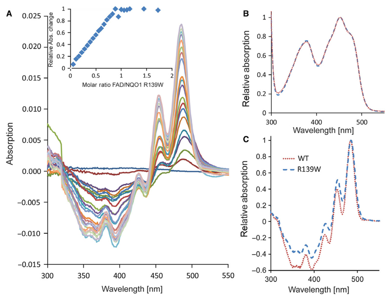
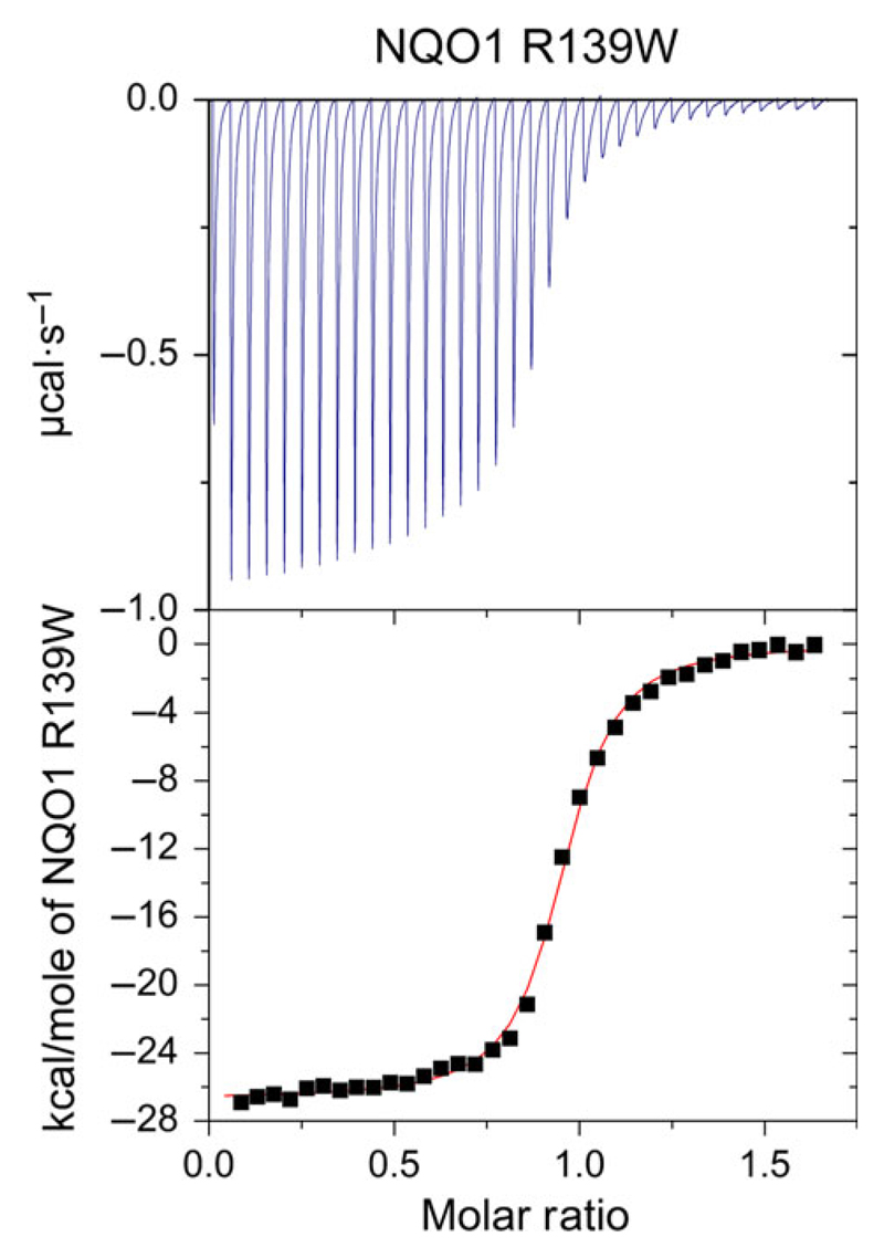
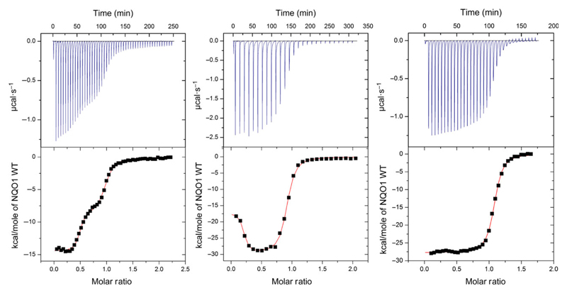
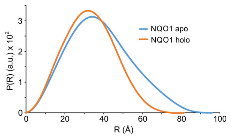
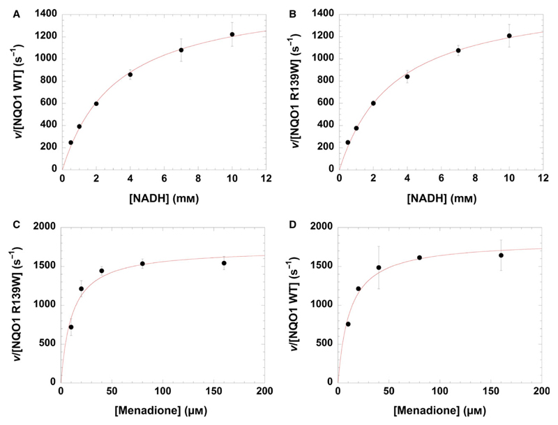
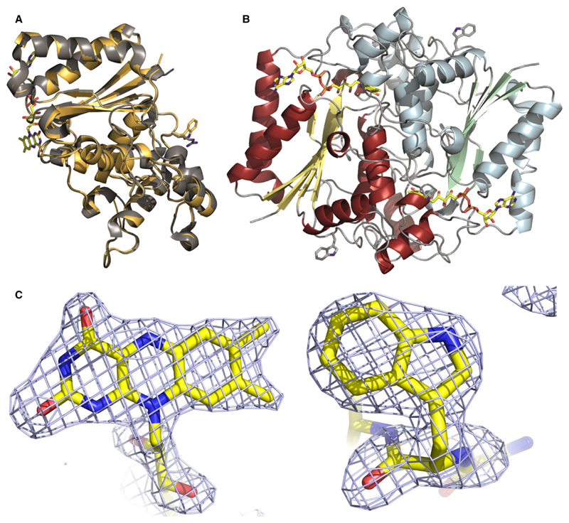
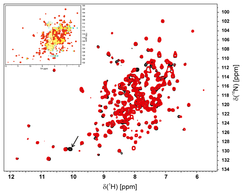
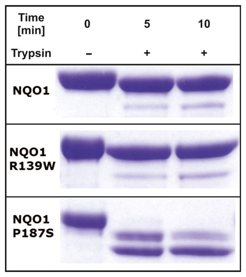
The human NAD(P)H:quinone oxidoreductase 1 (NQO1; EC1.6.99.2) is an essential enzyme in the antioxidant defence system. Furthermore, NQO1 protects tumour suppressors like p53, p33ING1b and p73 from proteasomal degradation. The activity of NQO1 is also exploited in chemotherapy for the activation of quinone-based treatments. Various single nucleotide polymorphisms are known, such as NQO1*2 and NQO1*3 yielding protein variants of NQO1 with single amino acid replacements, i.e. P187S and R139W, respectively. While the former NOQ1 variant is linked to a higher risk for specific kinds of cancer, the role, if any, of the arginine 139 to tryptophan exchange in disease development remains obscure. On the other hand, mitomycin C-resistant human colon cancer cells were shown to harbour the NQO1*3 variant resulting in substantially reduced enzymatic activity. However, the molecular cause for this decrease remains unclear. In order to resolve this issue, recombinant NQO1 R139W has been characterized biochemically and structurally. In this report, we show by X-ray crystallography and 2D-NMR spectroscopy that this variant adopts the same structure both in the crystal as well as in solution. Furthermore, the kinetic parameters obtained for the variant are similar to those reported for the wild-type protein. Similarly, thermostability of the variant was only slightly affected by the amino acid replacement. Therefore, we conclude that the previously reported effects in human cancer cells cannot be attributed to protein stability or enzyme activity. Instead, it appears that loss of exon 4 during maturation of a large fraction of pre-mRNA is the major reason of the observed lack of enzyme activity and hence reduced activation of quinone-based chemotherapeutics.

Keywords: NMR-spectroscopy; cancer; crystal structure; enzyme kinetics; microcalorimetry; single nucleotide polymorphism.


Figures

Fig. 1
Fig. 1
UV-visible and difference titration absorption spectra of wild-type NQO1 and NQO1 R139W. (A) Difference titration spectra of 800 μL NQO1 R139W (45 μm) with 0–42 μL in 2 μL intervals FAD (1 mm). The insert shows the change of the absolute absorption values at 436 nm and 455 nm against the FAD/protein molar ratio with two additional points (with additional 10 μL FAD each) compared to the main figure. (B) Absorption spectra of wild-type NQO1 and NQO1 R139W normalized to the maximum at 450 nm. (C) Difference titration spectra of wild-type NQO1 and NQO1 R139W with protein/FAD ratio of 1 normalized to the maximum at 480 nm.
Fig. 2
Fig. 2
Isothermal titration microcalorimetry measurement of NQO1 R139W. NQO1 R139W: 35 injections with 6 μL of 298 μm NQO1 R139W apoprotein solution in 29 μm FAD solution and 300 s spacing (top) at 25 °C. From three independent measurements under the same conditions, the dissociation constant was calculated to KD = 155 ± 27 nm. Data was fitted using the one binding site model.
Fig. 3
Fig. 3
Isothermal titration microcalorimetry measurements of NQO1 wild-type in HEPES buffer. The left and the middle measurements were conducted with apo-NQO1 in the sample cell of the microcalorimeter and FAD in the injection syringe. Data was fitted using the two binding site model. Left: First injection with 2 μL and 49 injections with 6 μL of 477 μm FAD solution in 49.4 μm NQO1 apoprotein solution and 300 s spacing at 25 °C. The determined KD and N values are: KD1 = 9.2 nm; KD2 = 780 nm; N1 = 0.47; N2 = 0.5. Middle: First injection with 2 μL and 27 injections with 10 μL of 457 μm FAD solution in 46.8 μm NQO1 apoprotein solution and 600 s spacing at 25 °C. The determined KD and N values are: KD1 = 4 nm; KD2 = 295 nm; N1 = 0.18; N2 = 0.71. The right measurement was conducted with FAD in the sample cell of the microcalorimeter and apo-NQO1 in the injection syringe. Data was fitted using the one binding site model. First injection with 2 μL and 34 injections with 6 μL of 284 μm NQO1 apoprotein solution in 29 μm FAD solution and 300 s spacing at 25 °C. From three independent measurements under the same conditions, the dissociation constant was calculated to KD = 64 ± 23 nm.
Fig. 4
Fig. 4
Small-angle X-ray scattering measurement of NQO1. SAXS data showing a comparison of the experimental radial density distribution (P(r)) of apo- and holo-NQO1 in cyan and orange, respectively.
Fig. 5
Fig. 5
Steady state kinetics of NQO1 WT and NQO1 R139W. In the case of variation of NADH, the velocity v over the enzyme concentration is plotted against NADH concentration for NQO1 WT (A) and NQO1 R139W (B). For the variation of menadione, the velocity v over the enzyme concentration is plotted against menadione concentration for NQO1 WT (C) and NQO1 R139W (D). Standard deviations are shown with error bars.
Fig. 6
Fig. 6
Crystal structure of NQO1 R139W. Panel A: Cartoon model of the superposition of NQO1 and NQO1 R139W with the arginine and tryptophan residue (right) and the FAD (left) shown as a stick model. Panel B: Cartoon model of the NQO1 R139W homodimer with the tryptophan residue and the FAD shown as a stick model. Panel C: Fo − Fc omit electron density contoured at 3σ for the isoalloxazine moiety of FAD bound to one of the subunits (left) and for tryptophan 139 (right).
Fig. 7
Fig. 7
2D 1H-15N HSQC spectra. Overlay of the 2D 1H-15N HSQC spectrum of NQO1 R139W (black) and wild-type (red). An additional tryptophan side chain NH signal is visible in the R139W variant and indicated by an arrow. All other signals are almost identical in the HSQC spectrum of the R139W variant and wild-type, respectively, indicative of a very similar structure. A few minor shift differences result from residues close to residue 139. The insert in the upper left corner shows an overlay of the NMR spectra of NQO1 WT (red) and NQO1 P187S (green) where overlapping areas are marked yellow.
Fig. 8
Fig. 8
Trypsin digestion of different NQO1 variants. Comparison of Trypsin digestion of three NQO1 variants without addition of Trypsin (0 min) and two measurements after addition of Trypsin (5 and 10 min).

References

    1. Lienhart WD, Gudipati V, Macheroux P. The human flavoproteome. Arch Biochem Biophys. 2013;535:150–162. - PMC - PubMed
    1. Ross D, Siegel D. NAD(P)H:quinone oxidoreductase 1 (NQO1, DT-diaphorase), functions and pharmacogenetics. Methods Enzymol. 2004;382:115–144. - PubMed
    1. Garate M, Wong RP, Campos EI, Wang Y, Li G. NAD(P)H quinone oxidoreductase 1 inhibits the proteasomal degradation of the tumour suppressor p33 (ING1b) EMBO Rep. 2008;9:576–581. - PMC - PubMed
    1. Asher G, Tsvetkov P, Kahana C, Shaul Y. A mechanism of ubiquitin-independent proteasomal degradation of the tumor suppressors p53 and p73. Genes Dev. 2005;19:316–321. - PMC - PubMed
    1. Kelsey KT, Ross D, Traver RD, Christiani DC, Zuo ZF, Spitz MR, Wang M, Xu X, Lee BK, Schwartz BS, et al. Ethnic variation in the prevalence of a common NAD(P)H quinone oxidoreductase polymorphism and its implications for anti-cancer chemotherapy. Br J Cancer. 1997;76:852–854. - PMC - PubMed

Publication types

Substances