Review
. 2017:123:365-397.
doi: 10.1016/bs.ctdb.2016.11.001. Epub 2016 Dec 5.

Regulation of Cell Polarity by PAR-1/MARK Kinase

Affiliations
Review

Regulation of Cell Polarity by PAR-1/MARK Kinase

Youjun Wu et al. Curr Top Dev Biol. 2017.

Abstract

PAR-1/MARK kinases are conserved serine/threonine kinases that are essential regulators of cell polarity. PAR-1/MARK kinases localize and function in opposition to the anterior PAR proteins to control the asymmetric distribution of factors in a wide variety polarized cells. In this review, we discuss the mechanisms that control the localization and activity of PAR-1/MARK kinases, including their antagonistic interactions with the anterior PAR proteins. We focus on the role PAR-1 plays in the asymmetric division of the Caenorhabditis elegans zygote, in the establishment of the anterior/posterior axis in the Drosophila oocyte and in the control of microtubule dynamics in mammalian neurons. In addition to conserved aspects of PAR-1 biology, we highlight the unique ways in which PAR-1 acts in these distinct cell types to orchestrate their polarization. Finally, we review the connections between disruptions in PAR-1/MARK function and Alzheimer's disease and cancer.

Keywords: C. elegans; Cell polarity; Drosophila; MARK; Microtubule-associated protein; PAR proteins; PAR-1.


Figures

Figure 1
Figure 1
Schematic of PAR-1/MARK kinase. The kinase and UBA domains are positioned near the N-terminus and a relatively long spacer domain separates the UBA domain from the C-terminal KA1 membrane binding domain. PAR-1 kinase activity is stimulated by phosphorylation on Thr208 in the activation loop by LKB1/PAR-4 and MARKK/TAO-1 kinases. GSK3β phosphorylation on Ser212 in the activation loop inhibits PAR-1 kinase activity. aPKC phosphorylation at residue Thr595 results in the association of the 14-3-3 protein PAR-5 (not depicted) and the sequestration of PAR-1 in the cytoplasm. MARK2 residues are indicated.
Figure 2
Figure 2
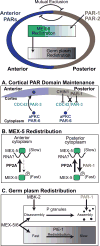
Establishment of the Anterior/Posterior axis in the C. elegans zygote. In the polarized C. elegans zygote, the Anterior PARs (Blue) are enriched at the anterior cortex and PAR-1 and PAR-2 (brown) are enriched at the posterior cortex. Antagonistic interactions between the PAR proteins mediates their mutual exclusion. PAR-1 directs the redistribution of MEX-5 to the anterior cytoplasm and MEX-5 contributes to the redistribution of germ plasm proteins to the posterior cytoplasm. All of these factors are symmetrically distributed before symmetry breaking. A. Asymmetric PAR domains are maintained in part by the recruitment of cytoplasmic PAR-6 and aPKC to the anterior, but not to the posterior, cortex. PAR-1 and CHIN-1 restrict PAR-3 and active CDC42 from the posterior cortex, thereby preventing the recruitment of PAR-6 and aPKC from the cytoplasm to the posterior cortex. Other mechanisms that contribute to PAR domain maintenance are described in the text and not illustrated for simplicity. B. The redistribution of MEX-5 to the anterior cytoplasm is controlled by PAR-1. PAR-1 phosphorylates MEX-5 and increases MEX-5 mobility in the posterior cytoplasm while PP2A reverses this effect. As a result, MEX-5 mobility is relatively fast in the posterior cytoplasm and MEX-5 redistributes to the anterior cytoplasm. MEX-5 association with RNA is likely to contribute to its slow diffusion. C. As MEX-5 and MEX-6 accumulate in the anterior, germ plasm factors segregate to the posterior cytoplasm. P granules partition to the posterior cytoplasm because MEX-5/6 and MBK-2 promote their disassembly in the anterior cytoplasm and PAR-1 promotes their stability in the posterior cytoplasm. MEX-5/6 act to increase PIE-1 mobility in the anterior cytoplasm, thereby stimulating the redistribution of PIE-1 to the posterior cytoplasm.
Figure 3
Figure 3
Establishment of the Anterior/Posterior axis in the Drosophila oocyte. A. Gurken (Grk) is secreted from the oocyte and signals to the neighboring follicle cells. The posterior follicle cells subsequently signal back to the oocyte, leading to the recruitment of dPar-1 to the posterior cortex. The signal from the follicle cells to the oocyte is not known. B. Microtubules grow from the oocyte anterior cortex from foci containing Shot and Patronin, which binds microtubule minus ends. C. At the oocyte posterior cortex, Par-6 is removed from the cortex by the E3 ubiquitin ligase, Slimb. Par-3 is removed from the posterior cortex by dPar-1 phosphorylation, which leads to interaction with Par-5 and sequestration of Par-3 in the cytoplasm. dPar-1 also acts through an unknown mechnanism to restrict Shot from the posterior cortex D. The oocyte is positioned next to the posterior follicle cells and is connected to the nurse cells by ring canals. dPar-1 (blue) forms a posterior cortical domain that is reciprocal to the anterior/lateral Anterior PAR domain (aPAR, yellow). Microtubules (Green) are nucleated from the anterior and lateral cortex and not from the posterior cortex. As a result, microtubule density is higher in the anterior are microtubules tend to point towards the posterior.

Similar articles

Cited by

References

    1. Aceto D, Beers M, Kemphues KJ. Interaction of PAR-6 with CDC-42 is required for maintenance but not establishment of PAR asymmetry in C. elegans. Developmental Biology. 2006;299:386–397. - PMC - PubMed
    1. Beatty A, Morton DG, Kemphues K. PAR-2, LGL-1 and the CDC-42 GAP CHIN-1 act in distinct pathways to maintain polarity in the C. elegans embryo. Development. 2013;140:2005–14. - PMC - PubMed
    1. Beghini A, Magnani I, Roversi G, Piepoli T, Di Terlizzi S, Moroni RF, Pollo B, Fuhrman Conti AM, Cowell JK, Finocchiaro G, Larizza L. The neural progenitor-restricted isoform of the MARK4 gene in 19q13.2 is upregulated in human gliomas and overexpressed in a subset of glioblastoma cell lines. Oncogene. 2003;22:2581–2591. - PubMed
    1. Benton R, Palacios IM, Johnston DS. Drosophila 14-3-3/PAR-5 Is an Essential Mediator of PAR-1 Function in Axis Formation. Developmental Cell. 2002;3:659–671. - PubMed
    1. Benton R, St Johnston D. Drosophila PAR-1 and 14-3-3 inhibit Bazooka/PAR-3 to establish complementary cortical domains in polarized cells. Cell. 2003;115:691–704. - PubMed

MeSH terms

Substances

LinkOut - more resources