

Review
doi: 10.1053/j.sult.2016.10.002.
Epub 2016 Oct 14.
Hereditary Renal Tumor Syndromes: Update on Diagnosis and Management
Affiliations
- PMID: 28237281
- PMCID: PMC5330200
- DOI: 10.1053/j.sult.2016.10.002
Item in Clipboard
Review
Hereditary Renal Tumor Syndromes: Update on Diagnosis and Management
Semin Ultrasound CT MR.
2017 Feb.
Abstract
Hereditary renal cancers account for approximately 5%-8% of all renal tumors. Over the past 2 decades, a number of syndromes have been identified that predispose patients to early renal cancer development, representing all the major histologic types of tumor pathology. In this article, we describe the current knowledge concerning the cell type, known mechanism of tumor development, other manifestations of the syndrome, imaging findings, genetic screening, and imaging surveillance recommendations for each of the major syndromes associated with hereditary renal cancers.
Published by Elsevier Inc.
Figures
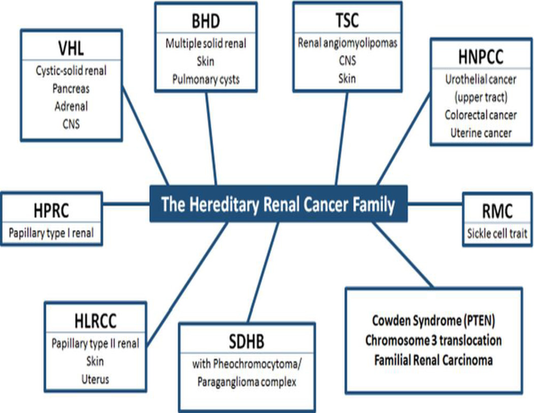
Hereditary renal cancer syndromes. Genetic syndromes with their most common manifestations are listed in each box.

Single renal mass in a VHL patient. Axial contrast enhanced CT of a VHL patient demonstrates a left-sided sponge-like renal mass with both solid and cystic components. The solid components show contrast enhancement (arrow).

Bilateral solid-cystic renal masses in a VHL patient. Axial T2W MRI of a VHL patient shows solid-cystic masses in both kidneys (arrows) (A); whereas axial fat saturated contrast enhanced T1W MRI at the same level reveals better differentiation of cystic versus solid components within these masses (arrows) (B).

Renal and extra-renal manifestations of VHL. Axial contrast enhanced CT of a VHL patient demonstrates a a dominantly solid lesion in the right kidney (long white arrow), a cystic lesion within the left kidney (asterisk), several cysts within the body and tail of the pancreas (short white arrows), additionally noted is an enhancing lesion within the spinal column suggestive of a hemangioblastoma (red arrow).

Bilateral renal tumors in a BHD patient. Axial contrast enhanced CT of a BHD patient demonstrates multiple homogenously enhancing tumors (arrows). Within these tumors, some higher density calcifications can be seen.

MRI findings of renal involvement in BHD. Axial contrast-enhanced T1W MRI of a BHD patient showing a uniformly appearing hypointense tumor in the left kidney (arrow).

CT findings of pulmonary involvement in BHD. Axial contrast-enhanced lung CT of a BHD patient showing multiple pulmonary cysts (asterisks). These cysts have the potential of leading to spontaneous pneumothorax.

Bilateral renal involvement in HPRC. Axial contrast-enhanced abdominal CT of a HPRC patient demonstrating tumors with uniform mild enhancement (arrows). This appearance is typical of type 1 papillary tumors and may sometimes resemble cystic lesions on imaging.

Bilateral type I papillary renal tumors in HPRC. Axial contrast-enhanced fat-saturated T1 MRI of HPRC patient demonstrating the uniform hypointense appearance of type I papillary renal tumors, present in this patient bilaterally (arrows).
Papillary type II tumor in HLRCC. Axial contrast enhanced CT scan of a HLRCC patient, demonstrating an enhancing large solitary tumor in the left kidney (arrows). This poor enhancement is characteristic of the appearance of renal papillary type II tumors. Some cystic change can also be seen within this tumor.

MRI findings of renal involvement in HLRCC. Axial contrast enhanced fat saturated T1W MRI of HLRCC patient demonstrating a solitary, poorly enhancing, cystic-appearing type II papillary tumor in the left kidney (arrow).

Local tumor invasion in HLRCC. Axial contrast enhanced abdominal CT scan in a HLRCC patient demonstrating the aggressive nature of the papillary type II renal tumors associated with this syndrome. The tumor in this patient shows local invasion into the adjacent left abdominal wall (arrows).

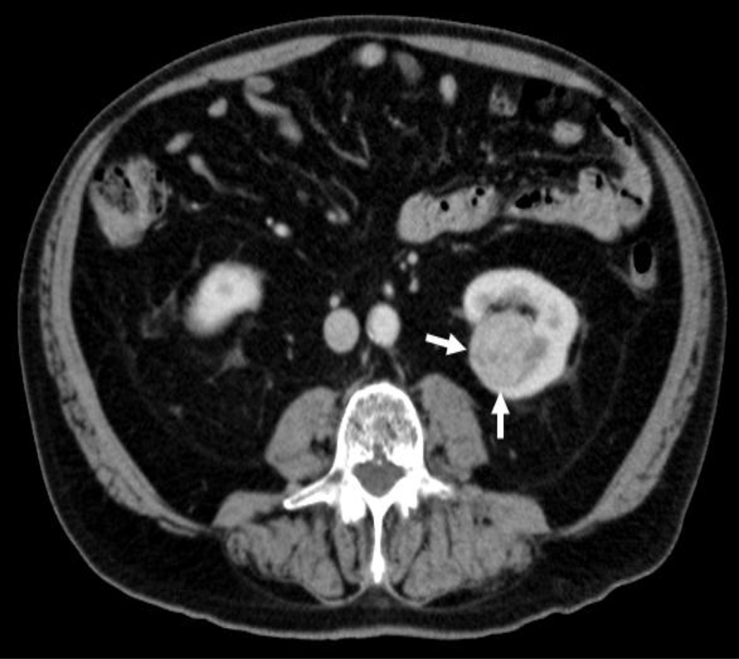
Renal involvement in SDH-B deficiency. Axial contrast enhanced CT scan of SDH-B deficiency patient, demonstrating both solid and cystic components within tumor in the left kidney (arrows). The appearance of these tumors varies, as there are several solid-cystic subtypes seen in this disease.

MRI findings of renal involvement in SDH-B deficiency. Axial contrast enhanced T1W MRI of SDH-B deficiency patient demonstrating 2 tumors in the left kidney, that are non-uniform in appearance and not particularly distinctive by imaging (arrows). The more hypointense tumor appears to contain more cystic component, while the other tumor appears less hypointense and predominantly solid.
References
-
- Linehan WM, Walther MM, Zbar B. The genetic basis of cancer of the kidney. J Urol. 2003;170(6 Pt 1):2163–2172. - PubMed
-
- Adeniran AJ, Shuch B, Humphrey PA. Hereditary Renal Cell Carcinoma Syndromes: Clinical, Pathologic, and Genetic Features. Am J Surg Pathol. 2015;39(12):e1–e18. - PubMed
-
- Richard S, Gardie B, Couve S, Gad S. Von Hippel-Lindau: how a rare disease illuminates cancer biology. Semin Cancer Biol. 2013;23(1):26–37. - PubMed
Publication types
MeSH terms
Grants and funding
LinkOut - more resources
Full Text Sources
Other Literature Sources
Medical
Miscellaneous
