Observational Study
. 2017 Jun;124(6):793-803.
doi: 10.1016/j.ophtha.2017.01.030. Epub 2017 Feb 23.

Aqueous Angiography in Living Nonhuman Primates Shows Segmental, Pulsatile, and Dynamic Angiographic Aqueous Humor Outflow

Affiliations
Observational Study

Aqueous Angiography in Living Nonhuman Primates Shows Segmental, Pulsatile, and Dynamic Angiographic Aqueous Humor Outflow

Alex S Huang et al. Ophthalmology. 2017 Jun.

Abstract

Purpose: To evaluate the feasibility of safely performing aqueous angiography in intact eyes of living nonhuman primates (NHPs) for evaluating aqueous humor outflow and segmental patterns.

Design: Cross-sectional, observational study.

Subjects: Six nonhuman primates.

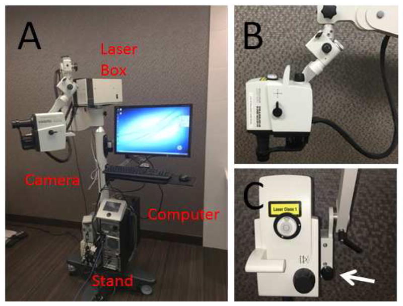
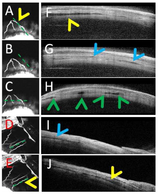
Methods: Aqueous angiography was performed in 6 nonhuman primates. After anesthesia, an anterior chamber (AC) maintainer was placed through a temporal 1-mm side-port wound. Indocyanine green (ICG; 0.4%) or 2.5% fluorescein was introduced (individually or in sequence) into the eye with a gravity-driven constant-pressure system. Aqueous angiography images were obtained with a Spectralis HRA+OCT (Heidelberg Engineering GmbH, Heidelberg, Germany) suspended over the NHP eye using a custom-designed surgical boom arm. Concurrent anterior segment optical coherence tomography (OCT) was performed on distally angiographically positive and negative regions.

Main outcome measures: Angiographic patterns described by location, time-course, choice of tracer, and anterior-segment OCT.

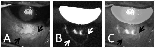
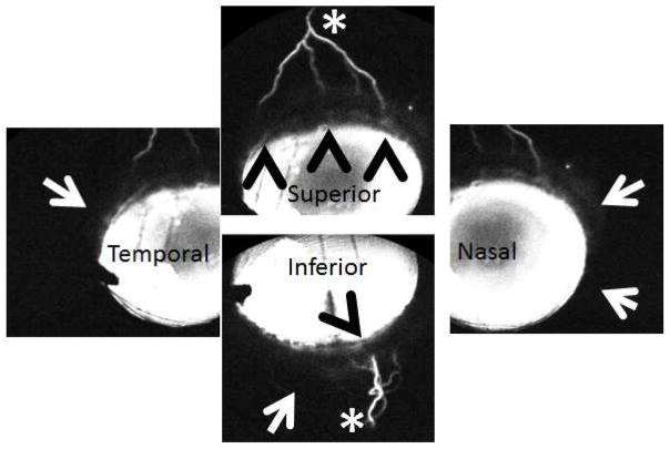
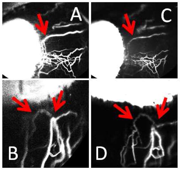
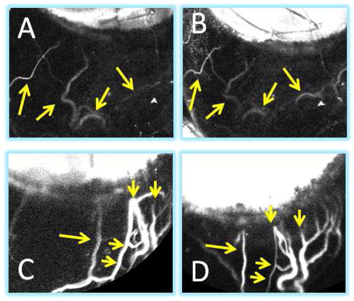
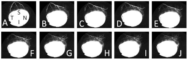
Results: Aqueous angiography in the living NHP eye demonstrated mostly stable angiographic patterns. With multimodal imaging, angiographically positive signal co-localized with episcleral veins as identified by infrared imaging and intrascleral lumens, as demonstrated by anterior segment OCT. Sequential aqueous angiography in individual eyes with ICG followed by fluorescein showed similar angiographic patterns. A pulsatile nature of aqueous angiographic outflow was sometimes observed. Aqueous angiographic patterns could also dynamically change. In some instances, positive angiographic flow suddenly arose in regions previously without an angiographic signal. Alternatively, an angiographic signal could suddenly disappear from regions in which an angiographic signal was initially documented.

Conclusions: Aqueous angiography in living NHPs demonstrated segmental and pulsatile patterns with a newly described ability to dynamically shift. These characteristics further the understanding of live aqueous humor outflow biology and may be useful in improving glaucoma surgeries aimed at trabecular meshwork bypass.


Figures

Figure 1
Figure 1. Heidelberg Engineering Flex Module
(A) The Flex Module suspended the camera head, disassembled from the standard table-top Spectralis, to allow for non-seated and non-upright image acquisition. (B) Seven pivot joints gave it maximum flexibility so that theoretically images could be taken sitting, supine, or prone with or without head down or up tilt. (C) A micro-manipulator allowed for precise z-axis movement (arrow). A 200-pound base in the stand provided stability and safety.
Figure 2
Figure 2. Aqueous Angiography in an Intact Living NHP Eye
(A) Infrared imaging of the inferior portion the NHP-A eye demonstrated surface episcleral veins (black arrows). (B) Aqueous angiography (fluorescein) in the same eye demonstrated three peri-limbal spots of angiographic signal that traced posterior (white arrows). (C) Overlay of images (A) and (B; 50% transparency setting using Photoshop CS5 v.12×32) showed good correspondence of angiographic signal to some of the episcleral vessels (black arrows).
Figure 3
Figure 3. Aqueous Angiography and Anterior Segment Optical Coherence Tomography
(A–C) Aqueous angiography (ICG) of the superior portion of the NHP-B eye showed an inferior pointing fork-like arrangement with 4 arms. (D/E) Aqueous angiography (ICG) of the superior-nasal portion of NHP-C eye showed a more linear arrangement. (F/H/J) Anterior segment OCT in angiographically positive areas (A/C/E) showed more intrascleral lumens (yellow and green arrowheads). (B/D) Angiographically negative regions showed (G/I; blue arrowheads) much less but still present lumens that many have corresponded to other intrascleral luminal structures such as arteries. Note that the horizontal anterior OCT scan pattern (C) cut across two angiographic arms on the left and near the root of another angiographic branch on the right corresponding to (H) four lumens on OCT (green arrowheads).
Figure 4
Figure 4. Aqueous Angiography Demonstrated Segmental Patterns (NHP-B)
Aqueous angiography looking at different quadrants of the eye demonstrated segmental patterns with some regions of peri-limbal angiographic signal (black arrowheads), intermixed with peri-limbal regions without signal (white arrows), leading to more distal angiographic signal (white asterisks).
Figure 5
Figure 5. Aqueous Angiography Demonstrated Segmental Patterns (NHP-C)
Aqueous angiography looking at different quadrants of the eye demonstrated segmental patterns with some regions of peri-limbal angiographic signal (black arrowheads), intermixed with peri-limbal regions without signal (white and black arrows), leading to more distal angiographic signal (white asterisks).
Figure 6
Figure 6. Sequential Aqueous Angiography with ICG and Fluorescein Demonstrated Similar Patterns
In one eye (NHP-C), sequential aqueous angiography was performed with ICG (A/B) followed by fluorescein (C/D). Like results in post-mortem human eyes, similar patterns were observed between the two dyes (red arrows).
Figure 7
Figure 7. Aqueous Angiography Patterns in a Living Intact NHP Eye Demonstrated Stability Over Time
For the most part, unlike post-mortem eyes where the episcleral veins were severed open, in intact eyes, aqueous angiography patterns usually remained stable. ICG aqueous angiography in NHP-F at (A) 20 second was similar to that at (B) 2.5 minutes (yellow arrows). ICG aqueous angiography in NHP-C at (C) 2 minutes was similar to that at (D) 8 minutes (yellow arrows). (C/D) Note that these two images are slightly rotated when comparing.
Figure 8
Figure 8. Aqueous Angiography Patterns in a Living Intact NHP Eye Were Sometimes Dynamic
(A–J) While aqueous angiography patterns were mostly stable, sometimes, the patterns would dynamically change. In NHP-B, superior signal moved superior-nasal over approximately 10 seconds. S = superior, N = nasal, T= temporal, I = inferior.

References

    1. GRANT WM. Experimental aqueous perfusion in enucleated human eyes. Arch Ophthalmol. 1963;69:783–801. - PubMed
    1. Johnson M. What controls aqueous humour outflow resistance? Exp Eye Res. 2006;82(4):545–57. - PMC - PubMed
    1. Swaminathan SS, Oh DJ, Kang MH, Rhee DJ. Aqueous outflow: segmental and distal flow. J Cataract Refract Surg. 2014;40(8):1263–72. - PMC - PubMed
    1. Van Buskirk EM. Trabeculotomy in the immature, enucleated human eye. Invest Ophthalmol Vis Sci. 1977;16(1):63–6. - PubMed
    1. Minckler D, Mosaed S, Dustin L, Ms BF. Trabectome (trabeculectomy-internal approach): additional experience and extended follow-up. Trans Am Ophthalmol Soc. 2008;106:149–59. discussion 59–60. - PMC - PubMed

Publication types