

Inattention and Reaction Time Variability Are Linked to Ventromedial Prefrontal Volume in Adolescents
- PMID: 28237458
- PMCID: PMC5509516
- DOI: 10.1016/j.biopsych.2017.01.003
Inattention and Reaction Time Variability Are Linked to Ventromedial Prefrontal Volume in Adolescents
Abstract
Background: Neuroimaging studies of attention-deficit/hyperactivity disorder (ADHD) have most commonly reported volumetric abnormalities in the basal ganglia, cerebellum, and prefrontal cortices. Few studies have examined the relationship between ADHD symptomatology and brain structure in population-based samples. We investigated the relationship between dimensional measures of ADHD symptomatology, brain structure, and reaction time variability-an index of lapses in attention. We also tested for associations between brain structural correlates of ADHD symptomatology and maps of dopaminergic gene expression.
Methods: Psychopathology and imaging data were available for 1538 youths. Parent ratings of ADHD symptoms were obtained using the Development and Well-Being Assessment and the Strengths and Difficulties Questionnaire (SDQ). Self-reports of ADHD symptoms were assessed using the youth version of the SDQ. Reaction time variability was available in a subset of participants. For each measure, whole-brain voxelwise regressions with gray matter volume were calculated.
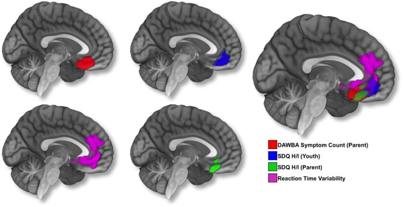
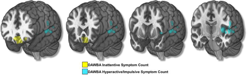
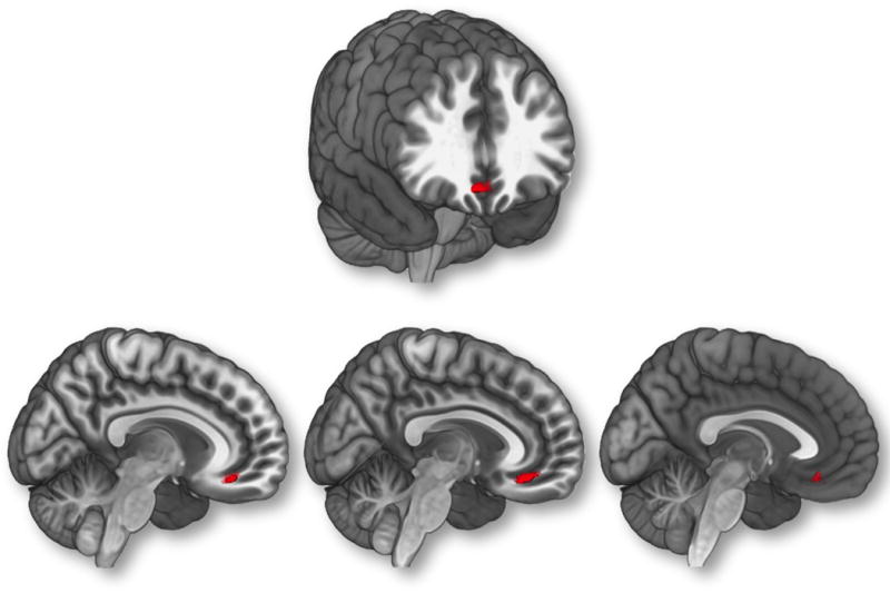
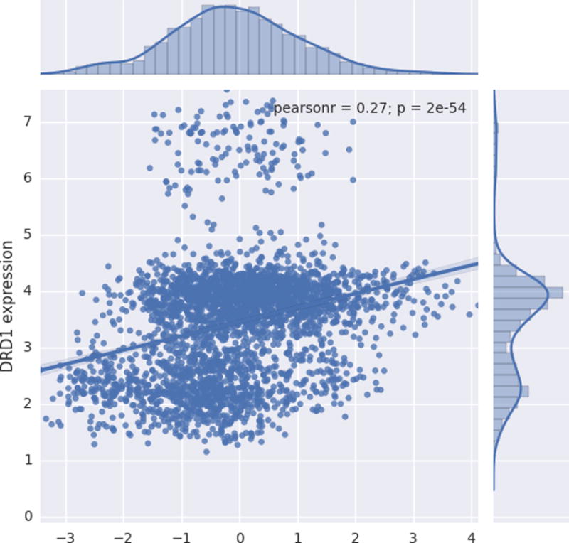
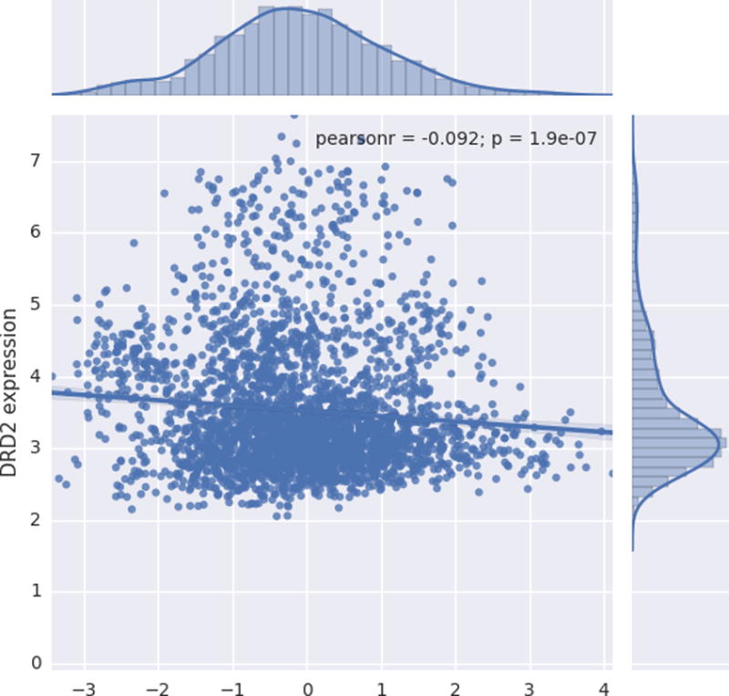
Results: Parent ratings of ADHD symptoms (Development and Well-Being Assessment and SDQ), adolescent self-reports of ADHD symptoms on the SDQ, and reaction time variability were each negatively associated with gray matter volume in an overlapping region of the ventromedial prefrontal cortex. Maps of DRD1 and DRD2 gene expression were associated with brain structural correlates of ADHD symptomatology.
Conclusions: This is the first study to reveal relationships between ventromedial prefrontal cortex structure and multi-informant measures of ADHD symptoms in a large population-based sample of adolescents. Our results indicate that ventromedial prefrontal cortex structure is a biomarker for ADHD symptomatology. These findings extend previous research implicating the default mode network and dopaminergic dysfunction in ADHD.
Keywords: Attention-deficit/hyperactivity disorder; Inattention; Multi-informant; Neuroimaging; Reaction time variability; Ventromedial prefrontal cortex.
Copyright © 2017 Society of Biological Psychiatry. Published by Elsevier Inc. All rights reserved.
Conflict of interest statement
The other authors report no biomedical financial interests or potential conflicts of interest.
Figures
References
-
- APA. Diagnostic and statistical manual of mental disorders : DSM-IV-TR. 4th. Washington, DC: American Psychiatric Association; 2000.
-
- Barkley RA, Fischer M, Edelbrock CS, Smallish L. The adolescent outcome of hyperactive children diagnosed by research criteria: I. An 8-year prospective follow-up study. J Am Acad Child Adolesc Psychiatry. 1990;29:546–557. - PubMed
-
- McGough JJ, Barkley RA. Diagnostic controversies in adult attention deficit hyperactivity disorder. Am J Psychiatry. 2004;161:1948–1956. - PubMed
-
- Almeida Montes LG, Ricardo-Garcell J, Barajas De La Torre LB, Prado Alcantara H, Martinez Garcia RB, Fernandez-Bouzas A, et al. Clinical correlations of grey matter reductions in the caudate nucleus of adults with attention deficit hyperactivity disorder. J Psychiatry Neurosci. 2010;35:238–246. - PMC - PubMed
MeSH terms
Grants and funding
LinkOut - more resources
Full Text Sources
Other Literature Sources
Medical
