. 2017 May:505:113-126.
doi: 10.1016/j.virol.2017.02.015. Epub 2017 Feb 23.

Evaluation of a novel multi-immunogen vaccine strategy for targeting 4E10/10E8 neutralizing epitopes on HIV-1 gp41 membrane proximal external region

Affiliations

Evaluation of a novel multi-immunogen vaccine strategy for targeting 4E10/10E8 neutralizing epitopes on HIV-1 gp41 membrane proximal external region

Saikat Banerjee et al. Virology. 2017 May.

Abstract

The membrane proximal external region (MPER) of HIV-1 gp41 is targeted by broadly neutralizing antibodies (bnAbs) 4E10 and 10E8. In this proof-of-concept study, we evaluated a novel multi-immunogen vaccine strategy referred to as Incremental, Phased Antigenic Stimulation for Rapid Antibody Maturation (IPAS-RAM) to induce 4E10/10E8-like bnAbs. Rabbits were immunized sequentially, but in a phased manner, with three immunogens that are progressively more native (gp41-28×3, gp41-54CT, and rVV-gp160DH12). Although nAbs were not induced, epitope-mapping analyses indicated that IPAS-RAM vaccination was better able to target antibodies towards the 4E10/10E8 epitopes than homologous prime-boost immunization using gp41-28×3 alone. MPER-specific rabbit monoclonal antibodies were generated, including 9F6. Although it lacked neutralizing activity, the target epitope profile of 9F6 closely resembled those of 4E10 and 10E8 (671NWFDITNWLWYIK683). B-cell repertoire analyses suggested the importance of co-immunizations for maturation of 9F6, which warrants further evaluation of our IPAS-RAM vaccine strategy using an improved priming immunogen.

Keywords: Antibody maturation; HIV-1; MPER; NGS; Neutralizing antibody; Next-generation sequencing; Rabbit; Vaccine; gp41.


Figures

Fig. 1
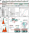
Fig. 1. Immunogens used in the study
(A) Schematic diagrams of gp41-28×3 and gp41-54CT. The entire gp41 ectodomain is shown on the top as a reference. To generate gp41-28×3, the T7Tag and HR1-HR2 portion of HR1-HR2-28×3 is removed with thrombin. (B) Expression and purification of HR1-HR2-28×3. Coomassie stained SDS-PAGE gels (U: uninduced; I: induced; P: purified). (C) Cleavage of HR1-HR2-28×3 with thrombin (O: original sample; T: thrombin cleaved; E: eluted from Ni-NTA column; F: flow through). (D) ELISA of gp41-28×3 with 2F5, 4E10, Z13e1 and 10E8. (E) Flow cytometry analyses of cell surface expression of gp41-54CT. 2F5, Z13e1 and 4E10 were used to probe the antigen. (F) Structural models of three immunogens are shown to illustrate their relative size. NMR structure of 28-mer peptide (2LP7; (Reardon et al., 2014)) and BG505 SOSIP gp140 structure (5C7K; (Kong et al., 2015)) were used. Structures were rendered using Chimera (Pettersen et al., 2004).
Fig. 2
Fig. 2. Immunization schedule and linear epitope mapping analyses
(A) Timeline for IPAS-RAM immunization and sampling. Rabbits were immunized on weeks 0, 4, 11 and 29. Pre-immune, as well as post-immune sera (two weeks post each immunization) were taken. PBMC were collected four days after immunization (except after 4th) for antibody repertoire analyses. To generate hybridomas, Rabbit R3 was immunized intravenously on week 35 with gp41-28×3 and spleen was collected four days later. (B) Immunogenic, linear epitopes were identified by ELISA using overlapping “10-mer” peptides. Serum samples collected two weeks after first (A1), second (A2), third (A3) and fourth (A4) immunizations were analyzed. A mixture of N- and C-terminally biotinylated peptides spanning the C-terminal 54 a.a. of gp41 ectodomain was used (BGGXXXXXXXXXX and XXXXXXXXXXGGKB, respectively; B=biotin, X=gp41 sequence). Pre-immune serum was used as a negative control. Horizontal brackets on top indicate the sequence for each peptide and core-binding epitopes for bnAbs 2F5, 4E10 and 10E8 are shown. The numbers on the X-axis indicate the starting a.a. position of “10-mer” peptides. Timeline (C) and epitope mapping analyses (D) for the homologous prime-boost immunization group with gp41-28×3.
Fig. 3
Fig. 3. Detailed epitope mapping analyses of immune sera
ELISAs were conducted using a set of C-(panels A and B) or N-terminally (panels C and D) biotinylated 13-mer alanine mutant peptides to assess impact of the mutation on antibody binding in comparison to the unmutated peptide. Serum samples collected after the fourth immunization were used. Peptides were biotinylated (shown as red spheres) at the primary amine of C-terminal lysine or N-terminal glycine. Graphic views of residues that affected binding when mutated are shown. An axial view of N671 to R683 segment of the 28-mer peptide co-crystalized with 10E8 is shown (PDB: 4G6F). Residues that showed <70% binding are shown. Residues that are more critical (<35%) are shown in darker tone. D674 is shown in grey.
Fig. 4
Fig. 4. Epitope mapping analyses of mAbs
(A) Linear epitope mapping of 6C10, 9F6, 21B5 and twelve 21B5-like mAbs using overlapping peptides. A structure of a 28-mer peptide co-crystallized with 10E8 (PDB: 4G6F) is shown to illustrate general locations of the epitopes. (B and C) Fine-epitope mapping analyses using C- or N-terminally biotinylated alanine mutant peptides. (D) Graphic presentations of residues important for 9F6 and 21B5 binding. 4E10 and 10E8 are included for comparison. (E) A summary of all residues targeted by antibodies induced by immunization with gp41-28×3 alone. Data from all three rabbits (R4, R5 and R6) are compiled.
Fig. 5
Fig. 5. Clonal and phylogenetic analysis of the heavy chain repertoire
(A) Heavy chain antibodyome: Each unique CDR3 sequence is represented as a point, colored by its sampling time (A1-A3: PBMC after the first, second and third immunizations; TP: terminal PBMC). Due to large amount of data, the analyses was limited to antibodies recovered from PBMC only. The same color-coding is used throughout the figure. Sequences forming clonal families are joined by lines. The approximate locations of 6C10, 9F6 and 21B5 mAb families are shown in expanded detail (Note: they are zoomed at different levels). The presence of multiple large clonal families indicates diverse responses to immunizations. Families remote from the isolated mAbs are dominated by reads from the A3 sample. (B) Heavy and kappa chain CDR3 spectratypes are shown for each sample. The distribution shows distinct and different skews in samples A2 and A3, and a composite of the two in sample TP, suggesting that clonal populations stimulated at the earlier timepoints have been integrated into longer term memory. (C) Antibody clone dynamics colored by sampling time and earliest identification of clonotypes. The overall band height at each sample point is proportional to the number of novel CDR3 nucleotide sequences in that sample, normalized to account for variation in sequencing depth. The height of each individual band (representing a single clonotype) is proportional to the number of novel CDR3 nucleotide sequences identified in that clonotype. Black bands indicate the 6C10 and 21B5 clonal families. The underlying table shows the percentage of novel CDR3s that are clonally related to the indicated mAb. In both cases, CDR3s found in multiple timepoints are counted only in the earliest (or leftmost) sample and clonal relationship is inferred by CDR3 sequence identity alone. (D) Dendrograms of mAb clonal families (inferred by CDR3 sequence identity and V-/J-gene origin), in which pie charts indicate by size the number of descendants at each node, and by color their timepoint origin (only PBMC samples are included). The dendrogram for 6C10 is shown for comparison in both the conventional and accumulated (pie chart) form. The limited representation of earlier timepoints in the 21B5 and 9F6 charts suggests that development of these clonal families occurred almost exclusively after the A3 sampling time. By contrast, the dendrogram for the Ab 6C10 clonal family shows development at each timepoint, suggesting that refinements have been introduced by successive immunizations. Dendrograms for the three mAbs are shown at different zoom levels. Thus, their size should not be compared between different mAbs.
Fig. 6
Fig. 6. Detailed analyses of 9F6 maturation
(A) Sequence variation of the HCDRs of the 9F6 clonal family. The germline sequence is shown at the top. The 9F6 sequence is shown at the bottom, with mutated residues indicated in red. (B) Lineage tree derived from representative sequences from the sample, selected to illustrate the inferred development of the antibody. Amino acid changes as a result of a possible gene conversion event are highlighted in blue. (C) Sequence identity/divergence plot of heavy chain sequences sharing the 9F6 germline. Clones shown in panel B are indicated, with inferred intermediates marked in red. The intermediates indicate good coverage of the development history. The contour plot encompasses all full-length productive sequences of the IGHV1S40 germline extracted from PBMC samples. The inferred 9F6 germline is marked by a blue cross.
Fig. 7
Fig. 7. Hypothetical comparison of two vaccine strategies
(A) A homologous prime-boost vaccine strategy using a whole, trimeric gp120/gp41 complex. Because MPER neutralizing epitopes (indicated by asterisks) are immunorecessive, B cells that target them are not stimulated sufficiently and are eventually eliminated. (B) In addition to sequential immunization with different immunogens (heterologous prime-boost), the unique and a novel feature of the IPAS-RAM vaccine strategy is co-immunization in a phased manner, which we expect will allow selective amplification of antibodies against common epitopes through immunological crosstalk and concurrent boosting. It is hypothesized that this could potentially rescue B cells against immunorecessive neutralizing epitopes.

Similar articles

Cited by

References

    1. Abascal F, Zardoya R, Telford MJ. TranslatorX: multiple alignment of nucleotide sequences guided by amino acid translations. Nucleic Acids Res. 2010;38:W7–13. doi: 10.1093/nar/gkq291. - DOI - PMC - PubMed
    1. Appella E, Chersi A, Rejnek J, Reisfeld R, Mage R. Rabbit immunoglobulin lambda chains: isolation and amino acid sequence of cysteine-containing peptides. Immunochemistry. 1974;11:395–402. - PubMed
    1. Banerjee S, Shi H, Habte HH, Qin Y, Cho MW. Modulating immunogenic properties of HIV-1 gp41 membrane-proximal external region by destabilizing six-helix bundle structure. Virology. 2016;490:17–26. doi: 10.1016/j.virol.2016.01.002. - DOI - PMC - PubMed
    1. Bastian M, Heymann S, Jacomy M. Gephi: an open source software for exploring and manipulating networks. ICWSM 2009
    1. Briney B, Sok D, Jardine JG, Kulp DW, Skog P, Menis S, Jacak R, Kalyuzhniy O, de Val N, Sesterhenn F, Le KM, Ramos A, Jones M, Saye-Francisco KL, Blane TR, Spencer S, Georgeson E, Hu X, Ozorowski G, Adachi Y, Kubitz M, Sarkar A, Wilson IA, Ward AB, Nemazee D, Burton DR, Schief WR. Tailored Immunogens Direct Affinity Maturation toward HIV Neutralizing Antibodies. Cell. 2016;166:1459–1470.e11. doi: 10.1016/j.cell.2016.08.005. - DOI - PMC - PubMed

LinkOut - more resources