

Retinal neurodegeneration in patients with type 1 diabetes mellitus: the role of glycemic variability
- PMID: 28238189
- PMCID: PMC5385321
- DOI: 10.1007/s00592-017-0971-4
Retinal neurodegeneration in patients with type 1 diabetes mellitus: the role of glycemic variability
Abstract
Aims: Recent studies have identified neuroretinal abnormalities in persons affected by diabetes mellitus, before the onset of microvascular alterations. However, the role of glycemic variability (GV) on early retinal neurodegeneration is still not clarified.
Methods: To explore the relationship between glycemic control and neuroretinal characteristics, 37 persons with Type 1 diabetes mellitus (Type 1 DM) divided into two groups with no signs (noRD) and with mild non-proliferative diabetic retinopathy (NPDR) compared to 13 healthy control participants (C) were recruited. All persons underwent an optical coherence tomography with automatic segmentation of all neuroretinal layers. Measurements of mean of nasal (N)/temporal (T)/superior (S)/inferior (I) macular quadrants for individual layer were also calculated. Metabolic control was evaluated by glycated hemoglobin (HbA1c), and indexes of GV were calculated from continuous glucose monitoring.
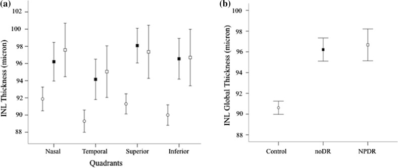
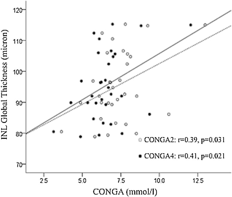
Results: The difference among the three groups in terms of RNFL thickness was significantly dependent on quadrant (F(6;132) = 2.315; p = 0.037). This interaction was due to a specific difference in RNFL-N thickness, where both Type 1 DM groups showed a similar reduction versus C (-3.9 for noDR and -4.9 for NPDR), without any relevant difference between them (-1.0). Inner nuclear layer (INL) was increased in all quadrants in the two Type 1 DM groups compared to C (mean difference = 7.73; 95% CI: 0.32-15.14, p = 0.043; mean difference = 7.74; 95% CI: 0.33-15.15, p = 0.043, respectively). A negative correlation between RNFL-N and low blood glucose index (r = -0.382, p = 0.034) and positive correlation between INL and continuous overall net glycemic action -1, -2, -4 h (r = 0.40, p = 0.025; r = 0.39, p = 0.031; r = 0.41, p = 0.021, respectively) were observed in Type 1 DM patients. The triglycerides were positively and significantly correlated to INL (r = 0.48, p = 0.011), in Type 1 DM subjects. GV and triglycerides resulted both independent predictors of increased INL thickness. No correlation was found with HbA1c.
Conclusions: Early structural damage of neuroretina in persons with Type 1 DM patients is related to glucose fluctuations. GV should be addressed, even in the presence of a good metabolic control.
Keywords: Glycemic variability; Retinal neurodegeneration; Type 1 diabetes mellitus.
Conflict of interest statement
Conflict of interest
The authors declare that they have no conflict of interest.
Ethical standards
All procedures performed in studies involving human participants were in accordance with the ethical standards of the institutional and/or national research committee and with the 1964 Helsinki Declaration and its later amendments or comparable ethical standards.
Informed consent
Informed consent was obtained from all individual participants included in the study.
Figures
Similar articles
-
Glucose variability and inner retinal sensory neuropathy in persons with type 1 diabetes mellitus.Eye (Lond). 2016 Jun;30(6):825-32. doi: 10.1038/eye.2016.48. Epub 2016 Apr 1. Eye (Lond). 2016. PMID: 27034201 Free PMC article.
-
Quantitative evaluation of early retinal changes in children with type 1 diabetes mellitus without retinopathy.Clin Exp Optom. 2018 Sep;101(5):680-685. doi: 10.1111/cxo.12667. Epub 2018 Feb 28. Clin Exp Optom. 2018. PMID: 29488254
-
Early Neurodegeneration of the Inner Retinal Layers in Type 1 Diabetes Mellitus.Ophthalmologica. 2016;235(3):125-32. doi: 10.1159/000442826. Epub 2015 Dec 17. Ophthalmologica. 2016. PMID: 26674204
-
Retinal Neurodegeneration in the Course of Diabetes-Pathogenesis and Clinical Perspective.Curr Neuropharmacol. 2016;14(8):805-809. doi: 10.2174/1570159x14666160225154536. Curr Neuropharmacol. 2016. PMID: 26915422 Free PMC article. Review.
-
Impact of glycemic variability on cardiovascular outcomes beyond glycated hemoglobin. Evidence and clinical perspectives.Nutr Metab Cardiovasc Dis. 2012 Sep;22(9):691-6. doi: 10.1016/j.numecd.2012.03.006. Epub 2012 Jun 4. Nutr Metab Cardiovasc Dis. 2012. PMID: 22673768 Review.
Cited by
-
Association between Early Neuroretinal Dysfunction and Peripheral Motor Unit Loss in Patients with Type 1 Diabetes Mellitus.J Diabetes Res. 2018 Oct 4;2018:9763507. doi: 10.1155/2018/9763507. eCollection 2018. J Diabetes Res. 2018. PMID: 30402503 Free PMC article.
-
Association of HbA1c With All-cause Mortality Across Varying Degrees of Glycemic Variability in Type 2 Diabetes.J Clin Endocrinol Metab. 2021 Oct 21;106(11):3160-3167. doi: 10.1210/clinem/dgab532. J Clin Endocrinol Metab. 2021. PMID: 34279663 Free PMC article.
-
Soluble CD163 and glycated haemoglobin were independently associated with the progression of diabetic retinopathy in adult patients with type 1 diabetes.BMJ Open Ophthalmol. 2023 Jul;8(1):e001314. doi: 10.1136/bmjophth-2023-001314. BMJ Open Ophthalmol. 2023. PMID: 37493689 Free PMC article.
-
Generation and quality control of lipidomics data for the alzheimer's disease neuroimaging initiative cohort.Sci Data. 2018 Nov 20;5:180263. doi: 10.1038/sdata.2018.263. Sci Data. 2018. PMID: 30457571 Free PMC article.
-
Complications of Diabetes and Metrics of Glycemic Management Derived From Continuous Glucose Monitoring.J Clin Endocrinol Metab. 2022 May 17;107(6):e2221-e2236. doi: 10.1210/clinem/dgac034. J Clin Endocrinol Metab. 2022. PMID: 35094087 Free PMC article. Review.
References
MeSH terms
Substances
LinkOut - more resources
Full Text Sources
Other Literature Sources
Medical
