Review
. 2018 Mar;125(3):547-563.
doi: 10.1007/s00702-017-1697-8. Epub 2017 Feb 25.

Advances in optogenetic and chemogenetic methods to study brain circuits in non-human primates

Affiliations
Review

Advances in optogenetic and chemogenetic methods to study brain circuits in non-human primates

Adriana Galvan et al. J Neural Transm (Vienna). 2018 Mar.

Abstract

Over the last 10 years, the use of opto- and chemogenetics to modulate neuronal activity in research applications has increased exponentially. Both techniques involve the genetic delivery of artificial proteins (opsins or engineered receptors) that are expressed on a selective population of neurons. The firing of these neurons can then be manipulated using light sources (for opsins) or by systemic administration of exogenous compounds (for chemogenetic receptors). Opto- and chemogenetic tools have enabled many important advances in basal ganglia research in rodent models, yet these techniques have faced a slow progress in non-human primate (NHP) research. In this review, we present a summary of the current state of these techniques in NHP research and outline some of the main challenges associated with the use of these genetic-based approaches in monkeys. We also explore cutting-edge developments that will facilitate the use of opto- and chemogenetics in NHPs, and help advance our understanding of basal ganglia circuits in normal and pathological conditions.

Keywords: Basal ganglia; Chemogenetics; DREADDs; Monkeys; Non-human primates; Opsins; Optogenetics.


Figures
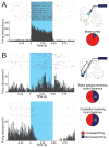
Figure 1
Figure 1
Effects of optogenetic stimulation of neurons in motor cortices and cortico-thalamic terminals in motor thalamus. A. Responses of cortical neurons to opsin activation. Left: Raster diagram and peri-stimulus time histogram (PSTH) of a single neuron in primary motor cortex, showing increased firing in responses to 500 ms light pulses. B. Responses of thalamic neurons to activation of opsins on corticothalamic axons. Top: Raster diagram and PSTH of a neuron in the basal ganglia-receiving territory of the motor thalamus that showed an increase in firing during the activation of ChR2. Bottom: A neuron in the cerebellar-receiving motor thalamus showing a decrease in firing during the activation of ChR2. For A and B, raster diagrams and PSTHs are aligned to the start of light pulses, each bin in the PSTH is 5 ms in duration; dashed horizontal lines indicate 2 SD above and below the baseline (based on the firing of the neuron during the 0.5 s prior to the start of each stimulus). The blue rectangles indicate the light pulses. At right: Experimental configuration to activate opsins expressed at the level of somas (top) or terminals (bottom) on cortico-thalamic neurons (green), with simultaneous extracellular recordings of cortical or thalamic neurons (top and bottom, respectively). Pie graphs represent the proportion of neurons in motor regions of cortex and thalamus that increased or decreased firing in response to the light stimulation. Numbers of cases are indicated (figure modified with permission from from Galvan et al. 2016).
Figure 2
Figure 2
GFP is selectively expressed in telencephalic GABAergic neurons of the HVC in zebra finch, as well as primary visual cortex (V1) of multiple mammalian species (including marmoset) following microinjections of rAAV-GFP placed under an enhancer sequence (mDLx) for the distalless homeobox 5/6 genes. These genes may be expressed in telencephalic GABergic interneurons during embryonic development. For each species, the degree of GFP colocalization with either GABA or GAD67 is provided on the right, in all cases confirming selective and robust GFP expression in GABAergic neurons. The use of well-designed short regulatory elements, as described here, may drive opsin or DREADD expression with exquisite cell type-specificity (figure reproduced with permission from Dimidschstein et al. 2016).

References

    1. Acker L, Pino EN, Boyden ES, Desimone R. FEF inactivation with improved optogenetic methods. Proc Natl Acad Sci U S A. 2016 doi: 10.1073/pnas.1610784113. - DOI - PMC - PubMed
    1. Afraz A, Boyden ES, DiCarlo JJ. Optogenetic and pharmacological suppression of spatial clusters of face neurons reveal their causal role in face gender discrimination. Proc Natl Acad Sci U S A. 2015;112:6730–6735. doi: 10.1073/pnas.1423328112. - DOI - PMC - PubMed
    1. Albin RL, Young AB, Penney JB. The functional anatomy of basal ganglia disorders. Trends Neurosci. 1989;12:366–375. - PubMed
    1. Alvarez L, Macias R, Pavon N, Lopez G, Rodriguez-Oroz MC, Rodriguez R, et al. Therapeutic efficacy of unilateral subthalamotomy in Parkinson’s disease: results in 89 patients followed for up to 36 months. J Neurol Neurosurg Psychiatry. 2009;80:979–985. doi: 10.1136/jnnp.2008.154948. - DOI - PubMed
    1. Armbruster BN, Li X, Pausch MH, Herlitze S, Roth BL. Evolving the lock to fit the key to create a family of G protein-coupled receptors potently activated by an inert ligand. Proc Natl Acad Sci U S A. 2007;104:5163–5168. doi: 10.1073/pnas.0700293104. - DOI - PMC - PubMed

Publication types

LinkOut - more resources