. 2017 Mar 16;24(3):281-292.
doi: 10.1016/j.chembiol.2017.02.003. Epub 2017 Feb 23.

Non-steroidal Anti-inflammatory Drugs Are Caspase Inhibitors

Affiliations

Non-steroidal Anti-inflammatory Drugs Are Caspase Inhibitors

Christina E Smith et al. Cell Chem Biol. .

Abstract

Non-steroidal anti-inflammatory drugs (NSAIDs) are among the most commonly used drugs in the world. While the role of NSAIDs as cyclooxygenase (COX) inhibitors is well established, other targets may contribute to anti-inflammation. Here we report caspases as a new pharmacological target for NSAID family drugs such as ibuprofen, naproxen, and ketorolac at physiologic concentrations both in vitro and in vivo. We characterize caspase activity in both in vitro and in cell culture, and combine computational modeling and biophysical analysis to determine the mechanism of action. We observe that inhibition of caspase catalysis reduces cell death and the generation of pro-inflammatory cytokines. Further, NSAID inhibition of caspases is COX independent, representing a new anti-inflammatory mechanism. This finding expands upon existing NSAID anti-inflammatory behaviors, with implications for patient safety and next-generation drug design.

Keywords: NSAIDs; anti-inflammation; caspase; pharmacology.


Figures

Figure 1
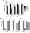
Figure 1. NSAIDs are multi-caspase inhibitors
Dose-response curves of five caspases treated with up to 66 μM of the indicated NSAIDs. Each caspase is denoted with a different color: caspase-1 in black, caspase-3 in pink, caspase-4 in red, caspase-5 in green, and caspase-9 in blue. The associated IC50 values are available in Table 2. 100%=substrate with solvent, 0%=z-VAD- FMK. Data are representative of the average and standard deviation of at least two independent replicates.
Figure 2
Figure 2. A biophysical and computational model of caspase binding interactions
(A) Titration with indoprofen or aspirin quenches caspase-3 tryptophan fluorescence. The apparent KD for indoprofen (purple circles) is 30 ± 2 μM. The apparent KD for aspirin (green squares) is 1290 ± 10 μM. Data are representative of the average and standard deviation of at least two independent experiments, normalized with the titrated solvent control as 100%, and buffer titrated with compound as 0%. (B) Caspase-3 (PDB 2DKO) is shown with a tetrapeptide substrate or NSAID (green). The catalytic cysteine denoted in pink, tryptophans are yellow, and other residues depict hydrophobicity (blue more polar, red more hydrophobic). The inset shows the surface of the binding pocket. (C) A representation of the top computationally predicted cluster of indoprofen bound to caspase-3. Predictions place indoprofen in the S1 pocket of the active site. (D) A representation of the top computationally predicted cluster of fenbufen bound to caspase-3. Predictions place indoprofen in the S1 pocket of the active site. (E) A representation of the top computationally predicted cluster of aspirin bound to caspase-3. Predictions place aspirin away from the active site.
Figure 3
Figure 3. NSAIDs inhibit catalysis of multiple caspases in cell culture
(A) HeLa cells treated with 1 μM staurosporine show dose-dependent caspase-3 inhibition when treated with 0–500 μM NSAIDs. 100%=staurosporine, 0%=z-VAD-FMK, *P≤0.05 compared to 100% control. (B) HeLa cells treated with 1 μM staurosporine and 100 μM NSAIDs show a reduction in caspase-9 activity. 100%=staurosporine, 0%=z-VAD-FMK, *P≤0.05 compared to 100% control. (C & D) THP-1 cells treated with 100 μM of selected NSAIDs in the presence of staurosporine (caspase-3, panel C) or 1 μM nigericin (caspase-1, panel D) show inhibited caspase catalytic activity. 100%=staurosporine or nigericin, 0%=z-VAD-FMK, *P≤0.05 compared to 100% control. See also Figure S3C.
Figure 4
Figure 4. NSAIDs inhibit caspase signaling in cell culture
(A) Treatment with 0–500 μM of NSAIDs improves viability of apoptotic THP-1 cells treated with 25 μM nigericin. 100%=untreated cells, 0%=nigericin, *P≤0.05 compared to 0% control. (B) THP-1 cells were treated with nigericin to activate caspase-1 and promote the inflammatory cytokine IL-1β. NSAID treatment from 0–500 μM demonstrates a dose-dependent reduction of IL-1β release. 100%=staurosporine, 0%=untreated cells, *P≤0.05 compared to 100% control. All data are the average and standard deviation of three biological replicates. The statistical significance was determined using a one-way ANOVA followed by Tukey’s test. See also Figure S3D.
Figure 5
Figure 5. Caspase inhibition by NSAIDs is COX-independent
(A) HCT116 cells treated with 10 μM staurosporine and 100 μM NSAIDs demonstrate inhibition of caspase-3 catalysis. HCT116 cells do not express COX-2, indicating that caspase inhibition is not contingent on canonical COX2 pharmacology. Data are representative of the average and standard deviation of three biological replicates. 100%=staurosporine, 0%=z-VAD-FMK, *P≤0.05 compared to 100% control. (B) Cell death assays in C. elegans embryos treated with fenbufen or indoprofen. L4-stage ced-1(e1735) animals were exposed to 100 μM fenbufen or indoprofen in 0.5% DMSO on NGM agar plates, and their progenies were analyzed. Embryonic cell corpses were scored at comma, 1.5-fold, 2-fold, 2.5-fold, 3-fold, and 4-fold stages. The y-axis represents the mean and standard deviation of cell corpses scored (n = 15). The statistical significance was determined using a one-way ANOVA followed by Tukey’s test. (*P≤0.05 compared to 0.5% DMSO control). See also Figure S4.

Similar articles

Cited by

References

    1. Ashburn TT, Thor KB. Drug repositioning: identifying and developing new uses for existing drugs. Nat Rev Drug Discov. 2004;3:673–683. - PubMed
    1. Barrett AJ. Fluorimetric assays for cathepsin B and cathepsin H with methylcoumarylamide substrates. Biochem J. 1980;187:909–912. - PMC - PubMed
    1. Conradt B, Wu YC, Xue D. Programmed Cell Death During Caenorhabditis elegans Development. Genetics. 2016;203:1533–1562. - PMC - PubMed
    1. Creagh EM. Caspase crosstalk: Integration of apoptotic and innate immune signalling pathways. Trends Immunol. 2014;35:631–640. - PubMed
    1. Denault J-B, Salvesen GS. Current Protocols in Protein Science. Hoboken, NJ, USA: John Wiley & Sons, Inc; 2002. Expression, Purification, and Characterization of Caspases; pp. 21.13.1–21.13.15. - PubMed

MeSH terms

LinkOut - more resources