. 2017 Mar 16;24(3):316-325.
doi: 10.1016/j.chembiol.2017.01.008. Epub 2017 Feb 23.

Functional and Structural Analysis of Programmed C-Methylation in the Biosynthesis of the Fungal Polyketide Citrinin

Affiliations

Functional and Structural Analysis of Programmed C-Methylation in the Biosynthesis of the Fungal Polyketide Citrinin

Philip A Storm et al. Cell Chem Biol. .

Abstract

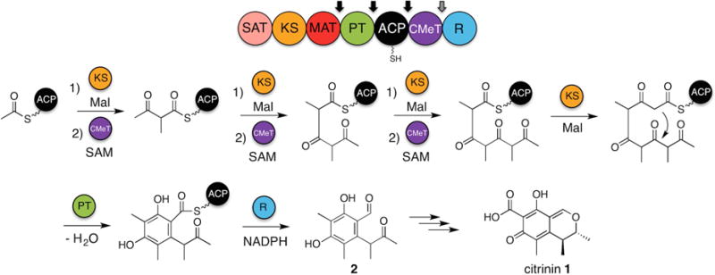
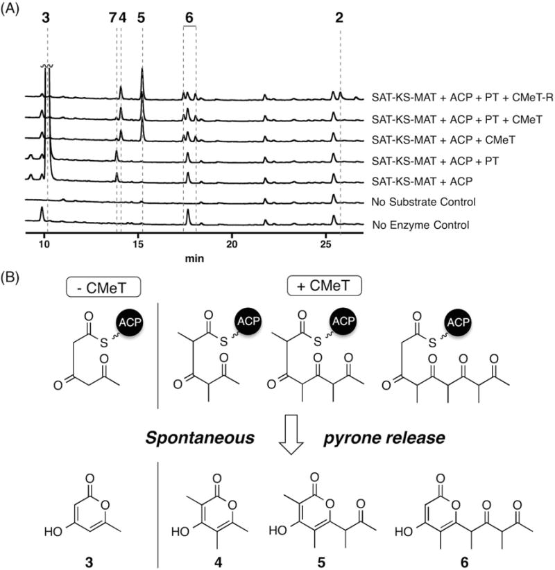
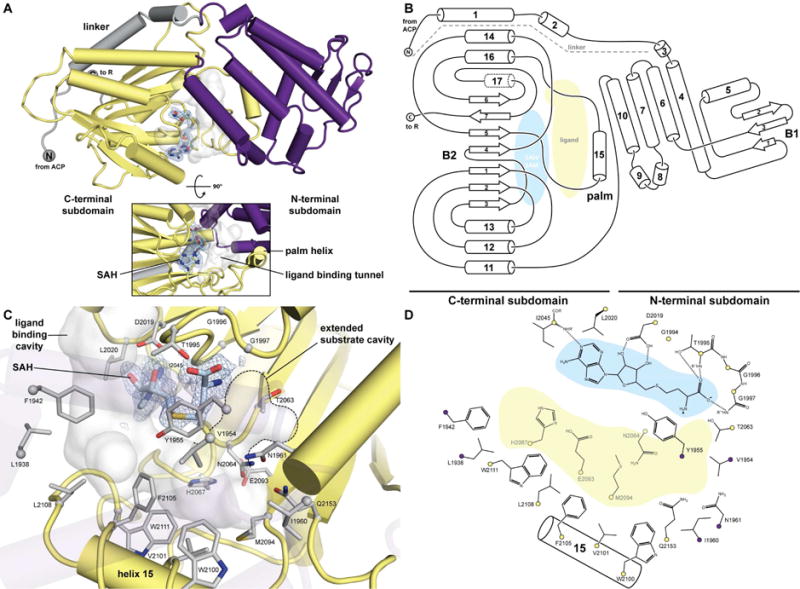
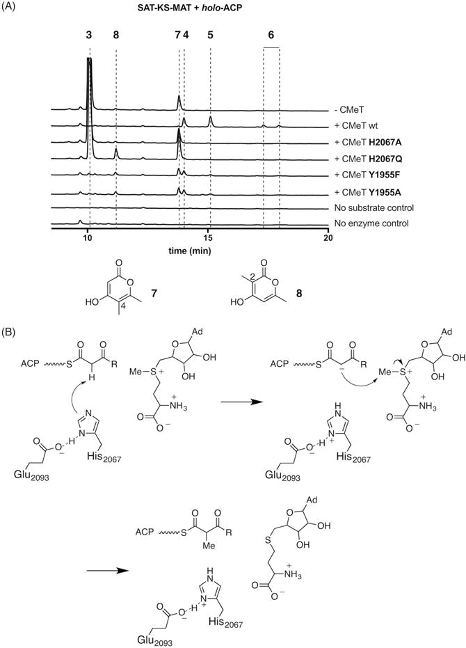
Fungal polyketide synthases (PKSs) are large, multidomain enzymes that biosynthesize a wide range of natural products. A hallmark of these megasynthases is the iterative use of catalytic domains to extend and modify a series of enzyme-bound intermediates. A subset of these iterative PKSs (iPKSs) contains a C-methyltransferase (CMeT) domain that adds one or more S-adenosylmethionine (SAM)-derived methyl groups to the carbon framework. Neither the basis by which only specific positions on the growing intermediate are methylated ("programming") nor the mechanism of methylation are well understood. Domain dissection and reconstitution of PksCT, the fungal non-reducing PKS (NR-PKS) responsible for the first isolable intermediate in citrinin biosynthesis, demonstrates the role of CMeT-catalyzed methylation in precursor elongation and pentaketide formation. The crystal structure of the S-adenosyl-homocysteine (SAH) coproduct-bound PksCT CMeT domain reveals a two-subdomain organization with a novel N-terminal subdomain characteristic of PKS CMeT domains and provides insights into co-factor and ligand recognition.

Keywords: C-methylation; PKS; catalytic programming; citrinin; domain deconstruction; iterative biosynthesis; polyketide; structure.


Figures

Figure 1
Figure 1. Proposed biosynthesis of 2 by PksCT
The domain architecture of PksCT is shown above, with arrows indicating points of domain dissection. Mal indicates a unit of ACP-bound malonyl used for decarboxylative condensation prior to methylation. Necessary domains and substrates are shown above and below the reaction arrows. See also Figure S1 and Item S1.
Figure 2
Figure 2. In vitro reconstitution of PksCT
(A) HPLC absorbance traces are shown at 280 nm for the combination of domains indicated to the right. Traces are vertically offset and peak 3 is truncated for clarity. See also Figure S1 and Table S2. (B) Spontaneous pyrone release of intermediates as tri-, tetra-, and pentaketide pyrones. High resolution UPLC-ESI-MS and UV-Vis data can be found in Table S4 and Figure S2.
Figure 3
Figure 3. Crystal structure and ligand binding site of CMeT
(A) CMeT is organized into an N-terminal linker (grey), an N-terminal subdomain (violet) and a C-terminal subdomain (yellow). The active site is located at the subdomain interface, Fo-Fc omit difference density at 2.5 σ level is shown for SAH. See also Table S3 and Figure S4. (B) Topology of CMeT highlighting domain organization and substrate binding sites. α-Helices are numbered, β-strands are numbered relative to their position in the respective β-sheet B1 or B2. Helix 17 (dotted) is a 310 helix. (C) Substrate interactions and ligand binding tunnel. The C-terminal subdomain laterally binds SAH and forms an active site tunnel (grey surface) along SAH. The back of this tunnel is lined with hydrophobic residues of the palm helix region and is closed by the N-terminal subdomain. The invariant residues Tyr1955 as well as His2067 together with Glu2093 face the ligand binding tunnel from opposite sides and are involved in catalysis. SAH contributes to the formation of an extended cavity for binding larger substrates. Difference density is depicted as in A. See also Figure S5. (D) Schematic active site representation. Hydrogen bonds are indicated by dotted lines. Cα atoms are shown as spheres in the color of their respective subdomain.
Figure 4
Figure 4. Phylogenetic analysis of 51 CMeT domains of PKS and FAS
38 active CMeT domains of PKS and 13 inactive ΨCMeT domains of HR-PKS and FAS were aligned and phylogenetically analyzed (see Figure S4). Multienzyme family classifications are indicated in colored groups. Units are given as amino-acid substitutions per site. All sequences are labelled as “protein name (organism abbreviation) Uniprot number”. The sequence of PksCT corresponds to Item S1 (°, endosymbiont of this org anism; , diketide synthase; *, inactive ΨCMeT domain)
Figure 5
Figure 5. PksCT CMeT His2067 is essential for methyl transfer and positioned to act as the catalytic base
(A) In vitro reconstitution of PksCT SAT-KS-MAT and holo-ACP with CMeT mutants show that methylated products 4, 5, and 6 are not produced by His2067 mutants. Tyr1955 mutants are capable of generating methylated triketides, but not pentaketides, suggesting a role for this residue in acceptor substrate binding. Absorbance traces are shown at 280 nm for the CMeT variant indicated on the right and vertically offset; peak 3 is truncated for clairty. For mass and UV-Vis absorption data, see Table S4 and Figure S2. (B) Proposed mechanism for methyl transfer. His2067 forms a catalytic dyad with Glu2093 and deprotonates the α-carbon to generate an enolate nucleophile capable of SN2-like attack at the methyl donor. Completion of the catalytic cycle by loss of the removed proton to solvent is not explicitly shown. R indicates the potential chain lengths described in Figure 1.

References

    1. Adams PD, Afonine PV, Bunkoczi G, Chen VB, Davis IW, Echols N, Headd JJ, Hung LW, Kapral GJ, Grosse-Kunstleve RW, et al. PHENIX: a comprehensive Python-based system for macromolecular structure solution. Acta Crystallogr D Biol Crystallogr. 2010;66:213–221. - PMC - PubMed
    1. Ahuja M, Chiang YM, Chang SL, Praseuth MB, Entwistle R, Sanchez JF, Lo HC, Yeh HH, Oakley BR, Wang CC. Illuminating the diversity of aromatic polyketide synthases in Aspergillus nidulans. J Am Chem Soc. 2012;134:8212–8221. - PMC - PubMed
    1. Belecki K, Townsend CA. Biochemical determination of enzyme-bound metabolites: preferential accumulation of a programmed octaketide on the enediyne polyketide synthase CalE8. J Am Chem Soc. 2013;135:14339–14348. - PMC - PubMed
    1. Cacho RA, Thuss J, Xu W, Sanichar R, Gao Z, Nguyen A, Vederas JC, Tang Y. Understanding Programming of Fungal Iterative Polyketide Synthases: The Biochemical Basis for Regioselectivity by the Methyltransferase Domain in the Lovastatin Megasynthase. J Am Chem Soc. 2015;137:15688–15691. - PMC - PubMed
    1. Conrad RS, Massey LK, Sokatch JR. D- and L-isoleucine metabolism and regulation of their pathways in Pseudomonas putida. J Bacteriol. 1974;118:103–111. - PMC - PubMed

MeSH terms