Review
. 2017 Jun:142:83-122.
doi: 10.1016/j.antiviral.2017.02.014. Epub 2017 Feb 24.

Current therapy for chronic hepatitis C: The role of direct-acting antivirals

Affiliations
Review

Current therapy for chronic hepatitis C: The role of direct-acting antivirals

Guangdi Li et al. Antiviral Res. 2017 Jun.

Abstract

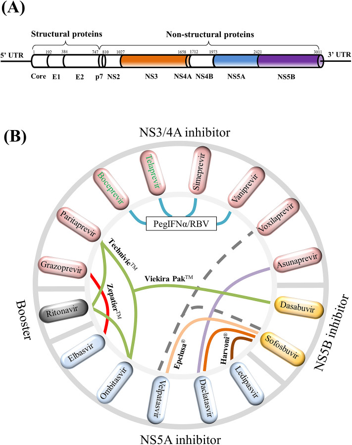
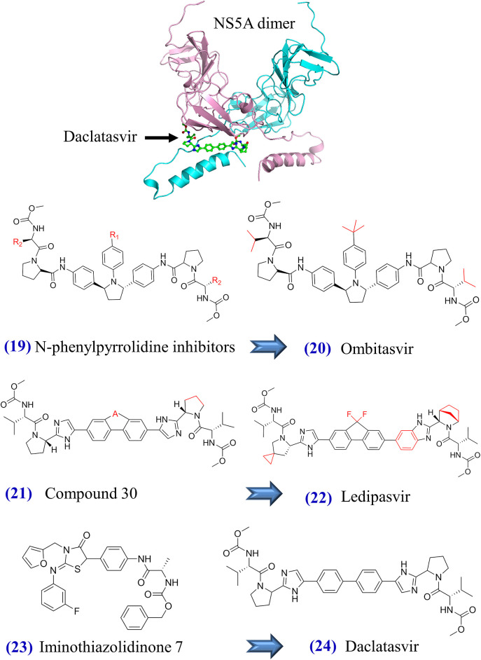
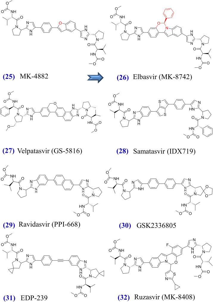
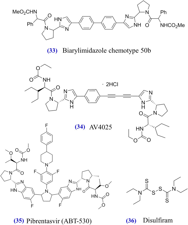
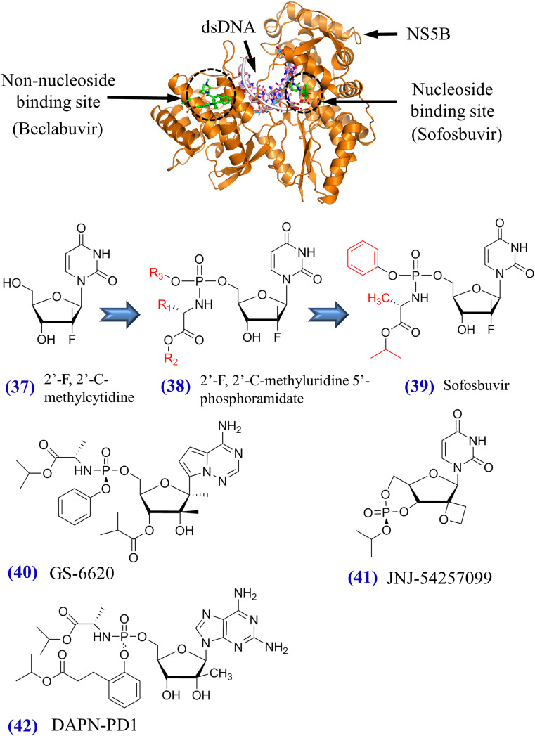
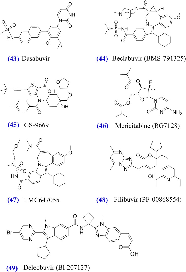
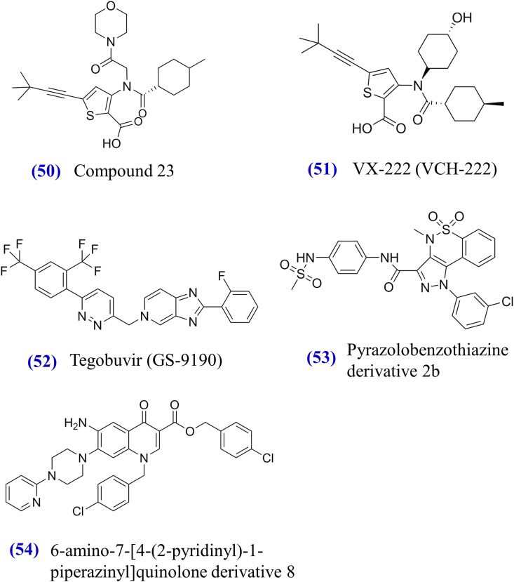
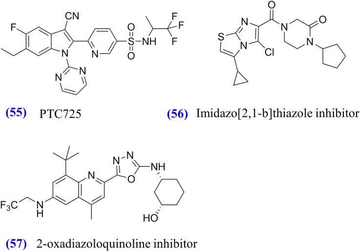
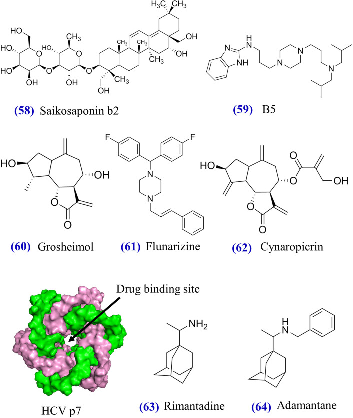
One of the most exciting developments in antiviral research has been the discovery of the direct-acting antivirals (DAAs) that effectively cure chronic hepatitis C virus (HCV) infections. Based on more than 100 clinical trials and real-world studies, we provide a comprehensive overview of FDA-approved therapies and newly discovered anti-HCV agents with a special focus on drug efficacy, mechanisms of action, and safety. We show that HCV drug development has advanced in multiple aspects: (i) interferon-based regimens were replaced by interferon-free regimens; (ii) genotype-specific drugs evolved to drugs for all HCV genotypes; (iii) therapies based upon multiple pills per day were simplified to a single pill per day; (iv) drug potency increased from moderate (∼60%) to high (>90%) levels of sustained virologic responses; (v) treatment durations were shortened from 48 to 12 or 8 weeks; and (vi) therapies could be administered orally regardless of prior treatment history and cirrhotic status. However, despite these remarkable achievements made in HCV drug discovery, challenges remain in the management of difficult-to-treat patients.

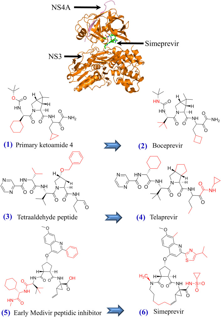
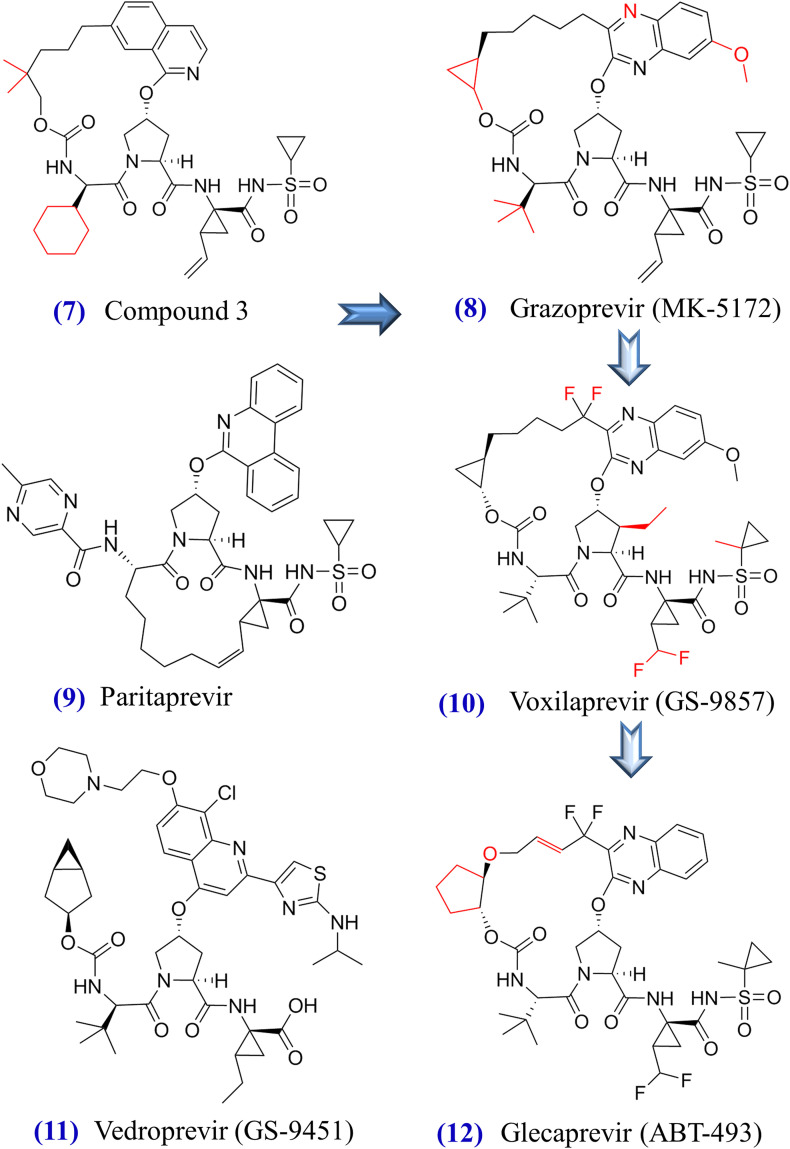
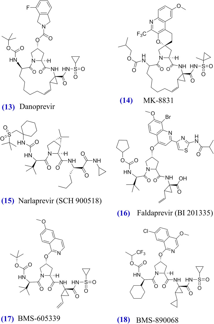
Keywords: Direct-acting antivirals; NS3/4A drugs; NS5A drugs; NS5B drugs.


Figures

Fig. 1
Fig. 1
HCV genome structure and schematic view of HCV combination drugs. (A) HCV genome structure. In the length of approximately 3011 amino acids, the HCV genome codes for three structural proteins (core, E1, E2) and seven non-structural proteins (p7, NS2, NS3, NS4A, NS4B, NS5A, NS5B) whose amino acid positions are mapped accordingly. The 5′ untranslated region (5′ UTR) and the 3′ untranslated region (3′ UTR) are also indicated. Approved antiviral agents directly target to NS3/4A, NS5A, and NS5B for effective inhibition of HCV replications. (B) A total of 15 NS3/4A, NS5A, NS5B compounds plus ritonavir are displayed in the circle. Colored links in the center visualize 13 drug combinations: (i) boceprevir (Victrelis®) + PegIFNα/RBV, (ii) telaprevir (Incivek®) + PegIFNα/RBV, (iii) sofosbuvir (Sovaldi®) + PegIFNα/RBV, (iv) simeprevir (Olysio®) + PegIFNα/RBV, (v) ledipasvir + sofosbuvir (Harvoni®), (vi) ombitasvir + paritaprevir + ritonavir + dasabuvir (Viekira Pak™), (vii) ombitasvir + paritaprevir + ritonavir (Technivie™), (viii) daclatasvir (Daklinza™) + sofosbuvir (Sovaldi®), (ix) elbasvir + grazoprevir (Zepatier™), (x) sofosbuvir + velpatasvir (Epclusa®), (xi) vaniprevir (Vanihep®) + PegIFNα/RBV, (xii) asunaprevir (Sunvepra®) + daclatasvir (Daklinza®), (xiii) voxilaprevir + velpatasvir + sofosbuvir. Notably, (i) to (x) were approved by the FDA and could be used with or without ribavirin; (xi) and (xii) were approved in Japan; and (xiii) is currently under assessment by the FDA. Two discontinued drugs boceprevir and telaprevir are indicated by green texts. This figure shows that HCV combination drugs are composed of anti-HCV inhibitors from different drug classes.
Fig. 2
Fig. 2
Tertiary structure of HCV NS3/4A protease and structural formulas of approved or experimental NS3/4A inhibitors from (1) to (18). The tertiary structure of NS3/4A protease in complex with simeprevir (PDB codes: 3KEE and 4B76) is shown on top. HCV NS3 and NS4A proteins are displayed in orange and pink, respectively. Beneath the NS3/4A protein, chemical structures of approved and experimental NS3/4A inhibitors (Table 20) are demonstrated. Blue arrows indicate the optimization from initial compounds to FDA-approved compounds such as boceprevir (Njoroge et al., 2008), telaprevir (Kwong et al., 2011), simeprevir (Rosenquist et al., 2014), and grazoprevir (Harper et al., 2012, Liverton et al., 2008). Red structures indicate the differences between initial compounds and FDA- approved compounds.
Fig. 2
Fig. 2
Tertiary structure of HCV NS3/4A protease and structural formulas of approved or experimental NS3/4A inhibitors from (1) to (18). The tertiary structure of NS3/4A protease in complex with simeprevir (PDB codes: 3KEE and 4B76) is shown on top. HCV NS3 and NS4A proteins are displayed in orange and pink, respectively. Beneath the NS3/4A protein, chemical structures of approved and experimental NS3/4A inhibitors (Table 20) are demonstrated. Blue arrows indicate the optimization from initial compounds to FDA-approved compounds such as boceprevir (Njoroge et al., 2008), telaprevir (Kwong et al., 2011), simeprevir (Rosenquist et al., 2014), and grazoprevir (Harper et al., 2012, Liverton et al., 2008). Red structures indicate the differences between initial compounds and FDA- approved compounds.
Fig. 3
Fig. 3
Tertiary structure of HCV NS5A and structural formulas of approved or experimental NS5A inhibitors from (19) to (36). Two units of an NS5A dimer are colored by pink and cyan, respectively. NS5A inhibitor daclatasvir is also illustrated. Blue arrows indicate the optimization from initial compounds to FDA-approved compounds such as ombitasvir (DeGoey et al., 2014), ledipasvir (Link et al., 2014), daclatasvir (Belema and Meanwell, 2014), and elbasvir (Coburn et al., 2013). Other NS5A inhibitors are summarized in Table 20.
Fig. 3
Fig. 3
Tertiary structure of HCV NS5A and structural formulas of approved or experimental NS5A inhibitors from (19) to (36). Two units of an NS5A dimer are colored by pink and cyan, respectively. NS5A inhibitor daclatasvir is also illustrated. Blue arrows indicate the optimization from initial compounds to FDA-approved compounds such as ombitasvir (DeGoey et al., 2014), ledipasvir (Link et al., 2014), daclatasvir (Belema and Meanwell, 2014), and elbasvir (Coburn et al., 2013). Other NS5A inhibitors are summarized in Table 20.
Fig. 4
Fig. 4
Tertiary structure of HCV NS5B and structural formulas of approved or experimental nucleoside inhibitors from (37) to (42). NS5B structure in complex with beclabuvir and sofosbuvir diphosphate (PDB codes: 4NLD and 4WTG) is visualized on top. The discovery of sofosbuvir undertakes the optimization path from 2′-F, 2′-C-methylcytidine to 2′-F, 2′-C-methyluridine 5’-phosphoramidate (Sofia et al., 2010). Structural formulas of GS-6620, JNJ-54257099, and DAPN-PD1 are also demonstrated.
Fig. 5
Fig. 5
Structural formulas of NS5B non-nucleoside inhibitors from (43) to (54). FDA-approved dasabuvir and experimental compounds target the non-nucleoside binding site in NS5B (see Fig. 4).
Fig. 5
Fig. 5
Structural formulas of NS5B non-nucleoside inhibitors from (43) to (54). FDA-approved dasabuvir and experimental compounds target the non-nucleoside binding site in NS5B (see Fig. 4).
Fig. 6
Fig. 6
Chemical formulas of experimental NS4B inhibitors fromSarrazin, 2016, AASLD/IDSA HCV Guidance Panel, 2015, European Association for Study of Liver, 2015.
Fig. 7
Fig. 7
Chemical formulas of experimental E1/E2 inhibitors from (58) to (62) and the tertiary structure of p7 and its experimental inhibitors from (63) to (64). Six units of the hexameric p7 channel are colored accordingly (PDB code: 2M6X). The drug binding site is located in the center of HCV p7 channel (OuYang et al., 2013).
Fig. 8
Fig. 8
Cellular protein inhibitors and immuno-stimulators from (65) to (72). Tertiary structure of host protein cyclophilin A in complex with phenyl–pyrrolidine 31 is visualized (PDB code: 3RDD). In addition, cyclophilin A inhibitors include alisporivir, bis-amide derivative 25, and NIM258 (Table 20). Other compounds target other host proteins to offer antiviral activity (see details in Section 5).
Fig. 8
Fig. 8
Cellular protein inhibitors and immuno-stimulators from (65) to (72). Tertiary structure of host protein cyclophilin A in complex with phenyl–pyrrolidine 31 is visualized (PDB code: 3RDD). In addition, cyclophilin A inhibitors include alisporivir, bis-amide derivative 25, and NIM258 (Table 20). Other compounds target other host proteins to offer antiviral activity (see details in Section 5).

References

    1. AASLD/IDSA HCV Guidance Panel Hepatitis C guidance: AASLD-IDSA recommendations for testing, managing, and treating adults infected with hepatitis C virus. Hepatology. 2015;62:932–954. - PubMed
    1. Abergel A., Metivier S., Samuel D., Jiang D., Kersey K., Pang P.S., Svarovskaia E., Knox S.J., Loustaud-Ratti V., Asselah T. Ledipasvir plus sofosbuvir for 12 weeks in patients with hepatitis C genotype 4 infection. Hepatology. 2016;64:1049–1056. - PubMed
    1. Abergel A., Asselah T., Metivier S., Kersey K., Jiang D., Mo H., Pang P.S., Samuel D., Loustaud-Ratti V. Ledipasvir-sofosbuvir in patients with hepatitis C virus genotype 5 infection: an open-label, multicentre, single-arm, phase 2 study. Lancet Infect. Dis. 2016;16:459–464. - PubMed
    1. Afdhal N., Zeuzem S., Kwo P., Chojkier M., Gitlin N., Puoti M., Romero-Gomez M., Zarski J.P., Agarwal K., Buggisch P., Foster G.R., Brau N., Buti M., Jacobson I.M., Subramanian G.M., Ding X., Mo H., Yang J.C., Pang P.S., Symonds W.T., McHutchison J.G., Muir A.J., Mangia A., Marcellin P., ION-1 Investigators Ledipasvir and sofosbuvir for untreated HCV genotype 1 infection. N. Engl. J. Med. 2014;370:1889–1898. - PubMed
    1. Afdhal N., Reddy K.R., Nelson D.R., Lawitz E., Gordon S.C., Schiff E., Nahass R., Ghalib R., Gitlin N., Herring R., Lalezari J., Younes Z.H., Pockros P.J., Di Bisceglie A.M., Arora S., Subramanian G.M., Zhu Y., Dvory-Sobol H., Yang J.C., Pang P.S., Symonds W.T., McHutchison J.G., Muir A.J., Sulkowski M., Kwo P., ION-2 Investigators Ledipasvir and sofosbuvir for previously treated HCV genotype 1 infection. N. Engl. J. Med. 2014;370:1483–1493. - PubMed

MeSH terms