. 2017 Apr:69:150-158.
doi: 10.1016/j.reprotox.2017.02.013. Epub 2017 Feb 24.

Mono-(2-ethylhexyl) phthalate-induced Sertoli cell injury stimulates the production of pro-inflammatory cytokines in Fischer 344 rats

Affiliations

Mono-(2-ethylhexyl) phthalate-induced Sertoli cell injury stimulates the production of pro-inflammatory cytokines in Fischer 344 rats

Angela R Stermer et al. Reprod Toxicol. 2017 Apr.

Abstract

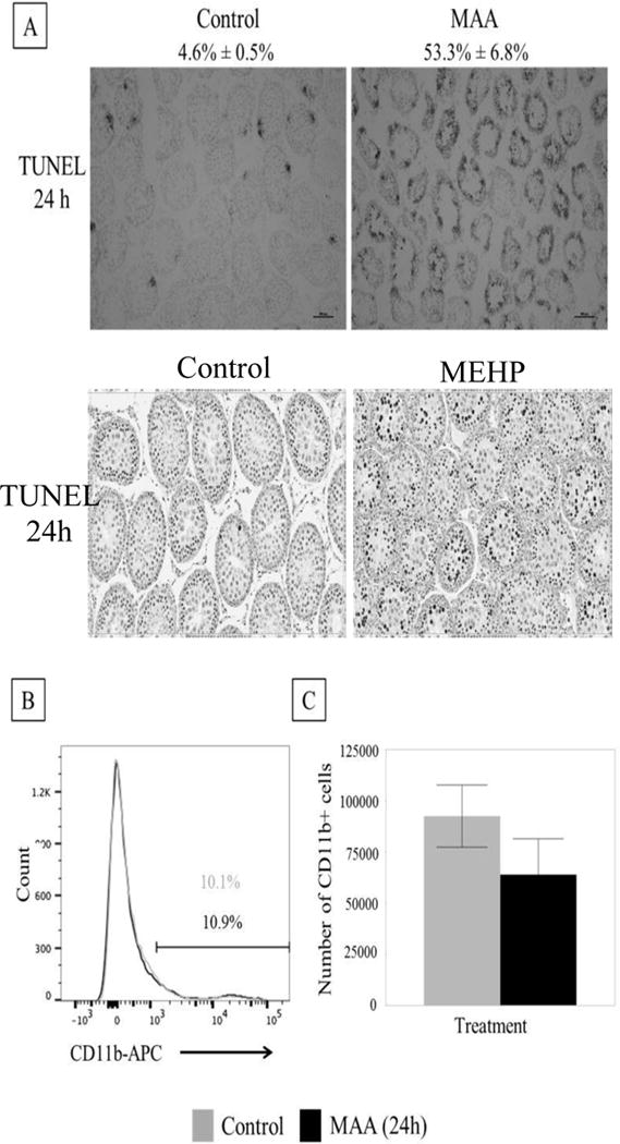
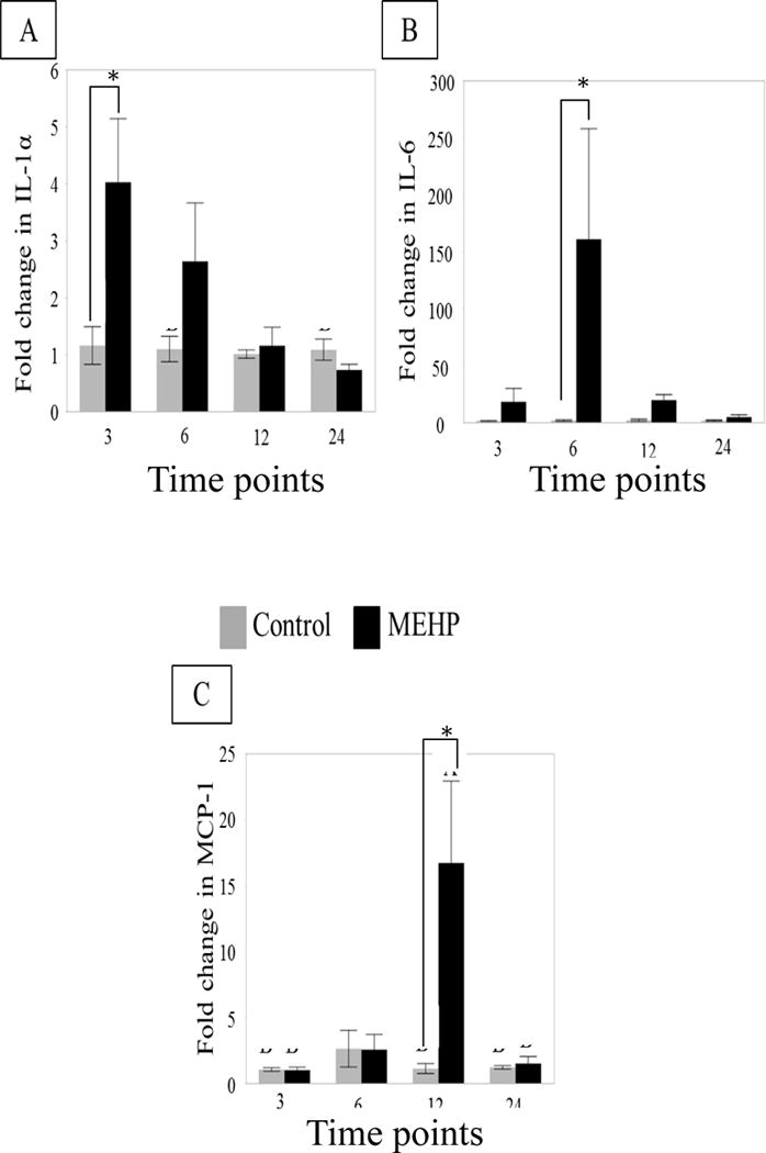
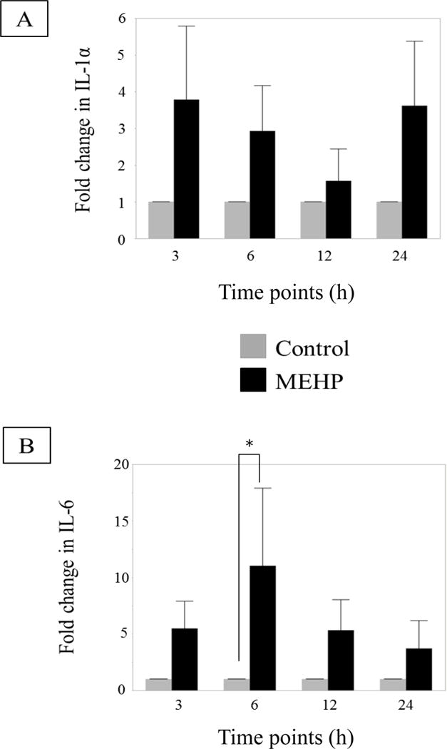
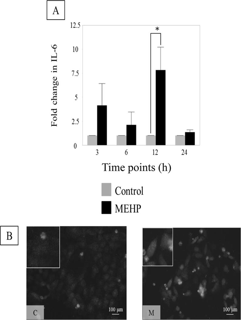
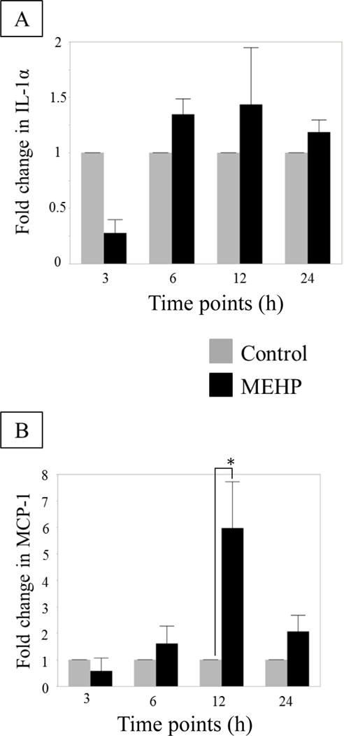
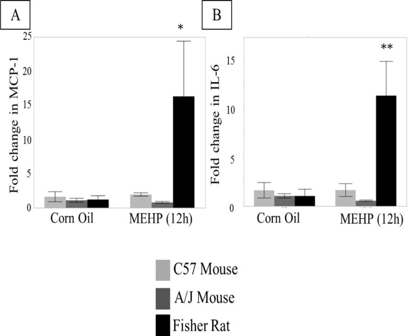
Exposure of rodents to the Sertoli cell (SC) toxicant mono-(2-ethylhexyl) phthalate (MEHP) has been reported to trigger an infiltration of macrophages into the testis in an age- and species-dependent manner. Here we challenge the hypothesis that the peripubertal rat-specific infiltration of macrophages after MEHP exposure is due, in part, to an increase in SC-specific inflammatory cytokine expression. To rule out that germ cell(GC) apoptosis itself is responsible for macrophage recruitment, rats were exposed to a direct GC toxicant, methoxyacetic acid (MAA), but no infiltration of macrophages was observed. Next, mRNA levels of inflammatory cytokines were evaluated after MEHP exposure. IL-1α, IL-6, and MCP-1 expression were increased in vivo and correlated with macrophage infiltration in a species-specific manner. Additionally, IL-6 and MCP-1 expression was increased in SC-GC co-cultures and ASC-17D SCs. These results indicate that MEHP-injury in pubertal rats specifically stimulates secretion of pro-inflammatory cytokines and alters the immune microenvironment.

Keywords: Cytokines; Mono-(2-ethylhexyl) phthalate; Sertoli cells; Testicular macrophages.


Figures

FIGURE 1
FIGURE 1. MAA causes extensive germ cell apoptosis but does not result in macrophage infiltration
(A) Top: Representative photomicrographs of TUNEL staining on frozen MAA (650 mg/kg, p.o.) or equivalent volume control (5.5 ml/kg, NaCl) treated PND 28 rat testis after 24h exposure (n=3 per treatment). MAA induced a significant increase in apoptotic index (% seminiferous tubules with >3 TUNEL positive germ cells/total number of seminiferous tubules) from 4.6% ± 0.5% to 53.3% ± 6.8%. Bottom: Representative photomicrographs of TUNEL staining on paraffin embedded MEHP (667 mg/kg, p.o.) or equivalent control (2ml/kg corn oil) treated PND 28 rat testis after 24 h exposure. MEHP induced a significant increase in apoptotic index nearly 100 fold [9]. (B) Histogram of MAA or control treated rat interstitial cells (after 24h exposure). The CD11b+ population has high intensity fluorescence and is on the right of the graph. This population does not change between treatment (black bar) and control (grey bar). The population on the left is the CD11b- population. (C) When the percentage CD11b+ found from flow were applied to the total number of cells collected from the interstitium, there were no differences in the total number of CD11b+ cells. Statistical analysis was performed using Student’s t-tests.
FIGURE 2
FIGURE 2. Cytokine RNA expression changes in in vivo seminiferous tubule preparations from PND 28 rat treated in vivo with MEHP
Changes in mRNA expression of (A) IL-1α and (B) IL-6 (C) MCP-1 in MEHP (667 mg/kg p.o, black bars) or control (2 ml/kg corn oil, grey bars) treated rat seminiferous tubules at 3, 6, 12, and 24 h (n=6 per treatment per time point). IL-1α expression was significantly increased at 3 h. IL-6 expression was found to be significantly increased at 6 h. MCP- mRNA expression was significantly increased at 12 h. Statistical analysis was performed using two-way ANOVA for unmatched data with Sidak’s multiple comparison test and each MEHP-treated sample was compared to its time point control.
FIGURE 3
FIGURE 3. Cytokine changes in primary rat co-cultures after in vitro exposure to MEHP
(A) Changes in IL-1α (B) and IL-6 mRNA levels primary SC-GC co-cultures after exposure to control (0.04% DMSO, grey bar), MEHP (200 μM, black bar), at selected time points (n=3 per treatment per time point, each n is a separate collection and pooling of at least 3 animals) (h). MEHP induced a significant increase of IL-6 mRNA levels at 6 h. Statistical analysis was performed using two-way ANOVA for matched pairs with Sidak’s multiple comparison test and each MEHP-treated sample was compared to its time point control.
FIGURE 4
FIGURE 4. IL-6 increases in Sertoli cell rat line ASC-17D cells after in vitro exposure to MEHP
(A) IL-6 expression in control (0.04% DMSO, control, grey) or MEHP (200 μM, black) treated ASC-17D cells at 3, 6, 12 and 24 h. MEHP induced a significant increase of IL-6 mRNA levels at 12 h. Statistical analysis was performed using two-way ANOVA for matched pairs with Sidak’s multiple comparison test and each MEHP-treated sample was compared to its time-point control. Representative microphotographs (B) of ASC-17D SC line immunostained with IL-6 (green, AlexaFlour488) after exposure to C (DMSO, control) or M (MEHP) for 24 h.
FIGURE 5
FIGURE 5. No change in IL-1α, but MCP-1 does increase in rat Sertoli cell line ASC-17D
(A) IL-1α and (B) MCP-1 mRNA expression by ASC-17D SC line at 3, 6, 12 and 24 h after MEHP (200 μM, black) or control (DMSO, grey) exposure (n=3 per treatment per time point). No statistical differences were found in IL-1α mRNA expression. MEHP induced a significant increase in MCP-1 mRNA compared to control at 12 h. Statistical analysis was performed using two-way ANOVA for matched pairs with Sidak’s multiple comparison test and each MEHP-treated sample was compared to its time-point control.
FIGURE 6
FIGURE 6. Differential expression of cytokines after in vivo exposure of Fisher rats, C57Bl/6J and A/J mice
(A) MCP-1 and (B) IL-6 mRNA expression in 12 h MEHP or control treated Fisher Rats (667 mg/kg p.o., light grey), C57BL/6J (800 mg/kg p.o., black) and A/J mice (800 mg/kg p.o., dark grey) (n=3 per treatment per species/strain). Only Fisher rats showed increased cytokine mRNA expression as a result of MEHP treatment. Statistical analysis was performed using two-way ANOVA for unmatched data with Sidak’s multiple comparison test and significance is indicated for each MEHP-treated animal compared to its control. * p < 0.05, ** p < 0.01.

Similar articles

Cited by

References

    1. O’Bryan MK, Hedger MP. Inflammatory networks in the control of spermatogenesis : chronic inflammation in an immunologically privileged tissue? Adv Exp Med Biol. 2008;636:92–114. - PubMed
    1. Hedger MP. Immunophysiology and pathology of inflammation in the testis and epididymis. J Androl. 2011 Nov-Dec;32(6):625–40. - PMC - PubMed
    1. Hedger MP, Winnall WR. Regulation of activin and inhibin in the adult testis and the evidence for functional roles in spermatogenesis and immunoregulation. Mol Cell Endocrinol. 2012 Aug 15;359(1–2):30–42. - PubMed
    1. Campese AF, Grazioli P, de Cesaris P, Riccioli A, Bellavia D, Pelullo M, et al. Mouse Sertoli cells sustain de novo generation of regulatory T cells by triggering the notch pathway through soluble JAGGED1. Biol Reprod. 2014 Mar;90(3):53. - PubMed
    1. Doyle TJ, Kaur G, Putrevu SM, Dyson EL, Dyson M, McCunniff WT, et al. Immunoprotective properties of primary Sertoli cells in mice: potential functional pathways that confer immune privilege. Biol Reprod. 2012 Jan;86(1):1–14. - PMC - PubMed

Publication types

LinkOut - more resources