

An atypical case of recurrent carotid body carcinoma in a young adult dog: Histopathological, immunohistochemical and electron microscopic study
- PMID: 28239052
- PMCID: PMC5402192
- DOI: 10.1292/jvms.16-0501
An atypical case of recurrent carotid body carcinoma in a young adult dog: Histopathological, immunohistochemical and electron microscopic study
Abstract
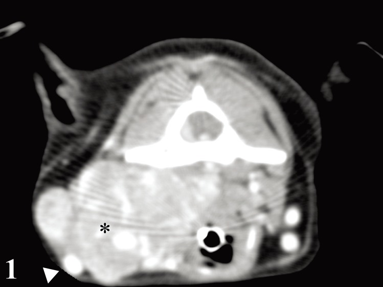
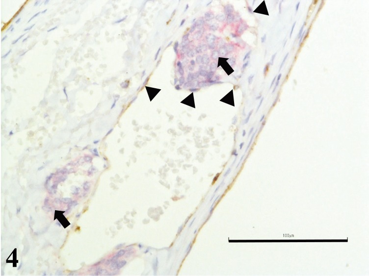
A 3.5-year-old female Chihuahua was presented with complaint of neck pain, intermittent cough and dysphagia. Physical examination and diagnostic imaging of neck region revealed a solid and highly vascularized mass involving the retropharyngeal region. Histologically, the mass showed an atypical zellballen pattern which comprised of high density of type I chief cells with high nuclear cytoplasmic ratio and separated by delicate fibrovascular stroma. Immunoreactivity for neuroendocrine markers was diffusely positive in cytoplasm of tumor cells. Disseminated tumor emboli in external jugular vein were detected 6 months after initial surgery. An electron microscopic study revealed numerous electron-dense intracytoplasmic neurosecretory granules. Based on these findings, carotid body carcinoma was diagnosed.
Figures


References
-
- Dean M. J., Strafuss A. C.1975. Carotid body tumors in the dog: a review and report of four cases. J. Am. Vet. Med. Assoc. 166: 1003–1006. - PubMed
Publication types
MeSH terms
LinkOut - more resources
Full Text Sources
Other Literature Sources
Medical