Review
. 2017 Apr;15(4):243-254.
doi: 10.1038/nrmicro.2016.197. Epub 2017 Feb 27.

Mimivirus: leading the way in the discovery of giant viruses of amoebae

Affiliations
Review

Mimivirus: leading the way in the discovery of giant viruses of amoebae

Philippe Colson et al. Nat Rev Microbiol. 2017 Apr.

Abstract

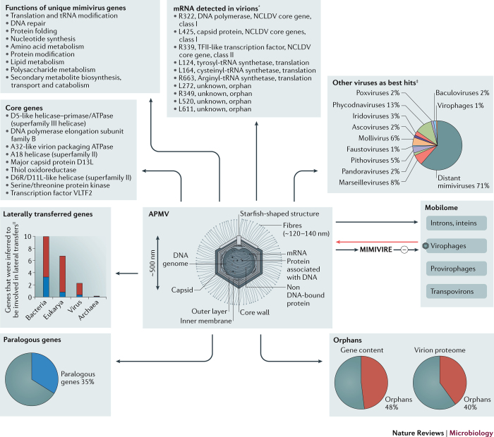
The accidental discovery of the giant virus of amoeba - Acanthamoeba polyphaga mimivirus (APMV; more commonly known as mimivirus) - in 2003 changed the field of virology. Viruses were previously defined by their submicroscopic size, which probably prevented the search for giant viruses, which are visible by light microscopy. Extended studies of giant viruses of amoebae revealed that they have genetic, proteomic and structural complexities that were not thought to exist among viruses and that are comparable to those of bacteria, archaea and small eukaryotes. The giant virus particles contain mRNA and more than 100 proteins, they have gene repertoires that are broader than those of other viruses and, notably, some encode translation components. The infection cycles of giant viruses of amoebae involve virus entry by amoebal phagocytosis and replication in viral factories. In addition, mimiviruses are infected by virophages, defend against them through the mimivirus virophage resistance element (MIMIVIRE) system and have a unique mobilome. Overall, giant viruses of amoebae, including mimiviruses, marseilleviruses, pandoraviruses, pithoviruses, faustoviruses and molliviruses, challenge the definition and classification of viruses, and have increasingly been detected in humans.


Conflict of interest statement

The authors declare no competing financial interests.

Figures

Figure 1
Figure 1. Particle and genome size of giant viruses of amoebae.
Families (namely, the Mimiviridae and Marseilleviridae; represented by dark blue circles) or putative families of giant viruses of amoebae (namely, those that include pandoraviruses, pithoviruses, faustoviruses and mollivirus; dark blue circles) are shown, along with other families in the proposed order Megavirales (namely, Poxviridae, Asfarviridae, Phycodnaviridae, Iridoviridae and Ascoviridae; light blue circles). Some families or genera of smaller viruses (grey circles) are shown in the inset, which magnifies a section of the larger graph and shows viruses that have a genome size ≤400 kb and a particle size ≤400 nm. Circle sizes are proportional to virus particle sizes. For each family or genus, the size of the largest member is shown. Viruses are referred to by their family or genus name unless there is no family or genus name. PowerPoint slide
Figure 2
Figure 2. Major genomic and structural features of APMV.
Major structural features of the Acanthamoeba polyphaga mimivirus (APMV) particle are shown in the middle of the figure. Black arrows link APMV to the major features of the gene repertoire and nucleic acid content of APMV, including its mobilome and mRNAs, which are shown in grey boxes. The red arrow indicates that virophages can infect mimivirus factories. The minus sign indicates that mimivirus virophage resistance element (MIMIVIRE) can protect against virophages. *Gene definitions and putative functions are given, as well as their relationship to other viral genes. Corresponds to mimivirus genes, the closest match to which in the NCBI GenBank protein sequence database is from a virus that does not belong in the family Mimiviridae (except distant mimiviruses). || Maximum and minimum proportions of genes that are inferred to be involved in lateral transfer are given, as assessed in three different studies,,. NCLDV, nucleocytoplasmic large DNA viruses; VLTF2, viral late transcription factor 2. PowerPoint slide
Figure 3
Figure 3. The mimivirus replication cycle and key replicative features of other giant viruses of amoebae.
a | Schematic of the replication cycle of Acanthamoeba polyphaga mimivirus (APMV). Virus particles can be seen at the surface of an Acanthamoeba sp. amoeba at the first stage of the cycle. Then, after virus entry through phagocytosis, the eclipse phase begins and the release of the APMV genome into the amoebal cytoplasm seeds the virus factory, which appears at a different location to the cell nucleus. Starting from ∼8 h post-infection, the assembly of virus particles can be seen at the periphery of the virus factory. Amoebal lysis occurs ∼12 h post-infection. b–e | Electron microscopy images of APMV particles and Acanthamoeba sp. cells that are infected with APMV. b | A mimivirus particle is shown. c | A mimivirus factory in the cytoplasm of an Acanthamoeba sp. amoeba 8 h post-infection. d | This image shows the edge and periphery of a virus factory, in which internal membrane biogenesis and assembly, capsid assembly and DNA packaging, and fibre acquisition of the APMV particles occur 8 h post-infection. e | This image shows an Acanthamoeba sp. amoeba ∼12 h post-infection with mimivirus, with the amoebal cytoplasm filled with mimivirus particles. f | The key replicative features of giant viruses of amoebae are shown in this table. Dashes indicate where there is no noticeable feature for a virus. A. castellanii, Acanthamoeba castellanii; V. vermiformis, Vermamoeba vermiformis. Microscopy images in parts b–e courtesy of I. Pagnier, Institut Hospitalo-Universitaire (IHU) Méditerranée Infection, Aix Marseille University, France. PowerPoint slide

References

    1. La Scola B, et al. A giant virus in amoebae. Science. 2003;299:2033. doi: 10.1126/science.1081867. - DOI - PubMed
    1. Raoult D, et al. The 1.2-megabase genome sequence of Mimivirus. Science. 2004;306:1344–1350. doi: 10.1126/science.1101485. - DOI - PubMed
    1. Raoult D. TRUC or the need for a new microbial classification. Intervirology. 2013;56:349–353. doi: 10.1159/000354269. - DOI - PubMed
    1. Scheid P, Balczun C, Schaub GA. Some secrets are revealed: parasitic keratitis amoebae as vectors of the scarcely described pandoraviruses to humans. Parasitol. Res. 2014;113:3759–3764. doi: 10.1007/s00436-014-4041-3. - DOI - PubMed
    1. La Scola B, et al. The virophage as a unique parasite of the giant mimivirus. Nature. 2008;455:100–104. doi: 10.1038/nature07218. - DOI - PubMed