Clinical Trial
. 2017 Feb 21:5:20.
doi: 10.1186/s40425-017-0220-y. eCollection 2017.

Analyses of the peripheral immunome following multiple administrations of avelumab, a human IgG1 anti-PD-L1 monoclonal antibody

Affiliations
Clinical Trial

Analyses of the peripheral immunome following multiple administrations of avelumab, a human IgG1 anti-PD-L1 monoclonal antibody

Renee N Donahue et al. J Immunother Cancer. .

Abstract

Background: Multiple anti-PD-L1/PD-1 checkpoint monoclonal antibodies (MAb) have shown clear evidence of clinical benefit. All except one have been designed or engineered to omit the possibility to mediate antibody-dependent cell-mediated cytotoxicity (ADCC) as a second potential mode of anti-tumor activity; the reason for this is the concern of lysis of PD-L1 positive immune cells. Avelumab is a fully human IgG1 MAb which has been shown in prior in vitro studies to mediate ADCC versus a range of human tumor cells, and clinical studies have demonstrated anti-tumor activity versus a range of human cancers. This study was designed to investigate the effect on immune cell subsets in the peripheral blood of cancer patients prior to and following multiple administrations of avelumab.

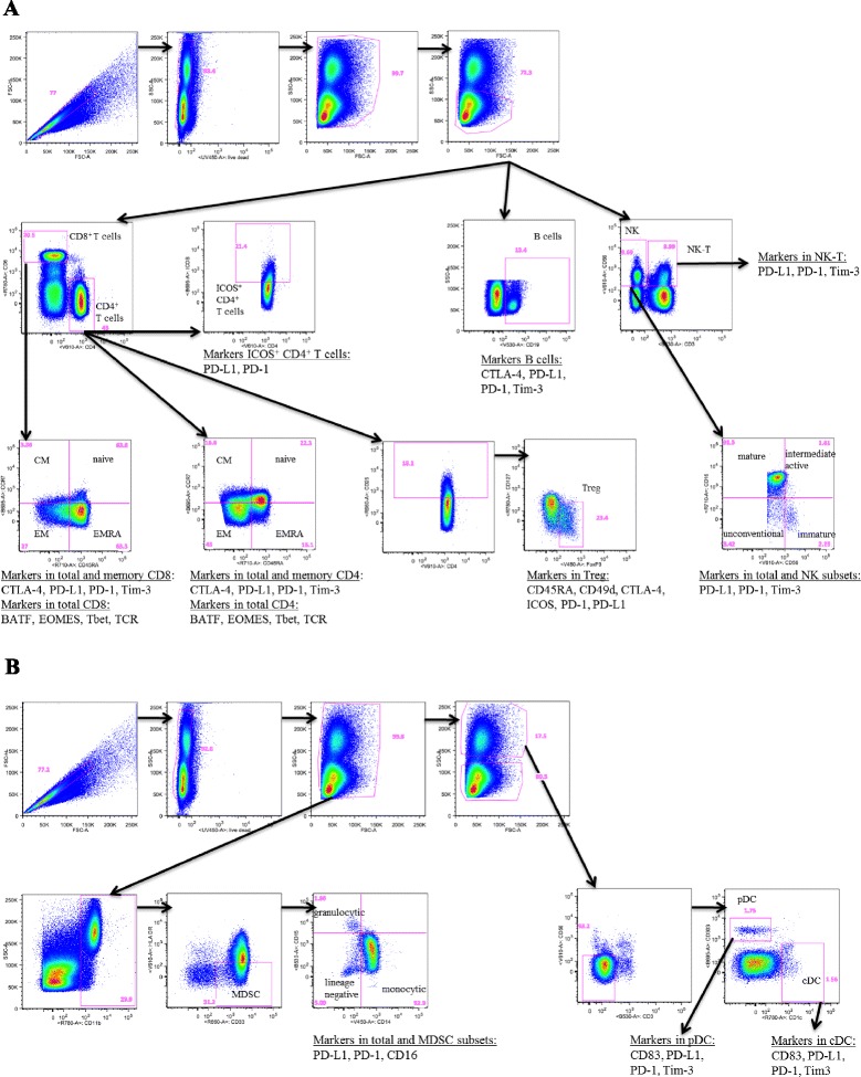
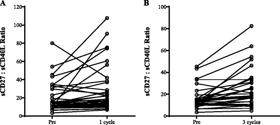
Methods: One hundred twenty-three distinct immune cell subsets in the peripheral blood of cancer patients (n = 28) in a phase I trial were analyzed by flow cytometry prior to and following one, three, and nine cycles of avelumab. Changes in soluble (s) CD27 and sCD40L in plasma were also evaluated. In vitro studies were also performed to determine if avelumab would mediate ADCC of PBMC.

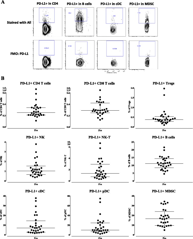
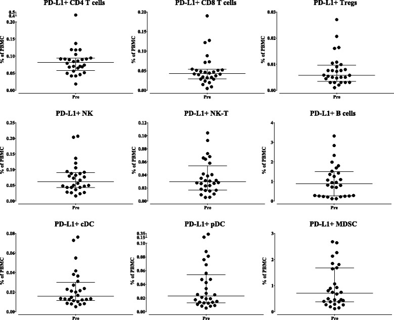
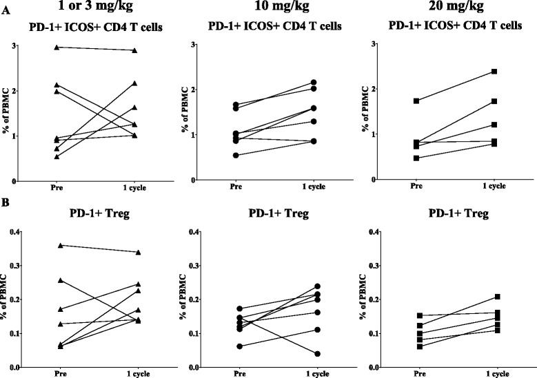
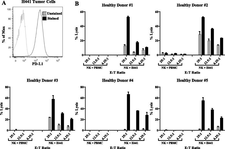
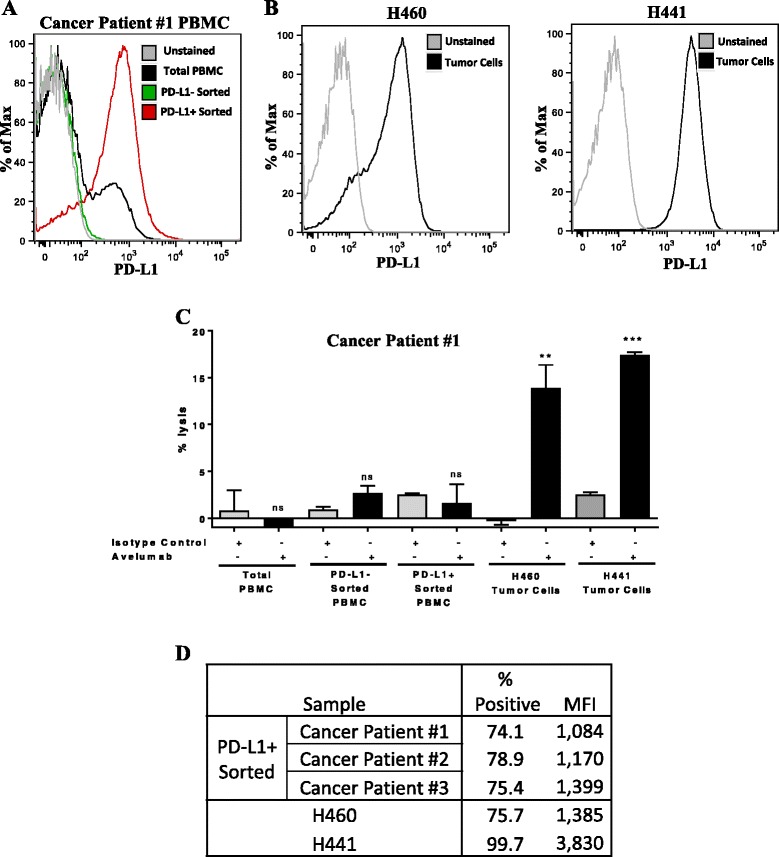
Results: No statistically significant changes in any of the 123 immune cell subsets analyzed were observed at any dose level, or number of doses, of avelumab. Increases in the ratio of sCD27:sCD40L were observed, suggesting potential immune activation. Controlled in vitro studies also showed lysis of tumor cells by avelumab versus no lysis of PBMC from five donors.

Conclusions: These studies demonstrate the lack of any significant effect on multiple immune cell subsets, even those expressing PD-L1, following multiple cycles of avelumab. These results complement prior studies showing anti-tumor effects of avelumab and comparable levels of adverse events with avelumab versus other anti-PD-1/PD-L1 MAbs. These studies provide the rationale to further exploit the potential ADCC mechanism of action of avelumab as well as other human IgG1 checkpoint inhibitors.

Trial registration: ClinicalTrials.gov identifier: NCT01772004 (first received: 1/14/13; start date: January 2013) and NCT00001846 (first received date: 11/3/99; start date: August 1999).

Keywords: ADCC; Anti-PD-L1; Antibody-dependent cell-mediated cytotoxicity; Avelumab; Checkpoint inhibitor; Immune subsets; Immunotherapy; Peripheral immunome.


Figures

Fig. 1
Fig. 1
Gating strategy to identity 123 peripheral immune cell subsets. Five immune flow cytometry panels using PBMC from a cancer patient following nine cycles of avelumab were used. Classic immune cell types included CD4+ T cells, CD8+ T cells, Tregs, B cells, natural killer (NK) and NK-T cells (panel a), and conventional dendritic cells (cDCs), plasmacytoid DCs (pDCs) and myeloid derived suppressor cells (MDSCs) (panel b)
Fig. 2
Fig. 2
Baseline (pre-treatment) expression of PD-L1 as a percentage of parental classic subset. a Representative flow cytometry plots of PD-L1 expression in CD4+ T cells, B cells, cDC, and MDSC. b In 28 patients prior to avelumab therapy, expression of PD-L1 was measured by flow cytometry for nine classic subsets as a percentage of total PBMC, with graphs displaying median and interquartile range
Fig. 3
Fig. 3
Baseline (pre-treatment) expression of PD-L1 as a percentage of total PBMC. In 28 patients prior to avelumab therapy, expression of PD-L1 was measured by flow cytometry for nine classic subsets as a percentage of total PBMC, with graphs displaying median and interquartile range
Fig. 4
Fig. 4
Immune cell subsets of a potentially biologic relevance following different doses of avelumab. Graphs display frequency as a percentage of PBMC for patients treated with 1 or 3 mg/kg (left panels, triangle), 10 mg/kg (middle panels, circle), and 20 mg/kg (right panels, square) of avelumab
Fig. 5
Fig. 5
ADCC assay using PBMC from healthy donors or H441 human lung tumor cells as targets. a PD-L1 expression in H441 cells. b NK cells were purified from PBMC from five healthy donors using negative magnetic selection. In vitro ADCC assays were performed at effector:target ratios of 25:1, 12.5:1, and 6.25:1, using an IgG1 isotype control antibody (gray bars, 1 ng/mL) or avelumab (black bars, 1 ng/mL). Results are displayed as mean + SEM of triplicate wells
Fig. 6
Fig. 6
ADCC assay using NK cells from a cancer patient with NSCLC against autologous PBMC sorted to enrich for PD-L1, or H460 or H441 human lung tumor cells. a PD-L1 expression in total PBMC, and PBMC magnetically sorted via negative and positive selection to enrich for PD-L1 negative and PD-L1 positive fractions, respectively. b PD-L1 expression in H460 and H441 cells. c NK cells were purified from PBMC from a cancer patient using negative magnetic selection. In vitro ADCC assays were performed at effector:target ratio of 25:1, using an IgG1 isotype control antibody (gray bars, 1 ng/mL) or avelumab (black bars, 1 ng/mL). Results are displayed as mean + SEM of triplicate wells. Data analyzed with unpaired T-test comparing avelumab vs isotype control; **p < 0.01, ***p < 0.001; ns: not significant. d Comparison of PD-L1 expression (% positivity and mean fluorescence intensity, MFI) in the PD-L1+ enriched PBMC fraction from three cancer patients compared to H460 and H441 cells
Fig. 7
Fig. 7
Effect of avelumab on soluble factors. Plasma levels of sCD27 and sCD40L were measured at baseline (pre-therapy) and following one cycle (a, n = 39) and three cycles (b, n = 33) of avelumab. Graphs display the ratio of sCD27 to sCD40L. Following one and three cycles of avelumab, there was a statistically significant increase in the ratio (p = 0.0087 and p = 0.0001, respectively, using the Wilcoxon matched-pairs signed rank test)

References

    1. Callahan MK, et al. Targeting T cell co-receptors for cancer therapy. Immunity. 2016;44:1069–1078. doi: 10.1016/j.immuni.2016.04.023. - DOI - PubMed
    1. Hoos A. Development of immuno-oncology drugs - from CTLA4 to PD1 to the next generations. Nat Rev Drug Discov. 2016;15:235–247. doi: 10.1038/nrd.2015.35. - DOI - PubMed
    1. Boyerinas B, et al. Antibody-dependent cellular cytotoxicity activity of a novel anti-PD-L1 antibody avelumab (MSB0010718C) on human tumor cells. Cancer Immunol Res. 2015;3:1148–1157. doi: 10.1158/2326-6066.CIR-15-0059. - DOI - PMC - PubMed
    1. Fujii R, et al. Enhanced killing of chordoma cells by antibody-dependent cell-mediated cytotoxicity employing the novel anti-PD-L1 antibody avelumab. Oncotarget. 2016;7:33498–5110. - PMC - PubMed
    1. Khanna S, et al. Malignant mesothelioma effusions are infiltrated by CD3+ T cells highly expressing PD-L1 and the PD-L1+ tumor cells within these effusions are susceptible to ADCC by the anti-PD-L1 antibody avelumab. J Thorac Oncol. 2016;11:1993–200. doi: 10.1016/j.jtho.2016.07.033. - DOI - PMC - PubMed

Publication types

MeSH terms

Associated data