. 2016 Dec;3(12):1496-1503.
doi: 10.1364/OPTICA.3.001496. Epub 2016 Dec 15.

Cubic meter volume optical coherence tomography

Affiliations

Cubic meter volume optical coherence tomography

Zhao Wang et al. Optica. 2016 Dec.

Abstract

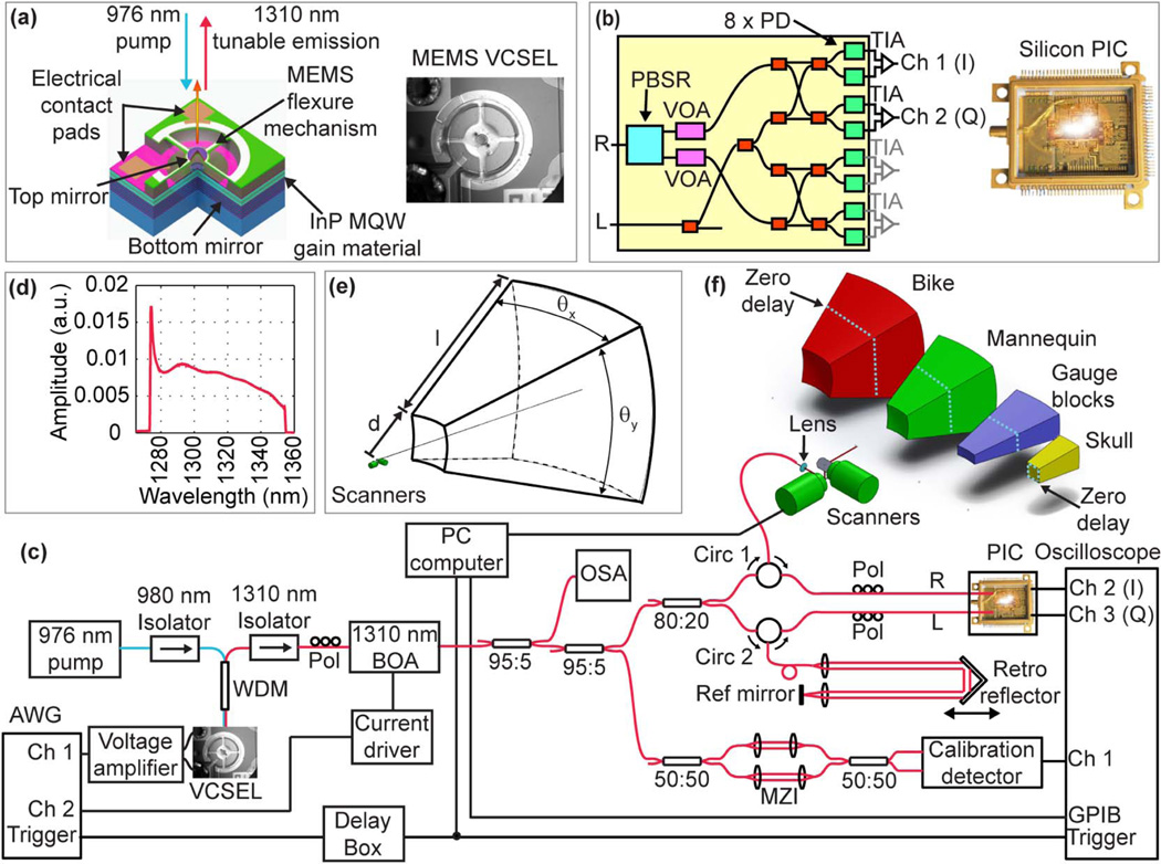
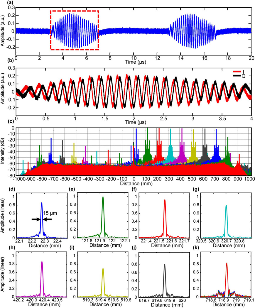
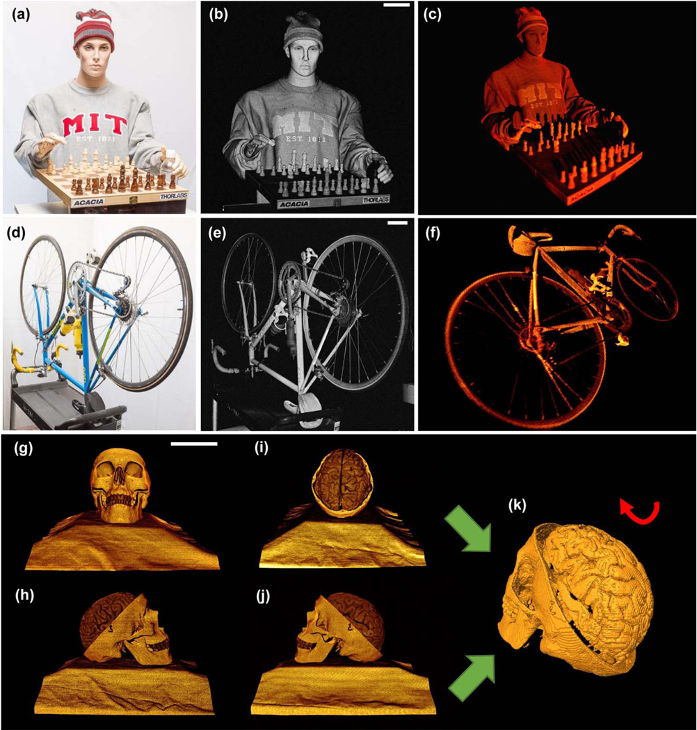
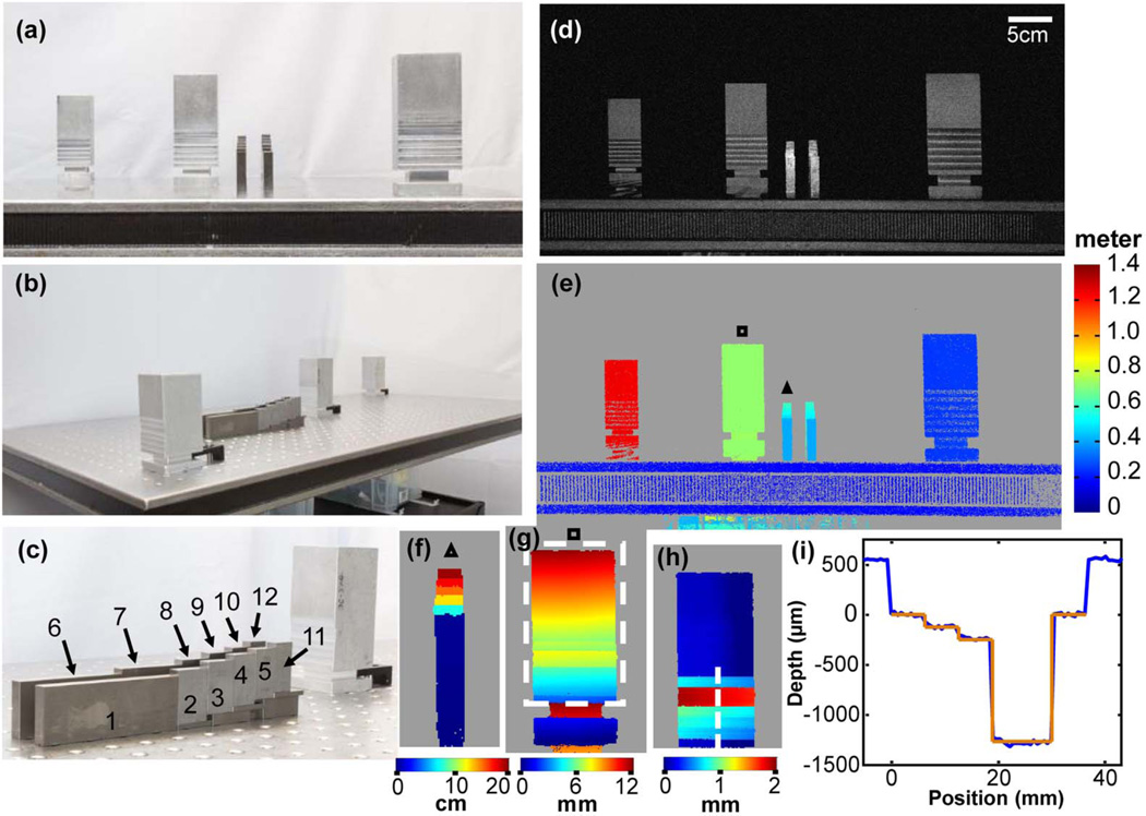
Optical coherence tomography (OCT) is a powerful three-dimensional (3D) imaging modality with micrometer-scale axial resolution and up to multi-GigaVoxel/s imaging speed. However, the imaging range of high-speed OCT has been limited. Here, we report 3D OCT over cubic meter volumes using a long coherence length, 1310 nm vertical-cavity surface-emitting laser and silicon photonic integrated circuit dual-quadrature receiver technology combined with enhanced signal processing. We achieved 15 µm depth resolution for tomographic imaging at a 100 kHz axial scan rate over a 1.5 m range. We show 3D macroscopic imaging examples of a human mannequin, bicycle, machine shop gauge blocks, and a human skull/brain model. High-bandwidth, meter-range OCT demonstrates new capabilities that promise to enable a wide range of biomedical, scientific, industrial, and research applications.


Figures

Fig. 1
Fig. 1
Details of the imaging system. (a) Schematic and photograph of the MEMS-tunable VCSEL swept laser source. (b) Schematic and photograph of the silicon photonic integrated circuit (PIC) IQ receiver. R, signal input; L, reference input; PBSR, polarization beam splitter; VOA, variable optical attenuators; TIA, trans-impedance amplifier. (c) Imaging system layout. AWG, arbitrary waveform generator; WDM, wavelength division multiplexer; Pol, polarization controller; BOA, booster optical amplifier; OSA, optical spectrum analyzer; Circ, circulator; MZI, Mach–Zehnder interferometer. (d) Spectrum of the BOA-amplified VCSEL emission recorded with the OSA. (e) Definition of scanning volume. (f) Representations of scanned volumes for the bicycle, mannequin, gauge blocks, and skull/brain that are proportionally accurate and show the position of the OCT zero delay.
Fig. 2
Fig. 2
System characterization of meter-range OCT. (a) Representative interferograms from a mirror showing two consecutive laser sweeps (I channel only). (b) I and Q channel signals from one sweep showing ~90-deg phase relationship between the two channels. (c) Signal roll-off measurement on a logarithmic scale with IQ processing. Negative axis shows suppressed complex conjugates. (d)–(k) Plots of PSFs on a linear scale at different depths. (k) 10 of 100 repeated PSFs at a depth of ~718.9 mm.
Fig. 3
Fig. 3
(a) Photograph, (b) maximum intensity projection, and (c) 3D OCT visualization of a life-size mannequin with a chess set consisting of 1000 × 1000 A-scans before scan correction. The volume size is 0.98 m3 (d = 89 cm, l = 150 cm, θx = 27.5 deg, and θy = 27.5 deg. For display, an intensity threshold of ~10 dB above the mean noise floor was applied. (d) Photograph, (e) maximum intensity projection, and (f) 3D OCT visualization of an adult bicycle more than 1.5 m in length consisting of 1000 × 1000 A-scans before scan correction. The volume size is 1.8 m3 (d = 97 cm, l = 150 cm, θx = 35.7 deg, and θy = 35.7 deg). (g)–(j) Human skull model imaged at 0 deg (g), 90 deg (h), 180 deg (i), and 270 deg (j). Each volume has 500 × 500 A-scans and a volume of 8000 cm3 (d = 97 cm, l = 75 cm, θx = 10.8 deg, and θy = 10.8 deg). (k) 3D skull surface reconstructed by segmenting and merging the individual object surface of (g)–(j) after scan correction. Scale bars are 10 cm. 3D visualization of the objects after scan correction can be found in Visualization 1, Visualization 2, and Visualization 3.
Fig. 4
Fig. 4
(a) and (b) Photographs of the aluminum posts and steel gauge blocks on an optics table from two perspectives. (c) Photograph of the gauge blocks. (d) OCT maximum intensity projection. The OCT volume has 500 × 1000 A-scans. The volume size is 0.288 m3 (d = 98 cm, l = 150 cm, θx = 20.4 deg, and θy = 9.7 deg). (e) Distance mapping of the objects in meter scale. (f) Distance mapping of the gauge blocks on a centimeter scale. (g) Visualization of the tilt of an aluminum post with respect to the incident OCT beam on a millimeter scale. (h) Topological mapping of the aluminum surface from the box in (g) after correcting for sample tilt. (i) Relative depths of milled steps in the aluminum post surface from dotted line in (h) on a micrometer scale showing good agreement between the OCT measured surface profile (blue) and nominal depths from the milling machine digital readout (orange). Depth scale was calibrated by measurement of gauge block 1, and the transverse scale was calibrated by the nominal milled widths.

References

    1. Huang D, Swanson EA, Lin CP, Schuman JS, Stinson WG, Chang W, Hee MR, Flotte T, Gregory T, Puliafito CA, Fujimoto JG. Optical coherence tomography. Science. 1991;254:1178–1181. - PMC - PubMed
    1. Wieser W, Draxinger W, Klein T, Karpf S, Pfeiffer T, Huber R. High definition live 3D-OCT in vivo: design and evaluation of a 4D OCT engine with 1 GVoxel/s. Biomed. Opt. Express. 2014;5:2963–2977. - PMC - PubMed
    1. Liang K, Traverso G, Lee H, Ahsen OO, Wang Z, Potsaid B, Giacomelli M, Jayaraman V, Barman R, Cable A, Mashimo H, Langer R, Fujimoto JG. Ultrahigh speed en face OCT capsule for endoscopic imaging. Biomed. Opt. Express. 2015;6:1146–1163. - PMC - PubMed
    1. Drexler W, Fujimoto JG. Optical coherence tomography: technology and applications. Springer Science & Business Media (Springer, 2015) Chap. 5:169.
    1. Leitgeb R, Hitzenberger C, Fercher AF. Performance of Fourier domain vs. time domain optical coherence tomography. Opt. Express. 2003;11:889–894. - PubMed