. 2017 Feb 23;2(4):e85687.
doi: 10.1172/jci.insight.85687.

HLA-DQ β 1 alleles associated with Epstein-Barr virus (EBV) infectivity and EBV gp42 binding to cells

Affiliations

HLA-DQ β 1 alleles associated with Epstein-Barr virus (EBV) infectivity and EBV gp42 binding to cells

Qingxue Li et al. JCI Insight. .

Abstract

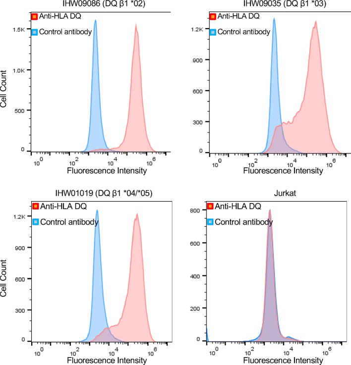
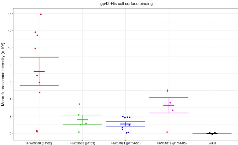
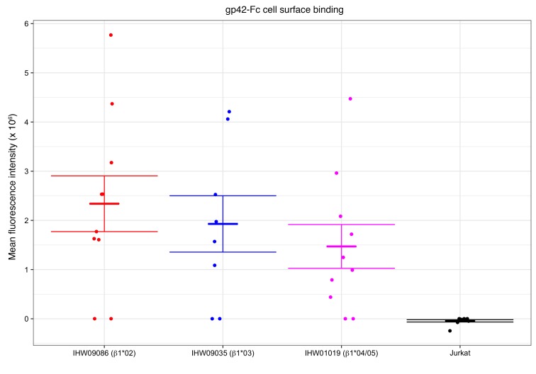
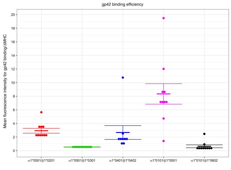
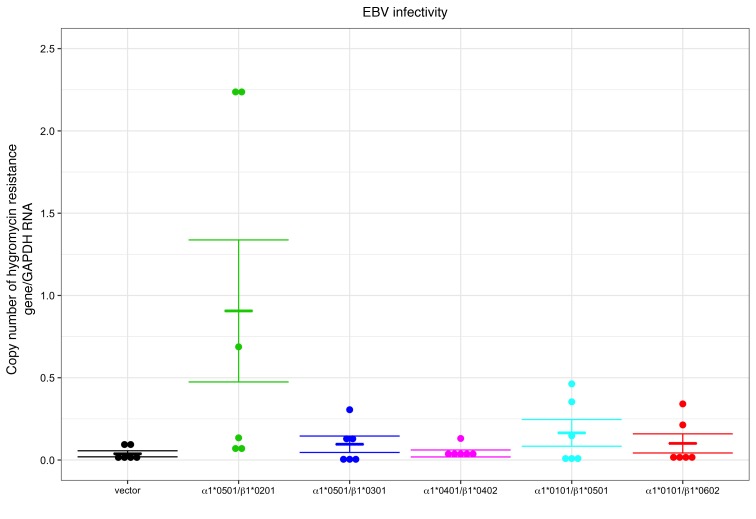
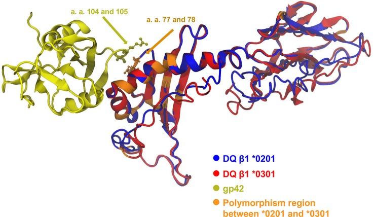
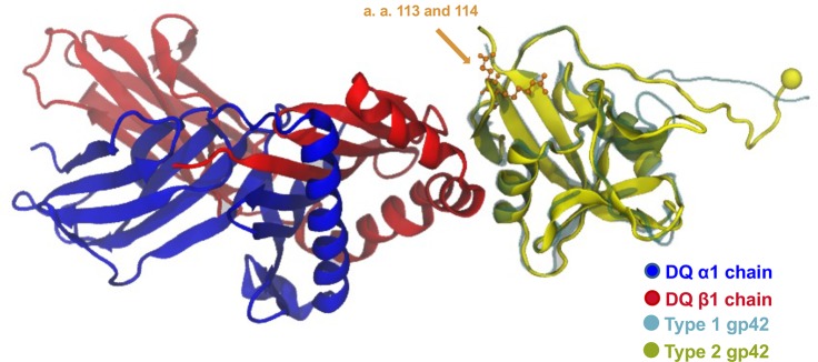
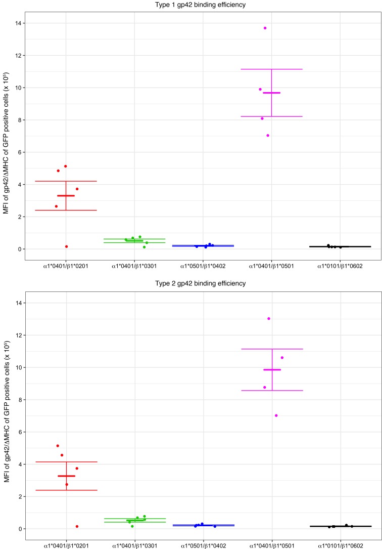
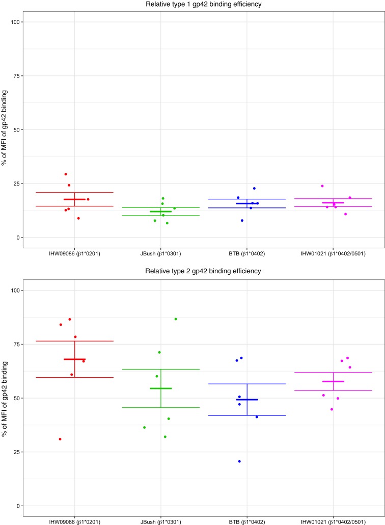
Epstein-Barr virus (EBV) infects B cells and ~95% of adults are infected. EBV glycoprotein gp42 is essential for entry of virus into B cells. EBV gp42 binds to the β1 chain of HLA-DQ, -DR, and -DP on B cells, and uses these molecules for infection. To investigate if certain HLA-DQ alleles are associated with EBV seronegativity, we recruited ~3,300 healthy adult blood donors, identified 106 EBV-seronegative individuals, and randomly selected a control group of EBV-seropositive donors from the donor pool. A larger than expected proportion of EBV-seronegative subjects were HLA-DQ β1 *04/*05 and *06/*06, and to a lesser extent, *02/*03, compared with the control group, while a larger than expected portion of EBV-seropositive persons were HLA-DQ β1 *02/*02. We examined the ability of EBV gp42 to bind to different HLA-DQ molecules using human and mouse cells stably expressing these alleles. EBV gp42 bound less effectively to cells expressing HLA-DQ β1 *04/*05, *06/*06, or *03/*03 than to cells expressing HLA-DQ β1 *02/*02. These data are consistent with our observations of increased EBV seronegativity with DQ β1 *04/*05 or *06/*06 alleles. These findings emphasize the importance of a single genetic locus (HLA-DQ β1) to influence infectivity with EBV.


Conflict of interest statement

Conflict of interest: The authors have declared that no conflict of interest exists.

Figures

Figure 1
Figure 1. Lymphoblastoid cell lines (LCLs) used in gp42 binding assays express similar levels of HLA-DQ on their cell surface.
LCLs or Jurkat T cells were incubated with mouse anti–HLA-DQ antibody Ia3 on ice followed by an anti-mouse secondary antibody conjugated with Alexa488. After fixation, fluorescence intensity was measured by flow cytometry.
Figure 2
Figure 2. Lymphoblastoid cell lines expressing HLA-DQ β1 *02/*02 bind more gp42-His than cell lines expressing DQ β1 *03 /*03 or *04/*05.
Cells were incubated with gp42-His on ice for 60 minutes and after washing with PBS, mouse anti-His antibody was added on ice for 60 minutes followed by Alexa488 fluorophore–conjugated anti-mouse antibody. After fixation of the cells with 2% paraformaldehyde, FACS was performed and the mean fluorescence intensity was measured. The data were obtained from 9 independent experiments. Means (short horizontal lines) ± SEM (long horizontal lines) are shown.
Figure 3
Figure 3. Lymphoblastoid cell lines expressing DQ β1 *02/*02 bind more dimerized gp42 than cells expressing DQ β1 *03/*03 or *04/*05 alleles.
Cells were incubated with purified gp42-Fc as described in the legend to Figure 2 for gp42-His protein. Human TruStain FcX Fc receptor blocking solution (Biolegend) was then added to the cells for 5 minutes before adding anti–human IgG Alexa488 antibody, and gp42 binding was quantified by FACS. The cells were also incubated with IgG-Fc and binding of Ig-Fc was measured. The net mean fluorescence intensity of gp42-Fc binding was calculated by subtracting the background binding from IgG-Fc. The data were derived from 10 independent experiments. Means (short horizontal lines) ± SEM (long horizontal lines) are shown.
Figure 4
Figure 4. Cells expressing HLA-DQ β1 *02/*02 bind more gp42 than cells expressing DQ*03/*03 or *06/*06 when expressed independently of other HLA molecules.
Mouse 3T3 cells expressing HLA-DQ αβ heterodimers were incubated with purified gp42-His followed by anti-His antibody as described in the legend to Figure 2. The amount of gp42 bound was quantified by FACS. Cell surface HLA-DQ β1 levels were also quantified in the same experiment by staining with Ia3 antibody followed by isotype-specific secondary antibody (Alexa647). The ratio of the mean fluorescence intensity (MFI) for gp42 binding (FL-4) divided by ΔMHC, which is the ratio of cell surface DQ expression to internal control GFP (24), is shown. The results were the same when we defined gp42 binding efficiency as the ratio of the MFI of gp42 binding divided by the MFI of HLA-DQ. The data were obtained from 10 independent experiments. Means (short horizontal lines) ± SEM (long horizontal lines) are shown.
Figure 5
Figure 5. HLA-DQ β1*02/*02 alleles facilitate EBV infectivity more efficiently than other alleles.
Human 721.174 cells were nucleofected with plasmids encoding DQ α1 and β1 alleles and infected with 293-EBV 48 hours later. RNA extraction was performed 3 days after infection, and copy numbers for the products of the hygromycin-resistance gene and GAPDH (internal control) were determined by real-time RT-qPCR in duplicate. The data were derived from 6 independent experiments. Means (short horizontal lines) ± SEM (long horizontal lines) are shown.
Figure 6
Figure 6. Predicted structures of DQ β1 *0201 (blue) and DQ β1 *0301 (red).
The polymorphic regions between the 2 DQ β1 alleles are shown in orange. The interface between DQ β1 *0201/*0301 and gp42 (yellow) was modeled by aligning the predicted structures with the β chain of the crystal structure of gp42 bound to HLA-DR1 (13). Amino acids 77 and 78 in DQ β1 are Gly and Glu in β1 *0201, respectively, and are Glu and Val in *0301, respectively; these 2 amino acids interact with amino acids 104 (Thr) and 105 (Arg) of EBV gp42.
Figure 7
Figure 7. Predicted structures of EBV type 2 gp42 (yellow) superimposed on the crystal structure of type 1 gp42 (gray) bound to DQ β1 (red) and DQ α1 (blue) (13).
Amino acids 113 and 114 near the DQ β1–binding interface in type 1 gp42 are Gly and Glu, respectively, and are Glu and Arg in type 2 gp42, respectively.
Figure 8
Figure 8. Type 1 and type 2 EBV gp42 bind to HLA-DQ with similar efficiency.
Mouse 3T3 cells expressing HLA-DQ αβ heterodimers were incubated with the same amount of purified type 1 (top) or type 2 (bottom) gp42-His protein followed by anti-His antibody staining and detection of gp42 by FACS as described in the legend to Figure 4. MFI, mean fluorescence intensity; ΔMHC, the ratio of cell surface DQ expression to internal control GFP (24). The data were obtained from 5 independent experiments. Means (short horizontal lines) ± SEM (long horizontal lines) are shown.
Figure 9
Figure 9. A neutralizing monoclonal antibody against gp42 blocks type 1 gp42 binding more efficiently than type 2 gp42 binding to human cells expressing different HLA class II alleles.
Type 1 (top) or type 2 (bottom) gp42 proteins were preincubated with monoclonal anti-gp42 Ab, F-2-1, or a control antibody at room temperature for 30 minutes, and added to human lymphoblastoid cell lines and binding was assayed as described in the legend to Figure 2 except that detection of His-tagged gp42 was performed using rabbit anti-His antibody (Cell Signaling Technology) followed by Alexa488-conjugated anti-rabbit secondary antibody. The relative percentage of gp42 binding to cells in the presence of F-2-1 antibody was calculated against that of the control antibody, which was set at 100%. The relative binding of gp42 in the presence of F-2-1 antibody is shown. MFI, mean fluorescence intensity. The data were derived from 6 independent experiments. Means (short horizontal lines) ± SEM (long horizontal lines) are shown.

Similar articles

Cited by

References

    1. Balfour HH, et al. Behavioral, virologic, and immunologic factors associated with acquisition and severity of primary Epstein-Barr virus infection in university students. J Infect Dis. 2013;207(1):80–88. doi: 10.1093/infdis/jis646. - DOI - PMC - PubMed
    1. Faulkner GC, Burrows SR, Khanna R, Moss DJ, Bird AG, Crawford DH. X-Linked agammaglobulinemia patients are not infected with Epstein-Barr virus: implications for the biology of the virus. J Virol. 1999;73(2):1555–1564. - PMC - PubMed
    1. Longnecker R, Kieff E, Cohen JI. Epstein-Barr Virus. In: Knipe DM, et al., eds. Fields Virology. Philadelphia, PA: Lippincott Williams and Wilkins; 2013:1898–1959.
    1. Nemerow GR, Mold C, Schwend VK, Tollefson V, Cooper NR. Identification of gp350 as the viral glycoprotein mediating attachment of Epstein-Barr virus (EBV) to the EBV/C3d receptor of B cells: sequence homology of gp350 and C3 complement fragment C3d. J Virol. 1987;61(5):1416–1420. - PMC - PubMed
    1. Ogembo JG, et al. Human complement receptor type 1/CD35 is an Epstein-Barr Virus receptor. Cell Rep. 2013;3(2):371–385. doi: 10.1016/j.celrep.2013.01.023. - DOI - PMC - PubMed

Publication types