. 2017 Feb 23;2(4):e91214.
doi: 10.1172/jci.insight.91214.

Tissue-resident macrophages can contain replication-competent virus in antiretroviral-naive, SIV-infected Asian macaques

Affiliations

Tissue-resident macrophages can contain replication-competent virus in antiretroviral-naive, SIV-infected Asian macaques

Sarah R DiNapoli et al. JCI Insight. .

Abstract

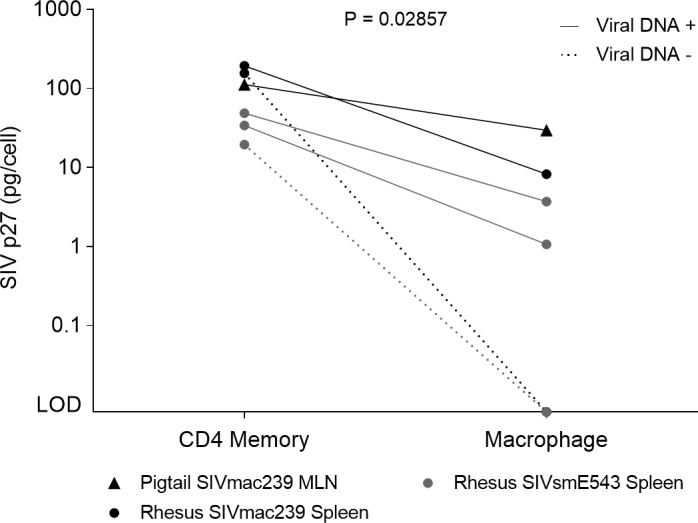
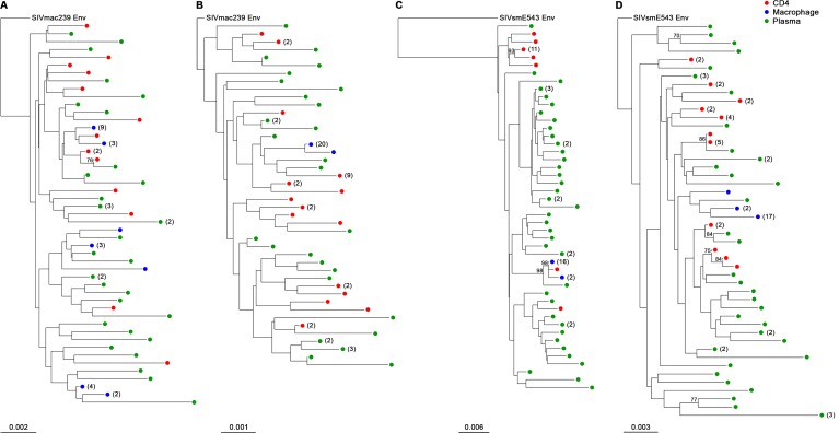
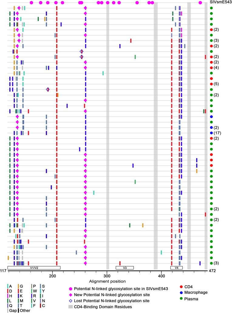
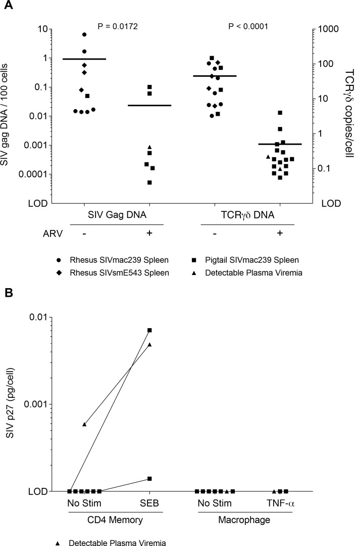
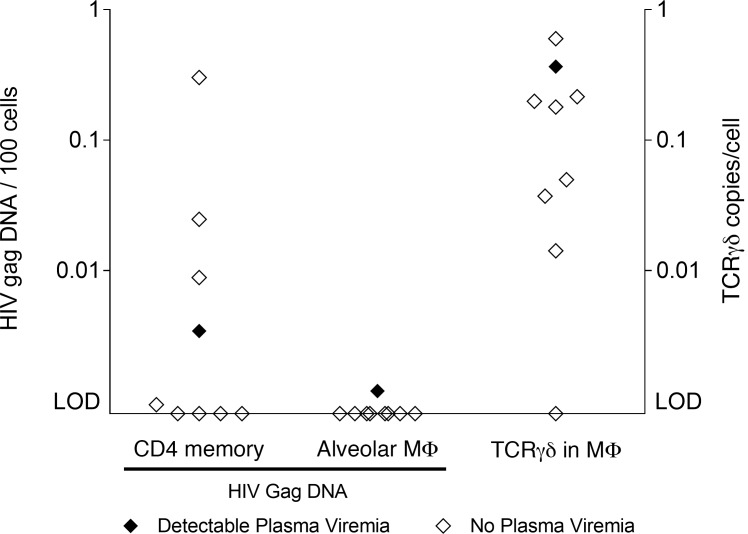
SIV DNA can be detected in lymphoid tissue-resident macrophages of chronically SIV-infected Asian macaques. These macrophages also contain evidence of recently phagocytosed SIV-infected CD4+ T cells. Here, we examine whether these macrophages contain replication-competent virus, whether viral DNA can be detected in tissue-resident macrophages from antiretroviral (ARV) therapy-treated animals and humans, and how the viral sequences amplified from macrophages and contemporaneous CD4+ T cells compare. In ARV-naive animals, we find that lymphoid tissue-resident macrophages contain replication-competent virus if they also contain viral DNA in ARV-naive Asian macaques. The genetic sequence of the virus within these macrophages is similar to those within CD4+ T cells from the same anatomic sites. In ARV-treated animals, we find that viral DNA can be amplified from lymphoid tissue-resident macrophages of SIV-infected Asian macaques that were treated with ARVs for at least 5 months, but we could not detect replication-competent virus from macrophages of animals treated with ARVs. Finally, we could not detect viral DNA in alveolar macrophages from HIV-infected individuals who received ARVs for 3 years and had undetectable viral loads. These data demonstrate that macrophages can contain replication-competent virus, but may not represent a significant reservoir for HIV in vivo.


Conflict of interest statement

Conflict of interest: The authors have declared that no conflict of interest exists.

Figures

Figure 1
Figure 1. Replication-competent virus in ARV-naive lymphoid tissues.
SIV p27 levels in cell culture media from cocultures of memory CD4+ T cells (left) or myeloid cells (right) with CEMx174 cells after 1 week. Cells were sorted from the spleen or mesenteric lymph node (MLN). SIV p27 levels in memory CD4+ T cell cocultures and macrophage cocultures (excluding viral DNA animals) were compared via Mann-Whitney test (P = 0.02857). Reported values are pg SIV p27 per primary cell added to the coculture (8 × 10³ sorted cells and 5 × 104 CEMx174 cells per well). LOD, limit of detection.
Figure 2
Figure 2. Phylogenetic distribution of Env sequences from co-culture and contemporaneous plasma.
Viral RNA from cell culture media and necropsy plasma was isolated and sequenced for the variable regions 1–4 (V1–V4) of Env. Phylogenies constructed using the neighbor-joining method and Kimura 2-parameter model illustrate the genetic distribution of viruses from cocultures and plasma for each animal: (A) PT99P052, (B) RhDB92, (C) Rh848, and (D) Rh591. Identical sequences are grouped with the number of sequences indicated in parentheses. Sequence source is indicated by color: macrophage coculture (blue), CD4+ T cell coculture (red), and contemporaneous plasma (green). Branch length is indicated per animal. Root SIV Env sequences were obtained from the NCBI Nucleotide database.
Figure 3
Figure 3. Alignment of translated Env sequences from Rh591.
Potential N-linked glycosylation sites and variation in amino acid residues were identified by Highlighter analysis. CD4 binding domain residues are indicated with shaded boxes. Identical sequences were grouped with the number of sequences given in parentheses. Amino acid changes and potential glycosylation sites depicted as indicated in the legend. Sequence source is indicated by color: macrophage (blue), CD4+ T cell (red), and plasma (green). The SIVsmE543 master sequence was obtained from the NCBI Nucleotide database.
Figure 4
Figure 4. Viral DNA, rearranged TCR DNA, and replication-competent SIV in ARV-treated macaques.
(A) Viral DNA (left) and rearranged TCR-γδ DNA (right) levels in splenic myeloid cells of antiretroviral-treated (ARV-treated) animals and ARV-naive animals via Mann-Whitney tests (ARV-treated n = 17, ARV-naive n = 28, qPCR in duplicate). (B) Memory CD4+ T cells (left) or myeloid cells (right) from ARV-treated animals were cocultured with CEMx174 cells and analyzed for SIV p27 with ELISA after 3 weeks. Reported values are pg SIV p27 per primary cell in coculture (7.5 × 10⁵ sorted cells per well).
Figure 5
Figure 5. Viral DNA and TCR DNA in BAL of HIV+ individuals on ARVs.
HIV Gag DNA levels in memory CD4+ T cells (left) and alveolar macrophages (MΦ, middle) sorted from bronchoalveolar lavage (BAL) of HIV+ individuals on antiretrovirals (ARVs) for 3 years. Alveolar macrophages were also analyzed for rearranged TCR DNA content via qPCR (right).

Similar articles

Cited by

References

    1. Murray AJ, Kwon KJ, Farber DL, Siliciano RF. The latent reservoir for HIV-1: how immunologic memory and clonal expansion contribute to HIV-1 persistence. J Immunol. 2016;197(2):407–417. doi: 10.4049/jimmunol.1600343. - DOI - PMC - PubMed
    1. Davey RT, et al. HIV-1 and T cell dynamics after interruption of highly active antiretroviral therapy (HAART) in patients with a history of sustained viral suppression. Proc Natl Acad Sci USA. 1999;96(26):15109–15114. doi: 10.1073/pnas.96.26.15109. - DOI - PMC - PubMed
    1. Chun TW, et al. Rebound of plasma viremia following cessation of antiretroviral therapy despite profoundly low levels of HIV reservoir: implications for eradication. AIDS. 2010;24(18):2803–2808. doi: 10.1097/QAD.0b013e328340a239. - DOI - PMC - PubMed
    1. Rothenberger MK, et al. Large number of rebounding/founder HIV variants emerge from multifocal infection in lymphatic tissues after treatment interruption. Proc Natl Acad Sci USA. 2015;112(10):E1126–E1134. doi: 10.1073/pnas.1414926112. - DOI - PMC - PubMed
    1. Jenkins SJ, et al. Local macrophage proliferation, rather than recruitment from the blood, is a signature of TH2 inflammation. Science. 2011;332(6035):1284–1288. doi: 10.1126/science.1204351. - DOI - PMC - PubMed

Publication types

MeSH terms