. 2017 Feb 23;2(4):e91456.
doi: 10.1172/jci.insight.91456.

α2-Adrenergic blockade rescues hypoglossal motor defense against obstructive sleep apnea

α2-Adrenergic blockade rescues hypoglossal motor defense against obstructive sleep apnea

Gang Song et al. JCI Insight. .

Abstract

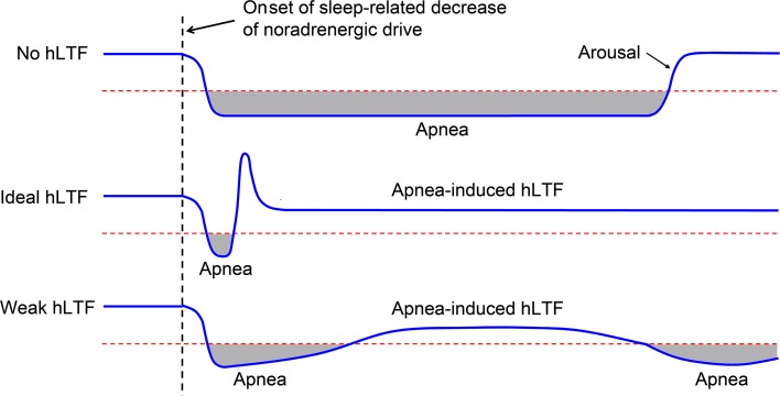
Decreased noradrenergic excitation of hypoglossal motoneurons during sleep causing hypotonia of pharyngeal dilator muscles is a major contributor to the pathogenesis of obstructive sleep apnea (OSA), a widespread disease for which treatment options are limited. Previous OSA drug candidates targeting various excitatory/inhibitory receptors on hypoglossal motoneurons have proved unviable in reactivating these neurons, particularly during rapid-eye-movement (REM) sleep. To identify a viable drug target, we show that the repurposed α2-adrenergic antagonist yohimbine potently reversed the depressant effect of REM sleep on baseline hypoglossal motoneuron activity (a first-line motor defense against OSA) in rats. Remarkably, yohimbine also restored the obstructive apnea-induced long-term facilitation of hypoglossal motoneuron activity (hLTF), a much-neglected form of noradrenergic-dependent neuroplasticity that could provide a second-line motor defense against OSA but was also depressed during REM sleep. Corroborating immunohistologic, optogenetic, and pharmacologic evidence confirmed that yohimbine's beneficial effects on baseline hypoglossal motoneuron activity and hLTF were mediated mainly through activation of pontine A7 and A5 noradrenergic neurons. Our results suggest a 2-tier (impaired first- and second-line motor defense) mechanism of noradrenergic-dependent pathogenesis of OSA and a promising pharmacotherapy for rescuing both these intrinsic defenses against OSA through disinhibition of A7 and A5 neurons by α2-adrenergic blockade.


Conflict of interest statement

Conflict of interest: The authors have declared that no conflict of interest exists.

Figures

Figure 1
Figure 1. Stylized illustration of the roles of baseline HM activity and hLTF as first- and second-line motor defenses against OSA.
Top: In the worst-case scenario where hLTF (hypoglossal long-term facilitation) is lacking, sustained decline of baseline HM activity (hypoglossal nerve activity, tracing in blue) secondary to decreased noradrenergic drive during sleep would result in protracted unremitting obstructive apnea (interval shaded in gray) until arousal. Horizontal dotted line (in red) denotes the hypothetical threshold of HM activity, below which obstructive apnea occurs. Middle: In the best-case scenario where hLTF is in full force, any lapse of baseline HM activity during sleep would be promptly reversed (<10 sec) by reflexive facilitation of HM activity and further apnea prevented by the ensuing hLTF memory, which (ideally) may be long-lasting after even a brief airway obstruction episode. Lower: In a real-life scenario where hLTF is in effect but significantly depressed secondary to decreased noradrenergic drive during sleep, a corresponding decline of baseline HM activity would be reversed much more slowly (>10 sec) due to weakened and delayed reflexive facilitation of HM activity in response to obstructive apnea. Additionally, the reversal (if at all prior to arousal) would be relatively short-lived (<12 min) due to a much shortened hLTF memory following each apnea episode, hence allowing full-blown obstructive apnea to occur and recur. In patients with severe OSA, the weakened hLTF in sleep is manifested as a short-term potentiation of genioglossus activity during an obstructive apnea episode with an “afterdischarge” afterward (25).
Figure 2
Figure 2. Obstructive apnea–induced hLTF was depressed during REM sleep.
(A) Obstructive apnea (lasting 10–15 seconds) in a urethane-anesthetized, paralyzed, and mechanically ventilated rat elicited time-dependent (reflexive) facilitation of the amplitude of integrated hypoglossal nerve activity (∫Hypoglossal, which comprised predominantly the inspiratory-phasic component) during each apnea episode (denoted by dots above the ∫Hypoglossal recording). Such obstructive apnea when applied repeatedly for 10–12 episodes induced sustained facilitation of ∫Hypoglossal amplitude with a long-term memory afterward, evidencing hypoglossal long-term facilitation (hLTF). (B) After microinjection of the cholinergic agonist carbachol at dorsomedial pons to induce a REM-like sleep state (indicated by increased hippocampal activity and decreased baseline ∫Hypoglossal amplitude) in one rat, baseline ∫Hypoglossal amplitude was markedly depressed, and obstructive apnea elicited relatively weak time-dependent facilitation of ∫Hypoglossal amplitude compared with the control state, seen in A. Also, episodic obstructive apnea no longer induced sustained hLTF. (C) Similar effects were seen during spontaneously occurring REM sleep (under urethane anesthesia, ref. 29) in another rat. (D) Bar graphs (overlaid with individual data points) showing the depressant effects (P < 0.05, 2-way ANOVA with repeated measures) of cholinergic-induced REM-like sleep (n = 6) and spontaneous REM sleep (n = 5) vs. no REM sleep (n = 12) on baseline ∫Hypoglossal amplitude before episodic obstructive apnea (left panel), as well as the facilitation of ∫Hypoglossal amplitude during the first and last apnea episodes (middle panel) and at 5 and 20 minutes after the last apnea episode (right panel). Values are normalized to control baseline at 100% (dashed line) and shown as means ± SEM. *P < 0.05 between values as indicated, Tukey post-hoc test.
Figure 3
Figure 3. Systemic yohimbine reversed the depressions of baseline hypoglossal activity and obstructive apnea–induced hLTF during REM sleep.
(A) Depression of baseline ∫Hypoglossal amplitude (amplitude of integrated hypoglossal nerve activity) during cholinergic-induced REM-like sleep was promptly reversed by systemic administration of yohimbine (0.75 mg/kg i.v.) in one rat. Systemic yohimbine also restored the facilitation of ∫Hypoglossal amplitude during and after episodic obstructive apnea (compare Figure 2, A and B). (B) Bar graphs (overlaid with individual data points) showing the efficacy (P < 0.05, 2-way ANOVA with repeated measures) of systemic yohimbine (0.75–1 mg/kg i.v., n = 7) in reversing the depressant effects of cholinergic-induced REM-like sleep (vs. cholinergic REM only, n = 6) on baseline ∫Hypoglossal amplitude before episodic obstructive apnea (left panel), as well as the facilitation of ∫Hypoglossal amplitude during the first and last apnea episodes (middle panel) and at 5 and 20 minutes after the last apnea episode (right panel). *P < 0.05 between values as indicated, Tukey post-hoc test. (C and D) Similar to A and B showing efficacy of system yohimbine (P < 0.05, 2-way ANOVA with repeated measures) but with a lower dose of 0.5 mg/kg i.v. (n = 6) under spontaneous REM sleep (vs. spontaneous REM only, n = 5). *P < 0.05 between values as indicated, Tukey post-hoc test.
Figure 4
Figure 4. Pontine A7 and A5 noradrenergic neuronal groups were activated by episodic obstructive apnea.
(A) Photomicrographs showing A7 and A5 noradrenergic neurons identified by dopamine β-hydroxylase (DBH) immunofluorescent labeling that were immunopositive to c-Fos in a rat exposed to episodic obstructive apnea, compared with one that was not exposed (control). “×” indicates artifacts; arrows indicate doubly labeled neurons. (B) Percentage of c-Fos immunopositive neurons among A7, A5, or A6 noradrenergic neurons in the experimental group (n = 6) and control group (n = 3). LC, locus coeruleus; SubC, subcoeruleus. *P < 0.05 between values as indicated, 2-tailed Student’s t test.
Figure 5
Figure 5. Episodic optogenetic stimulation at the A7 or A5 regions induced hLTF after stimulation.
(A) Immunohistological imaging showing that most A7 and A5 neurons that expressed Enhanced Yellow Fluorescent Protein (EYFP) after transduction with herpes simplex virus (HSV) were also immunopositive to the catecholamine marker tyrosine hydroxylase (TH). Similar selectivity of Channelrhodopsin-2 (ChR2-EYFP) expression in pontine noradrenergic neurons after transduction with traditional Cre-dependent AAV vector has been previously reported (66). (B) Episodic photostimulation (10 square-wave light pulses at 1 pulse per minute, each lasting 15 seconds) at the A5 region expressing ChR2-EYFP induced hLTF (hypoglossal long-term facilitation) after stimulation. (C) Similar to B but with episodic photostimulation at the A7 region. (D) After systemic application of the α1-adrenergic antagonist prazosin, episodic photostimulation at the A7 region no longer induced hLTF after stimulation. (E) Bar graphs (overlaid with individual data points) showing corresponding responses during (left panel) and after (right panel) episodic photostimulation of A7 in 5 rats. Optogenetics data for the HSV and AAV vectors were similar and were merged for statistical analysis. ∫Hypoglossal amplitude (amplitude of integrated hypoglossal nerve activity) is normalized relative to baseline (dashed line). *P < 0.05 vs. baseline, 2-tailed Student’s t test.
Figure 6
Figure 6. Yohimbine applied systemically or at bilateral A7 and A5 regions increased baseline hypoglossal activity and enhanced obstructive apnea–induced hLTF.
Bar graphs (overlaid with individual data points) showing the efficacy of yohimbine microinjected at bilateral A7 and A5 regions (50 nl at 2.5 mM at each injection site, n = 5) or administered systemically (0.75 mg/kg i.v., n = 5) versus control (n = 12) in augmenting baseline ∫Hypoglossal amplitude (amplitude of integrated hypoglossal nerve activity) before episodic obstructive apnea (left panel), as well as facilitation of ∫Hypoglossal amplitude during the first and last apnea episodes (middle panel) and at 5 and 20 minutes after the last apnea episode (right panel). *P < 0.05 between values as indicated, 2-way ANOVA with repeated measures and Tukey post-hoc test. See Supplemental Figure 1 for microinjection loci.
Figure 7
Figure 7. Activation of α2-adrenoceptors on A7 or A5 neurons impaired obstructive apnea–induced hLTF.
(AC) Tracings of integrated genioglossus electromyogram (∫GG EMG) and diaphragm EMG (∫Dia EMG) in 1 urethane-anesthetized, spontaneously breathing rat. (A) Microinjection of the α2-adrenoceptor agonist clonidine at bilateral A7 region (~50 nl at 5 mM at each injection site) reduced baseline GG activity (∫GG EMG amplitude). See Supplemental Figure 1 for microinjection loci. (B) After microinjection of clonidine, episodic obstructive apnea (denoted by dots above the ∫GG EMG recording) elicited relatively weak reflexive facilitation of GG activity during each apnea episode with no evidence of hLTF (hypoglossal long-term facilitation) afterward. (C) After washout of clonidine (2–3 hours after microinjection), baseline GG activity returned to control level. Episodic obstructive apnea elicited much stronger reflexive facilitation of GG activity during each apnea episode, with pronounced long-term facilitation of the inspiratory-phasic component of GG activity afterward. Note that, in B and C, a tonic component of GG activity (shaded in light blue) was recruited during obstructive apnea, but this tonic component decayed rapidly after each obstructive apnea episode and did not exhibit long-term facilitation afterward. (D) Bar graphs (overlaid with individual data points) showing the significant adverse effects (P < 0.05, 2-way ANOVA with repeated measures) of clonidine injection at bilateral A7 (n = 8) or A5 (n = 6) region (vs. corresponding control values after washout) on baseline GG activity before episodic obstructive apnea (left panel), as well as the facilitation of GG activity during the first and last apnea episodes (middle panel) and at 5 and 40 minutes after the last apnea episode (right panel). In the middle panel, areas of the bar graphs shaded in light blue indicate the magnitudes of the tonic component that contributed to GG activity; balance of the same bar graphs (not shaded in light blue) indicate the magnitudes of the corresponding inspiratory-phasic component. *P < 0.05 between values as indicated, Tukey post-hoc test.
Figure 8
Figure 8. Neural network diagram showing the proposed 2-tier mechanism of noradrenergic-dependent pathogenesis of OSA and corresponding mechanism of action of α2-adrenergic blocker therapy.
During REM sleep, pontine A7 and A5 noradrenergic neurons are inhibited by extrapontine noradrenergic/adrenergic inputs, including those possibly from the C1 adrenergic cell group in ventrolateral medulla (VLM), which become active during this state. Resultant disfacilitation (possibly together with concurrently increased inhibition) of the hypoglossal motoneurons and blunting of hypoglossal long-term facilitation (hLTF) precipitate obstructive sleep apnea (OSA). An α2-adrenergic blocker such as yohimbine reverses these depressant effects of REM sleep by disinhibiting A7 and A5 neurons, hence restoring the excitatory modulations of central noradrenergic drive on hypoglossal motoneuron activity and hLTF (upward arrows) in defense against OSA. How restoration of central noradrenergic drive by yohimbine may simultaneously gate off (downward arrow) inhibitory influences on hypoglossal motoneurons during REM sleep is unknown. GG, genioglossus muscle.

Similar articles

Cited by

References

    1. Young T, Peppard PE, Gottlieb DJ. Epidemiology of obstructive sleep apnea: a population health perspective. Am J Respir Crit Care Med. 2002;165(9):1217–1239. doi: 10.1164/rccm.2109080. - DOI - PubMed
    1. Punjabi NM, et al. Sleep-disordered breathing and mortality: a prospective cohort study. PLoS Med. 2009;6(8):e1000132. doi: 10.1371/journal.pmed.1000132. - DOI - PMC - PubMed
    1. Jordan AS, McSharry DG, Malhotra A. Adult obstructive sleep apnoea. Lancet. 2014;383(9918):736–747. doi: 10.1016/S0140-6736(13)60734-5. - DOI - PMC - PubMed
    1. White DP. Pathogenesis of obstructive and central sleep apnea. Am J Respir Crit Care Med. 2005;172(11):1363–1370. doi: 10.1164/rccm.200412-1631SO. - DOI - PubMed
    1. Weaver TE, Grunstein RR. Adherence to continuous positive airway pressure therapy: the challenge to effective treatment. Proc Am Thorac Soc. 2008;5(2):173–178. doi: 10.1513/pats.200708-119MG. - DOI - PMC - PubMed

Publication types

Substances