. 2017 Apr;250(2):183-193.
doi: 10.1007/s00232-017-9946-1. Epub 2017 Feb 26.

α-Synuclein's Uniquely Long Amphipathic Helix Enhances its Membrane Binding and Remodeling Capacity

Affiliations

α-Synuclein's Uniquely Long Amphipathic Helix Enhances its Membrane Binding and Remodeling Capacity

Anthony R Braun et al. J Membr Biol. 2017 Apr.

Erratum in

Abstract

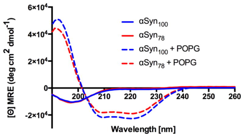
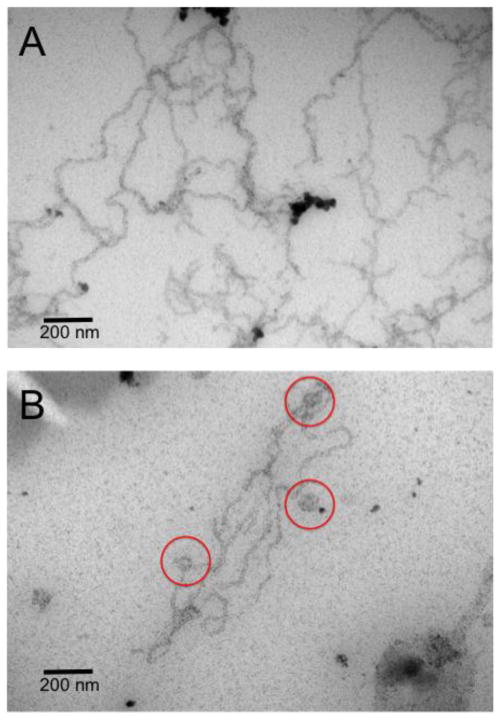
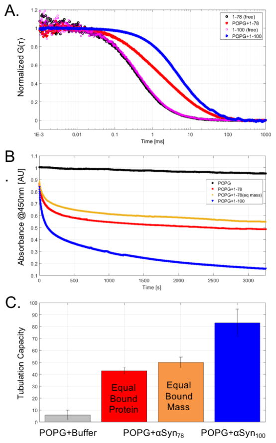
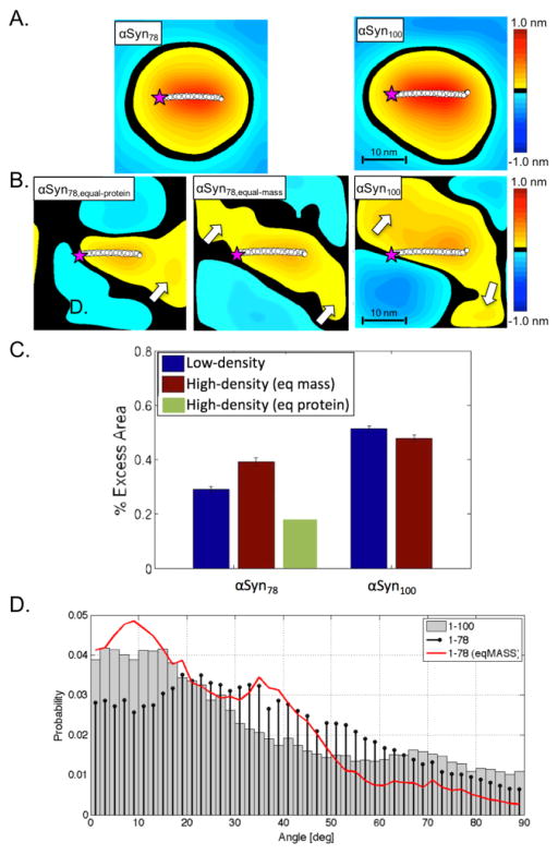
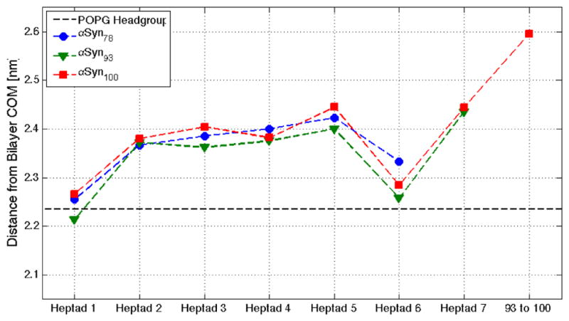
α-Synuclein is the primary protein found in Lewy bodies, the protein and lipid aggregates associated with Parkinson's disease and Lewy body dementia. The protein folds into a uniquely long amphipathic α-helix (AH) when bound to a membrane, and at high enough concentrations, it induces large-scale remodeling of membranes (tubulation and vesiculation). By engineering a less hydrophobic variant of α-Synuclein, we previously showed that the energy associated with binding of α-Synuclein's AH correlates with the extent of membrane remodeling (Braun et al. in J Am Chem Soc 136:9962-9972, 2014). In this study, we combine fluorescence correlation spectroscopy, electron microscopy, and vesicle clearance assays with coarse-grained molecular dynamics simulations to test the impact of decreasing the length of the amphipathic helix on membrane binding energy and tubulation. We show that truncation of α-Synuclein's AH length by approximately 15% reduces both its membrane binding affinity (by fivefold) and membrane remodeling capacity (by nearly 50% on per mole of bound protein basis). Results from simulations correlate well with the experiments and lend support to the idea that at high protein density there is a stabilization of individual, protein-induced membrane curvature fields. The extent to which these curvature fields are stabilized, a function of binding energy, dictates the extent of tubulation. Somewhat surprisingly, we find that this stabilization does not correlate directly with the geometric distribution of the proteins on the membrane surface.

Keywords: Alpha-Synuclein; Membrane remodeling; Tubulation.


Figures

Figure 1
Figure 1
Circular dichroism spectra for αSyn78 (red) and αSyn100 (blue) free in solution (solid lines) and incubated with POPG vesicles (dashed lines). Both αSyn78 and αSyn100 fold into an α-helical conformation when bound to POPG vesicles.
Figure 2
Figure 2
Representative EM images of tubules formed by αSyn100 (A) and αSyn78 (B). Scale bar is 200 nm. Red circles highlight vesicles in the αSyn78 system.
Figure 3
Figure 3
(A) FCS traces for αSyn78 and αSyn100 in the absence (black or magenta) or presence of POPG (red or blue). (B) Vesicle clearance absorbance traces for POPG + buffer (black), POPG + αSyn78 at equal protein concentration (red), POPG + αSyn78 at equal mass of protein (orange), and POPG + αSyn100 (blue). (C) Tubulation capacities for αSyn constructs relative to buffer.
Figure 4
Figure 4
Time-averaged height surfaces, h(x,y), for αSyn78 (left) and αSyn100 (right) for low-density (A) and high-density (B) systems. Purple star indicates N-terminus of protein. The white arrows highlight local region of on-average positive curvature suggesting an alignment of proximal proteins. (C) Excess area per protein for αSyn78 (left) and αSyn100 (right) systems [low-density, blue; high-density (equal-mass, brown; equal-protein, green)]. (D) Protein angle distributions for αSyn78 and αSyn100 high-density systems (αSyn100, grey-bars; αSyn78,eq-protein, black-stem; αSyn78,eq-mass, red-curve).
Figure 5
Figure 5
Heptad depth profile for αSyn constructs. Depth profiles for αSyn78 (blue), αSyn93 (green), and αSyn100 (red) are parsed by heptad and illustrate differential penetration relative to the POPG headgroup (black dashed-line).

References

    1. Braun AR, Lacy MM, Ducas VC, Rhoades E, Sachs JN. α-Synuclein-Induced Membrane Remodeling Is Driven by Binding Affinity, Partition Depth, and Interleaflet Order Asymmetry. J Am Chem Soc. 2014;136:9962–72. - PMC - PubMed
    1. Jao CC, Hegde BG, Chen J, Haworth IS, Langen R. Structure of membrane-bound alpha-synuclein from site-directed spin labeling and computational refinement. Proc Natl Acad Sci U S A. 2008;105:19666–19671. - PMC - PubMed
    1. Trexler AJ, Rhoades E. Alpha-synuclein binds large unilamellar vesicles as an extended helix. Biochemistry. 2009;48:2304–2306. - PMC - PubMed
    1. Nakamura K. alpha-Synuclein and mitochondria: partners in crime? Neurotherapeutics. 2013;10:391–399. - PMC - PubMed
    1. Kamp F, Exner N, Lutz AK, Wender N, Hegermann J, Brunner B, Nuscher B, Bartels T, Giese A, Beyer K, Eimer S, Winklhofer KF, Haass C. Inhibition of mitochondrial fusion by alpha-synuclein is rescued by PINK1, Parkin and DJ-1. EMBO J. 2010;29:3571–3589. - PMC - PubMed

Publication types

LinkOut - more resources