. 2017 Feb 27:7:43207.
doi: 10.1038/srep43207.

Mammalian TRAPPIII Complex positively modulates the recruitment of Sec13/31 onto COPII vesicles

Affiliations

Mammalian TRAPPIII Complex positively modulates the recruitment of Sec13/31 onto COPII vesicles

Shan Zhao et al. Sci Rep. .

Abstract

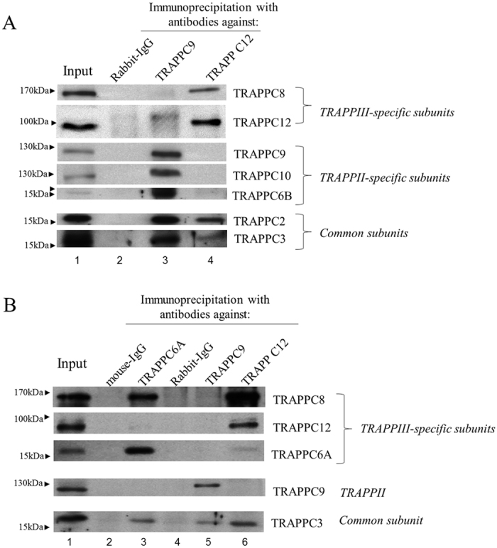
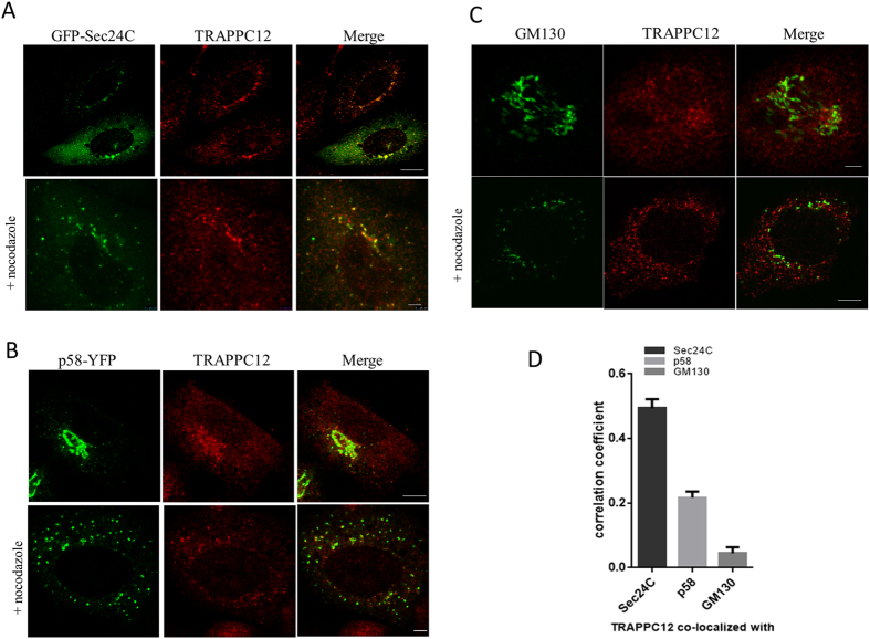
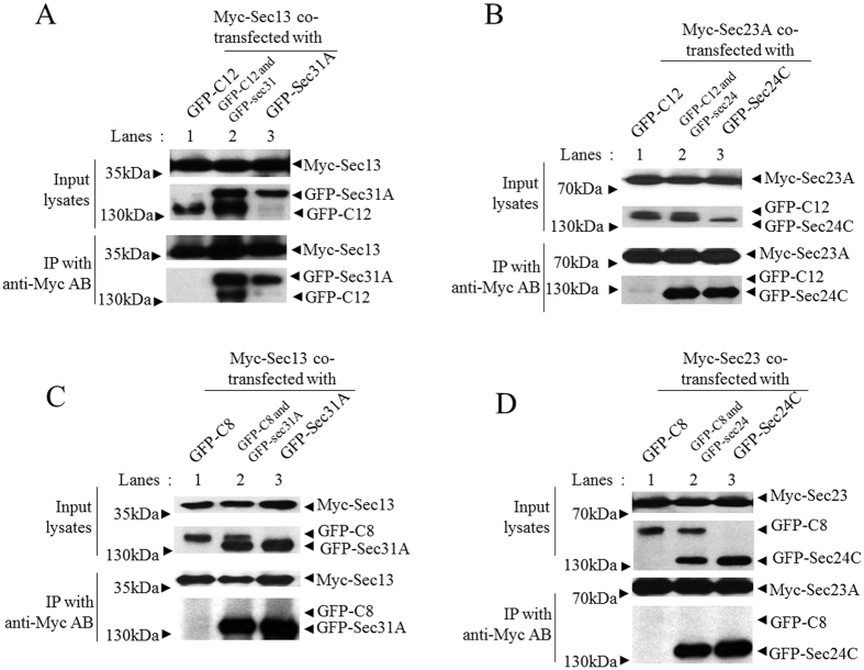
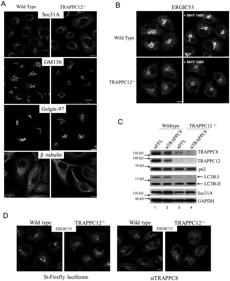
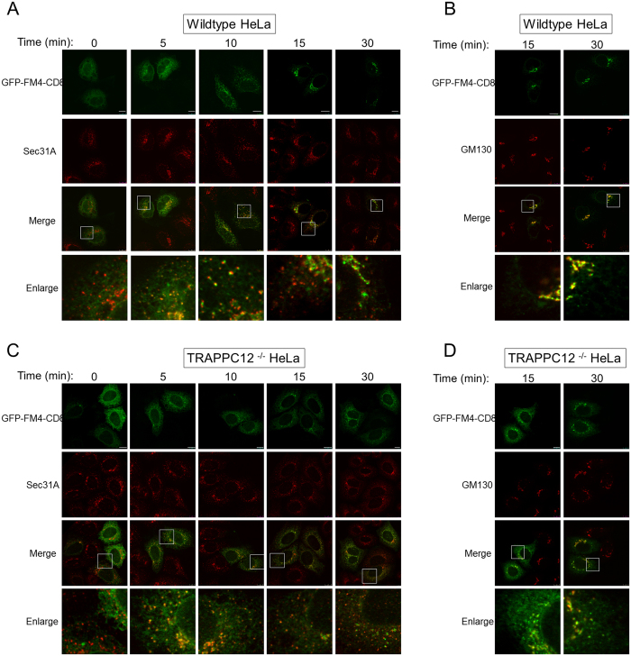
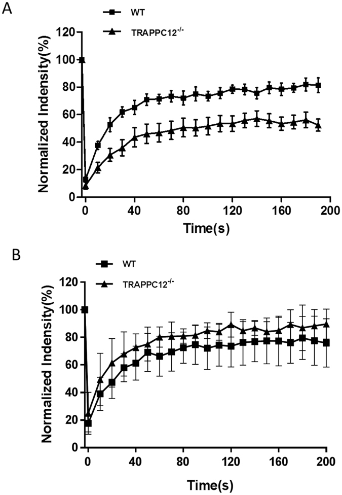
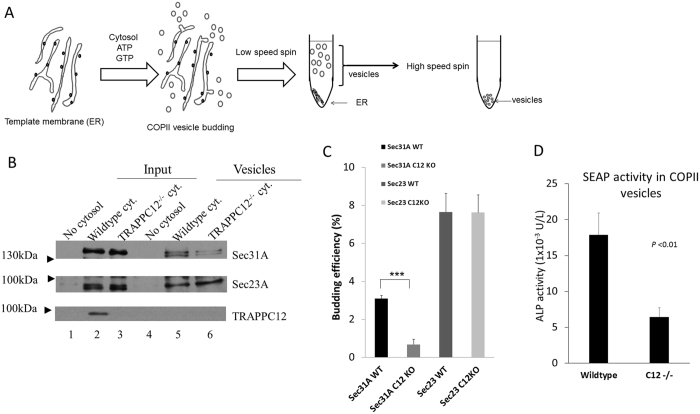
The Transport protein particle (TRAPP) complex is a tethering factor for COPII vesicle. Of three forms of TRAPP (TRAPPI, II and III) complexes identified so far, TRAPPIII has been largely considered to play a role in autophagy. While depletion of TRAPPIII specific subunits caused defects in the early secretory pathway and TRAPPIII might interact with components of the COPII vesicle coat, its exact role remains to be determined. In this study, we studied the function of TRAPPIII in early secretory pathway using a TRAPPIII-specific subunit, TRAPPC12, as starting point. We found that TRAPPC12 was localized to the ER exit sites and ERGIC. In cells deleted with TRAPPC12, ERGIC and to a lesser extent, the Golgi became dispersed. ER-to-Golgi transport was also delayed. TRAPPC12, but not TRAPPC8, bound to Sec13/Sec31A tetramer but each Sec protein alone could not interact with TRAPPC12. TRAPPIII positively modulated the assembly of COPII outer layer during COPII vesicle formation. These results identified a novel function of TRAPPIII as a positive modulator of the outer layer of the COPII coat.


Conflict of interest statement

The authors declare no competing financial interests.

Figures

Figure 1
Figure 1. TRAPPC12 and TRAPPC8 were TRAPPIII-specific subunits.
(A) HeLa cell lysates were subjected to immunoprecipitation using antibody specific to TRAPPC9 and TRAPPC12 and rabbit-IgG as negative control. The different subunits of TRAPP complexes in the immunoprecipitates were detected by immunoblotting using the indicated antibodies specific to these proteins. (B) A comparison of subunit composition between TRAPPC6A IP, and TRAPPC9 and TRAPPC12 IP’s. Immunoprecipitations with mouse monoclonal TRAPPC6A antibody was performed and mouse IgG was used as negative control. IP’s with TRAPPC9 and TRAPPC12 were similar to those shown in (A). In both panels, approximately 1% of the input lysates and 20% of the immunoprecipitates were loaded onto the SDS-PAGE.
Figure 2
Figure 2. Immunofluorescence colocalization studies on TRAPPC12.
Colocalization of TRAPPC12 with various markers by immunofluorescence. (A) TRAPPC12 was colocalized with ER exit site marker GFP-Sec24C; (B) TRAPPC12 was colocalized with ERGIC marker p58-YFP. (C) TRAPPC12 was colocalized with Golgi markers GM130. TRAPPC12 signal was detected by incubating the cell samples purified antibody against TRAPPC12 followed by AF568-conjugated secondary antibodies (A,C), or AF635-conjugated secondary antibodies (B). ER exit sites and ERGIC were detected by the fluorescence signals of transfected GFP-Sec24C and p58-YFP, respectively (A,B). Golgi was detected by staining the samples with monoclonal antibody against GM130, followed by AF488-conjugated secondary antibody (C). In samples where nocodazole was used, 10 μg/ml of nocodazole was added to the cells for 1 hour before fixation and staining. Scale bar is 10 μm (upper) and 2.5 μm (lower). (D) Quantitative analysis of TRAPPC12 co-localized with different organelle markers in nocodazole-treated cells. Pearson’s correlation coefficients were 0.49 (TRAPPC12 & Sec24C), 0.22 (TRAPPC12 & p58) and 0.04 (TRAPPC12 & GM130). The coefficients were generated using ImageJ software and specific plugins. The data were shown Mean ± S.D., n > 3.
Figure 3
Figure 3. TRAPPC12 directly binds to the outer coat complex of COPII vesicle.
GFP-TRAPPC12 or GFP-TRAPPC8 was overexpressed with indicated combinations of plasmids expressing Myc- or GFP-tagged COPII coat subunit(s) in 293T cells. Immunoprecipitations were performed using 9E10 anit-c-Myc antibody and detections of co-precipitated proteins were detected by immunoblottings using the indicated antibodies (anti-c-Myc or anti-GFP). Approximately 1% of the input lysates and 20% of the immunoprecipitates were loaded. (A) GFP-TRAPPC12 (GFP-C12) was tested for interaction with Myc-Sec13 alone or with GFP-Sec31A. (B) GFP-TRAPPC12 was tested for interaction with Myc-Sec23A alone or with GFP-Sec24C. (C) GFP-TRAPPC8 (GFP-C8) was tested for interaction with Myc-Sec13 alone or with GFP-Sec31A. (D) GFP-TRAPPC8 was tested for interaction with Myc-Sec23A alone or with GFP-Sec24C.
Figure 4
Figure 4. Analysis of the early secretory pathway in TRAPPC12−/− cells.
(A) Wildtype and TRAPPC12−/− HeLa cells were investigated with indicated markers of the early secretory pathway. (B) Wildtype and TRAPPC12−/− HeLa cells were stained with ERGIC53 after the cells were treated with or without autophagy inhibitor MHY 1485 at 10 μM for 2 hours. (C) Lysates of wildtype and TRAPPC12−/− HeLa cells were immunoblotted to detect the indicated proteins. (D) Wildtype and TRAPPC12−/− HeLa cells were depleted with siRNA specific to Firefly luciferase (control) or TRAPPC8 sequence and then stained with antibody against ERGIC-53. Scale bars = 10 μm.
Figure 5
Figure 5. ER-to-Golgi transport in wildtype and TRAPPC12−/− HeLa cells.
ER-to-Golgi transport was monitored by the transfected GFP-FM4-CD8 as transport marker. GFP-FM4-CD8 accumulated in the ER lumen in aggregated form until D/D solubilizer was applied to the cells for the indicated time. ER-to-Golgi traffic was monitored by co-staining with ERES marker Sec31A (A,C), or with Golgi marker GM130 (B,D). Scale bars = 10 μm.
Figure 6
Figure 6. FRAP analysis of GFP-Sec31A wildtype and TRAPPC12−/− HeLa cells.
(A) Wildtype (square) or TRAPPC12−/− (triangle) HeLa cells transiently transfected with GFP-Sec31A was bleached by laser and the recoveries of GFP fluorescence were recorded. Examples of detailed FRAP operations were documented in Supplementary Figure 5. (B) Wildtype (square) or TRAPPC12−/− (triangle) HeLa cells transiently transfected with GFP-Sec24C were bleached by laser and the recoveries of GFP fluorescence were recorded. n = 7; Error bars = S.D.
Figure 7
Figure 7. In vitro COPII budding reaction supported by wildtype or TRAPPC12−/− cytosol.
(A) Schematic drawing of the procedures of in vitro COPII budding assay. (B) A representative experiment of COPII budding reaction. ER membranes from wildtype HeLa cells were mixed with no cytosol (lanes 1 and 4), wildtype cytosol (lanes 2 and 5), or TRAPPC12−/− cytosol (lanes 3 and 6) to allow COPII vesicles budding to occur. 10% input and 100% of budded vesicles were loaded on SDS-PAGE. (C) Quantitative analysis of COPII budding assay. Sec31A- and Sec23A-specific bands were quantified by ImageJ software. The budding efficiencies were 3.08% (Sec31 WT), 0.68% (Sec31, C12 KO), 7.64% (Sec23 WT), and 7.62% (Sec23, C12 KO) of the corresponding input signals (i.e., comparing signals of lane 5 to lane 2, and lane 6 to lane 3). The data were shown Mean ± S.D., n = 3, P < 0.01. (D) COPII budding reactions were carried using ER template membranes isolated from a HeLa cell line stably transfected with SEAP as transport cargo. The budded COPII vesicles were measured with alkaline phosphatase activity as an indication of cargo transport. The data represented averages of above-background alkaline phosphatase activities of the two samples, using no cytosol control as background. Error bars = S.D., n = 3, P < 0.01.

References

    1. Aridor M., Bannykh S. I., Rowe T. & Balch W. E. Sequential coupling between COPII and COPI vesicle coats in endoplasmic reticulum to Golgi transport. J Cell Biol 131, 875–893 (1995). - PMC - PubMed
    1. Budnik A. & Stephens D. J. ER exit sites–localization and control of COPII vesicle formation. FEBS letters 583, 3796–3803 (2009). - PubMed
    1. D’Arcangelo J. G., Stahmer K. R. & Miller E. A. Vesicle-mediated export from the ER: COPII coat function and regulation. Biochim Biophys Acta 1833, 2464–2472 (2013). - PMC - PubMed
    1. Sacher M. et al. TRAPP I implicated in the specificity of tethering in ER-to-Golgi transport. Mol Cell 7, 433–442 (2001). - PubMed
    1. Cai H. et al. TRAPPI tethers COPII vesicles by binding the coat subunit Sec23. Nature 445, 941–944 (2007). - PubMed

Publication types

LinkOut - more resources