. 2017 Apr 3;127(4):1338-1352.
doi: 10.1172/JCI89857. Epub 2017 Feb 27.

Anticancer kinase inhibitors impair intracellular viral trafficking and exert broad-spectrum antiviral effects

Anticancer kinase inhibitors impair intracellular viral trafficking and exert broad-spectrum antiviral effects

Elena Bekerman et al. J Clin Invest. .

Abstract

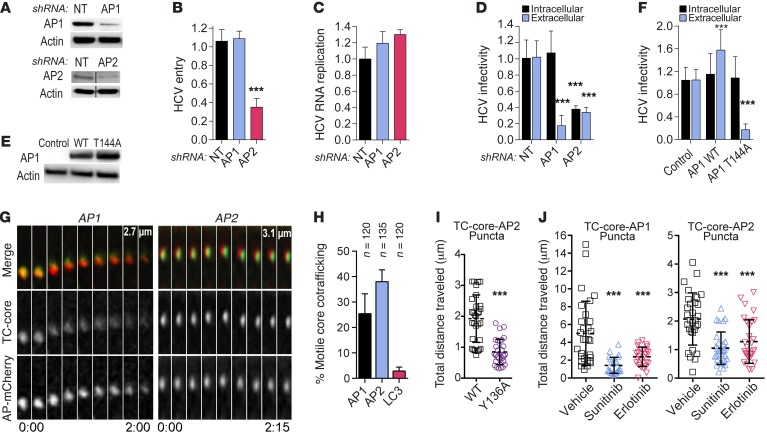
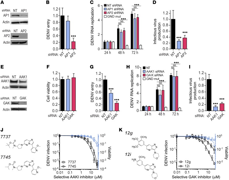
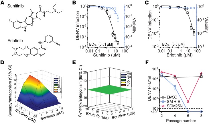
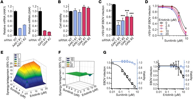
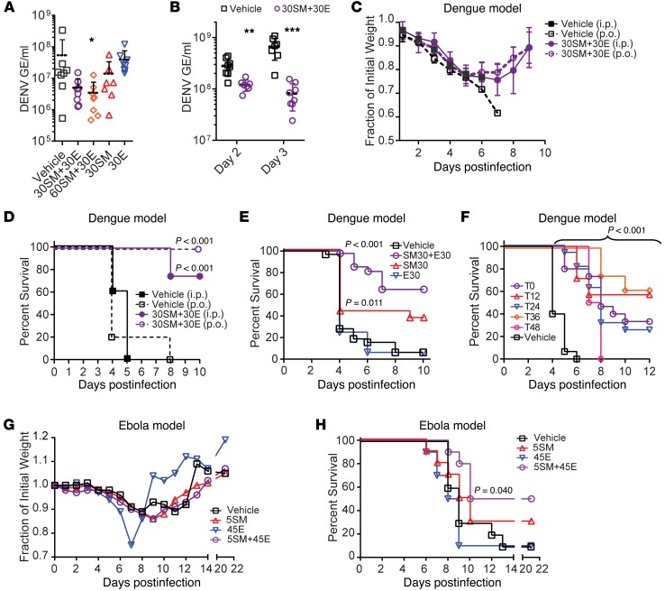
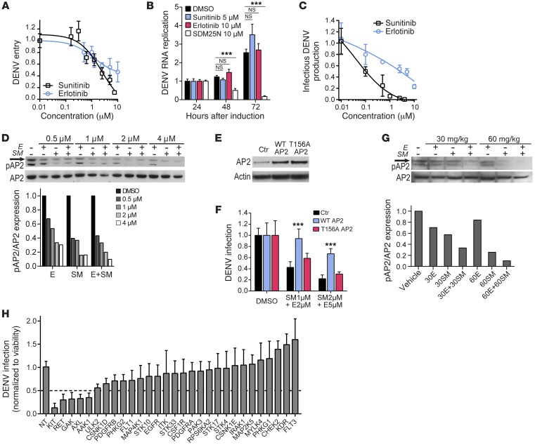
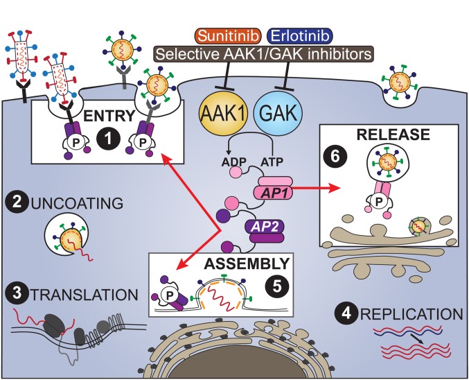
Global health is threatened by emerging viral infections, which largely lack effective vaccines or therapies. Targeting host pathways that are exploited by multiple viruses could offer broad-spectrum solutions. We previously reported that AAK1 and GAK, kinase regulators of the host adaptor proteins AP1 and AP2, are essential for hepatitis C virus (HCV) infection, but the underlying mechanism and relevance to other viruses or in vivo infections remained unknown. Here, we have discovered that AP1 and AP2 cotraffic with HCV particles in live cells. Moreover, we found that multiple viruses, including dengue and Ebola, exploit AAK1 and GAK during entry and infectious virus production. In cultured cells, treatment with sunitinib and erlotinib, approved anticancer drugs that inhibit AAK1 or GAK activity, or with more selective compounds inhibited intracellular trafficking of HCV and multiple unrelated RNA viruses with a high barrier to resistance. In murine models of dengue and Ebola infection, sunitinib/erlotinib combination protected against morbidity and mortality. We validated sunitinib- and erlotinib-mediated inhibition of AAK1 and GAK activity as an important mechanism of antiviral action. Additionally, we revealed potential roles for additional kinase targets. These findings advance our understanding of virus-host interactions and establish a proof of principle for a repurposed, host-targeted approach to combat emerging viruses.


Conflict of interest statement

Conflict of interest: The authors have declared that no conflict of interest exists.

Figures

Figure 1
Figure 1. AP1 and AP2 cotraffic with HCV and orchestrate infection.
(A) Confirmation of gene expression knockdown by Western blot in Huh7.5 cells stably expressing AP shRNA or nontargeting control (NT). (B) Entry of HCV pseudoparticles (HCVpp) was measured by luciferase assays at 48 hours after infection. (C) HCV RNA replication measured via luciferase assays 72 hours after HCV RNA electroporation. (D) HCV infectivity measured via luciferase assays by inoculation of naive cells with lysates (intracellular) and supernatants (extracellular) from electroporated cells. (E) AP1 ectopic expression following transfection of Huh7.5 cells with GLuc-tagged WT, T144A AP1, or an empty control; blotted with anti-GLuc antibody. (F) HCV intra- and extracellular infectivity in AP1-overexpressing cells versus control. Shown are means ± SD (n = 3–10). (G) Representative live cell fluorescence microscopy montages of TC-core HCV (green) cotrafficking with AP1- and AP2-mCherry (red). Distance traveled (μm) and time elapsed (min:s) during video acquisition are indicated. (H) Quantification of motile TC-core puncta cotrafficking with AP1, AP2, and LC3. (I) Quantification of distance traveled per acquisition of WT or Y136A mutant TC-core HCV associated with AP2. (J) Quantification of distance traveled per acquisition of TC-core HCV associated with AP1 or AP2 upon treatment with sunitinib (4 μM) and erlotinib (10 μM). Results in BD and F represent data pooled from at least 2 independent experiments each with 6–10 biological replicates. HJ are representative experiments out of at least 3 conducted. Shown are means ± SD; ***P < 0.001 relative to corresponding NT (BD), empty vector control (F), WT TC-core (I), or vehicle control (J) by 1-way ANOVA, followed by Dunnett’s (B, D, and J) or Tukey’s (F) multiple comparisons test or 2-tailed unpaired t test (I).
Figure 2
Figure 2. AP1, AP2, and their regulatory kinases, AAK1 and GAK, are essential for DENV infection in vitro.
(A and E) Confirmation of stable shRNA-mediated (A) or transient siRNA-mediated (E) gene expression silencing by Western blot in Huh7 cells. (B and G) DENV entry measured via luciferase assay 6 hours after infection. (C and H) DENV RNA replication monitored by luciferase activity every 24 hours following transfection of Huh7 cells with a Tet-inducible DNA-launched DENV replicon and induction by doxycycline for 6 hours (GND is a replication-incompetent DENV). Data are normalized to signal at 24 hours. (D and I) Infectious DENV production measured via luciferase assays by inoculation of naive cells with supernatants from stable or siRNA-transfected cells 48 hours after electroporation with DENV RNA. (F) Relative cell viability following gene expression knockdown measured by alamarBlue assays. (J and K) Cell viability (blue) and dose response of DENV infection (black) to more selective AAK1 (J) and GAK (K) inhibitors (structures shown) measured by luciferase assays 48 hours after infection. Data are plotted relative to vehicle control. Shown are representative experiments from at least 2 conducted. Individual experiments in BD and FK had 8–10 biological replicates; shown are means ± SD; ***P < 0.001 relative to corresponding NT control by 1-way ANOVA (B, D, F, G, and I) or 2-way ANOVA (C and H), followed by Dunnett’s multiple comparisons test.
Figure 3
Figure 3. Sunitinib and erlotinib have a synergistic anti-DENV effect and a high genetic barrier to resistance in vitro.
(A) Chemical structures of the indicated drugs. (B and C) Cellular viability (blue) and dose response of overall DENV infection (black) to sunitinib and erlotinib measured by luciferase assays at 48 hours after infection. Data are plotted relative to vehicle control. (D and E) Synergy/antagonism at the 95% CI of sunitinib/erlotinib combination treatment on antiviral effect (D) and cellular viability (E) computed by MacSynergy II. (F) DENV4 was used to infect Huh7 cells and passaged every 72 hours by inoculation of naive cells with equal volumes of viral supernatants under DMSO treatment or selection with sunitinib and erlotinib (SM + E) or SDM25N (DENV NS4B inhibitor) increasing from 0.5 to 2.5 μM over 8 passages. Viral titers were measured by plaque assays at every other passage. Dashed line represents assay detection limit. Results in B and C represent data pooled from at least 2 independent experiments. Data in DF are representative of at least 2 experiments. Shown in B, C, and F are means ± SD. Individual experiments in BE and F had 5–10 and 2 biological replicates, respectively.
Figure 4
Figure 4. Inhibition of AAK1 and GAK suppresses EBOV infection.
(A) Confirmation of siRNA-mediated gene expression silencing by quantitative PCR in Vero cells. Shown is normalized gene expression relative to GAPDH at 48 hours after transfection. (B and C) Relative cell viability measured by alamarBlue assays (B) and rVSV-GP EBOV infection of Vero cells measured by flow cytometry (C) at 20 hours after infection of AAK1- and GAK-depleted cells. ***P < 0.001 relative to NT (1-way ANOVA followed by Dunnett’s multiple comparisons test). (D) Dose response of rVSV-GP EBOV infection to 4-hour treatment with inhibitors measured by flow cytometry at 20 hours after infection in Vero cells. (E and F) Synergy/antagonism of sunitinib/erlotinib combination treatment on rVSV-GP EBOV infection (E) and cell viability (F). (G) Dose response to drug treatment in Huh7 cells under biosafety level 4 containment following 48 hours with EBOV infection (black) measured by immunostaining with an anti-GP antibody, and cell viability (blue) measured by Hoechst counterstain and quantified by a high-content imager. Data are plotted relative to vehicle control. All data shown are representative of at least 2 experiments; BG have 3 biological replicates each. Shown in BD and G are means ± SD.
Figure 5
Figure 5. AAK1 and GAK inhibitors are protective in murine models of dengue and Ebola.
(A) DENV viremia in AG-B6 mice measured by qRT-PCR on day 2 postinfection following once-daily administration of vehicle, sunitinib (SM), and/or erlotinib (E). (B) DENV viremia in AG-B6 mice on days 2 and 3 postinfection following twice-daily drug administration. (C and D) Weight loss (C) and mortality (D) of DENV-infected AG-B6 mice treated once daily for 5 days with vehicle or sunitinib/erlotinib combination (n = 8 per treatment group). (E) Mortality of DENV-infected AG-129 mice treated once daily for 5 days with vehicle, sunitinib, and/or erlotinib (data are pooled from 2 independent experiments, n = 8–16 per treatment group). (F) Mortality of DENV-infected AG-B6 mice treated once daily with vehicle or sunitinib/erlotinib combination beginning at the indicated hour after inoculation, T0–T48 (data are pooled from 2 independent experiments, n = 8–16 per treatment group). (G and H) Weight loss (G) and mortality (H) of EBOV-infected C57BL/6 mice treated once daily for 10 days with vehicle, sunitinib, and/or erlotinib (n = 10 per treatment group). Doses are in mg/kg. Administration was i.p., except when denoted p.o. (C and D), at inoculation (AE, G, and H) or after inoculation (F). AD, G, and H are representative of 2 or more independent experiments. *P < 0.05, **P < 0.01, ***P < 0.001 relative to vehicle control by nonparametric Kruskal-Wallis test with Dunn’s multiple comparisons post-test (A) or nonparametric Mann-Whitney test (B). Survival analysis (DF and H) was done with log-rank (Mantel-Cox) test; P values are relative to vehicle control. GE, genomic equivalents.
Figure 6
Figure 6. Mechanisms underlying the antiviral effect of sunitinib and erlotinib in vitro and in vivo.
(AC) Huh7 cells were treated with the inhibitors and monitored for DENV entry (A) at 6 hours after infection, DENV RNA replication (B) after induction of replication of DNA-launched DENV replicon, and infectious virus production (C) at 48 hours after electroporation with DENV RNA. SDM25N is an inhibitor of DENV RNA replication. (D) Effect of 1-hour treatment with erlotinib (E) and/or sunitinib (SM) on phosphorylation of AP2 in DENV-infected Huh7 cells measured by Western blotting. Arrow indicates approximately 50 kDa. The ratio of phospho-AP2 (pAP2) to total AP2 was quantified. (E) Level of AP2 and actin expression measured by Western blot following lentiviral transduction with control or AP2-expressing constructs. (F) Rescue of DENV infection in the presence of inhibitors upon overexpression of WT or T156A AP2 versus vector control measured by luciferase assays 48 hours after infection. Micromolar concentration of each inhibitor is noted on the x axis. (G) Effect of 3-hour i.p. treatment with erlotinib (E) and/or sunitinib (SM) on phosphorylation of AP2 in liver tissue of AG-B6 mice measured by Western blotting and quantified as the ratio of pAP2 to total AP2. (H) DENV infection relative to NT control following siRNA-mediated knockdown of kinases targeted by sunitinib and erlotinib measured by luciferase assays at 48 hours and normalized to cell viability. Data in A, C, and I are pooled from 2 independent experiments with 4–8 replicates each. Data in the other panels are representative of 2 or more independent experiments. B and F have at least 5 replicates each. ***P < 0.001 relative to DMSO by 2-way ANOVA followed by Dunnett’s multiple comparisons test (B) or relative to vector control by 1-way ANOVA followed by Dunnett’s multiple comparisons test (F).
Figure 7
Figure 7. Model: AAK1 and GAK in infection and as broad-spectrum antiviral targets.
Host kinases AAK1 and GAK regulate entry, assembly, and/or release of multiple RNA viruses through phosphorylation of the membrane trafficking adaptors AP1 (pink) and AP2 (purple). Sunitinib, erlotinib, and selective inhibitors of AAK1 and GAK disrupt these temporally distinct steps of the viral life cycle and act as broad-spectrum antivirals.

Comment in

References

    1. Bhatt S, et al. The global distribution and burden of dengue. Nature. 2013;496(7446):504–507. doi: 10.1038/nature12060. - DOI - PMC - PubMed
    1. Flipse J, Smit JM. The complexity of a Dengue vaccine: a review of the human antibody response. PLoS Negl Trop Dis. 2015;9(6):e0003749. doi: 10.1371/journal.pntd.0003749. - DOI - PMC - PubMed
    1. Priyamvada L, et al. Human antibody responses after dengue virus infection are highly cross-reactive to Zika virus. Proc Natl Acad Sci U S A. 2016;113(28):7852–7857. doi: 10.1073/pnas.1607931113. - DOI - PMC - PubMed
    1. Dejnirattisai W, et al. Dengue virus sero-cross-reactivity drives antibody-dependent enhancement of infection with Zika virus. Nat Immunol. 2016;17(9):1102–1108. doi: 10.1038/ni.3515. - DOI - PMC - PubMed
    1. Stettler K, et al. Specificity, cross-reactivity, and function of antibodies elicited by Zika virus infection. Science. 2016;353(6301):823–826. doi: 10.1126/science.aaf8505. - DOI - PubMed

MeSH terms