. 2017 Feb 27;11(2):e0005413.
doi: 10.1371/journal.pntd.0005413. eCollection 2017 Feb.

Cytokines and microbicidal molecules regulated by IL-32 in THP-1-derived human macrophages infected with New World Leishmania species

Affiliations

Cytokines and microbicidal molecules regulated by IL-32 in THP-1-derived human macrophages infected with New World Leishmania species

Jéssica Cristina Dos Santos et al. PLoS Negl Trop Dis. .

Abstract

Background: Interleukin-32 (IL-32) is expressed in lesions of patients with American Tegumentary Leishmaniasis (ATL), but its precise role in the disease remains unknown.

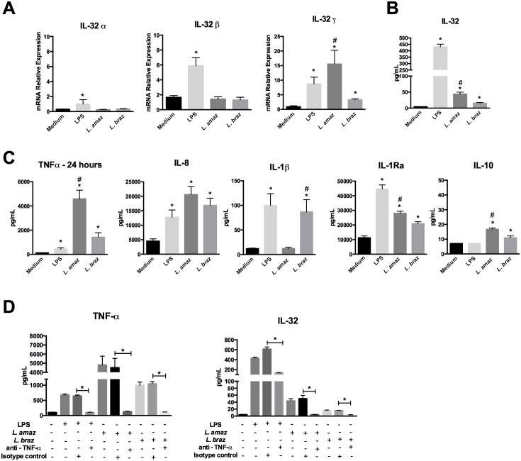
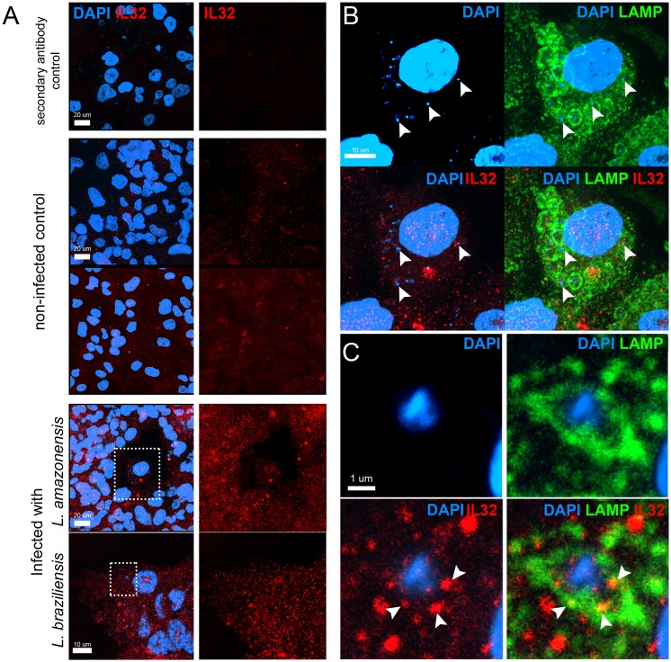
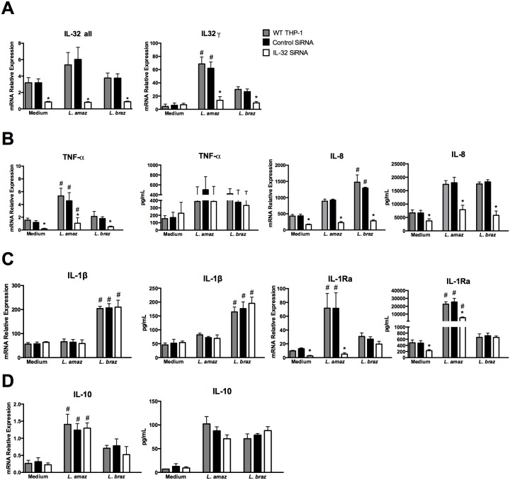
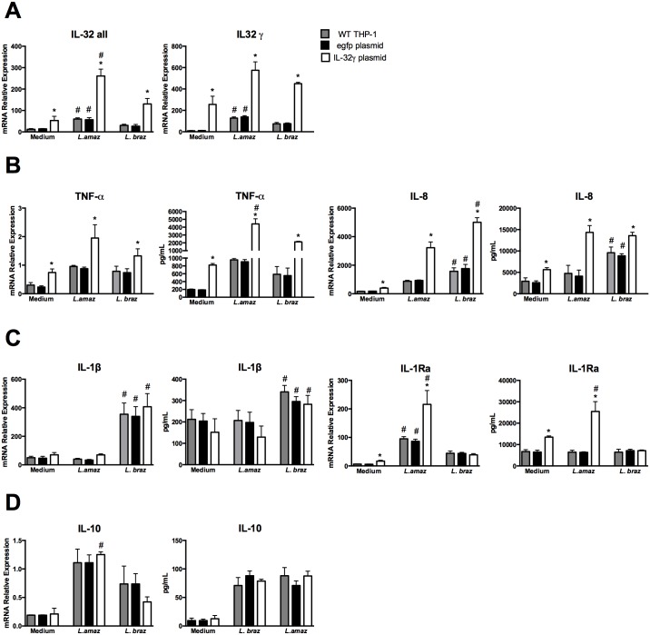
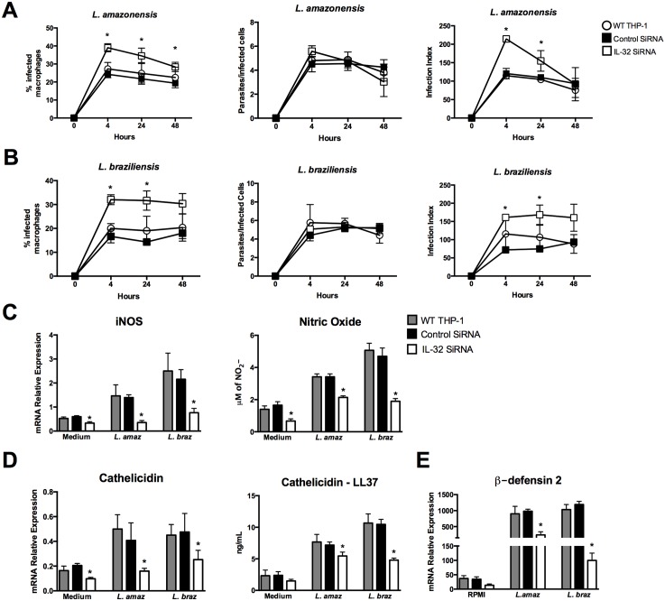
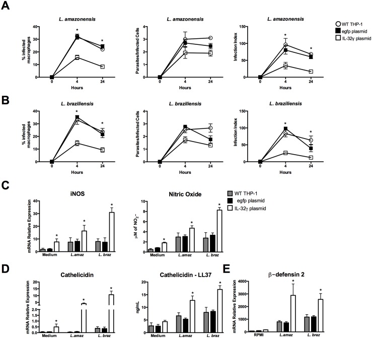
Methodology/principal findings: In the present study, silencing and overexpression of IL-32 was performed in THP-1-derived macrophages infected with Leishmania (Viannia) braziliensis or L. (Leishmania) amazonensis to investigate the role of IL-32 in infection. We report that Leishmania species induces IL-32γ, and show that intracellular IL-32γ protein production is dependent on endogenous TNFα. Silencing or overexpression of IL-32 demonstrated that this cytokine is closely related to TNFα and IL-8. Remarkably, the infection index was augmented in the absence of IL-32 and decreased in cells overexpressing this cytokine. Mechanistically, these effects can be explained by nitric oxide cathelicidin and β-defensin 2 production regulated by IL-32.

Conclusions: Thus, endogenous IL-32 is a crucial cytokine involved in the host defense against Leishmania parasites.


Conflict of interest statement

The authors have declared that no competing interests exist.

Figures

Fig 1
Fig 1. Leishmania induces IL-32γ expression in a TNFα-dependent manner.
PMA-differentiated THP-1 cells (1 x 10⁶ cells/mL) were infected with promastigote forms (5 x 10⁶ parasites) in the growth stationary phase of L. (L.) amazonensis (L. amaz), metacyclic promastigote forms (5 x 10⁶ parasites) of L. (V.) braziliensis (L. braz) or LPS (100 ng/mL) as a positive control during 4 h. Cells were washed to remove non-internalized parasites and incubated for 24 h. (A) mRNA expression of isoforms α, β and γ of IL-32 was determined by quantitative real-time PCR. (B) Intracellular IL-32 protein levels were determined by ELISA in cell lysates. (C) TNFα, IL-8, IL-1β, IL-1Ra, IL-10 productions were determined by ELISA in culture supernatants. (D) Antibodies to TNFα (anti-TNFα, 5 μg/ml) or isotype control (5 μg/mL) were added 30 min before addition of LPS (100 ng/mL) or Leishmania sp. TNFα levels (left panel) and intracellular IL-32 (right panel) were determined by ELISA in supernatants and cell lysates, respectively. Values are expressed as means ± SEM of three independent experiments. *p < 0.05 (Medium vs LPS, L. amaz, L. braz); #p < 0.05 (L. amaz, L. braz).
Fig 2
Fig 2. Intracellular distribution of IL-32 after Leishmania species infection.
PMA-differentiated THP-1 cells (2 x 10⁵ cells/0.5 mL) were infected with promastigote forms (10 x 105 parasites) in stationary phase of growth of L. (L.) amazonensis or metacyclic promastigote forms (10 x 105 parasites) of L. (V.) braziliensis during 4 h. Afterwards, cells were washed to remove non-internalized parasites and incubated for 24 h. Cells were stained for IL-32 (rabbit polyclonal antibody; red), lysosomal-associated membrane protein, LAMP1 (mouse monoclonal antibody; green) and dapi (blue) for confocal microscopy. Yellow colour indicates colocalization of IL-32 and LAMP1. (A) In the eight first images, bar = 20 μm; two inferior images, bar = 10 μm (L. (V.) braziliensis). (B) L. (L.) amazonensis, white arrow heads indicate parasites present in LAMP1+-parasitophorous vacuoles; bar = 10 μm; (C) L. (V.) braziliensis, white arrow heads indicate colocalization of IL-32 and LAMP1 in a LAMP1+-parasitophorous vacuole containing one amastigote (blue), bar = 1 μm; images were took from (A) (left bottom, dashed white squares).
Fig 3
Fig 3. Decrease of cytokine production after IL-32 silencing in THP-1-derived macrophages infected with Leishmania species.
PMA-differentiated THP-1 cells (2.5 x 10⁶ cells/800 μL) were electroporated by using Amaxa Nucleofector Technology with IL-32 siRNA (for IL-32 knockdown) and control siRNA according to the protocol described in [38]. The final concentration per well was 3 x 10⁵ cells/100 μL. After 24 h of transfection, cells were infected with promastigote forms (1.5 x 10⁶ parasites) in the growth stationary phase of L. (L.) amazonensis (L. amaz) or metacyclic promastigote forms (1.5 x 10⁶ parasites) of L. (V.) braziliensis (L. braz). After 4 h, non-internalized parasites were washed out and cells were incubated for 24 h. (A) mRNA expression of IL-32γ isoform and all isoforms were determined by quantitative real-time PCR. mRNA expression and protein levels of (B) TNFα and IL-8, (C) IL-1β and IL-1Ra and (D) IL-10 were determined by quantitative real-time PCR and ELISA in supernatants, respectively. Values are expressed as means ± SEM of three independent experiments. *p < 0.05 (Control SiRNA vs IL-32 SiRNA); #p < 0.05 (L. amaz, L. braz).
Fig 4
Fig 4. Increased cytokine production after overexpression of IL-32 in human THP-1-derived macrophages infected with Leishmania species.
PMA-differentiated THP-1 cells (2.5 x 10⁶ cells/800 μL) were electroporated by using Amaxa Nucleofector Technology with IL-32 plasmid (for IL-32 overexpression) and egfp plasmid (as a control) according to the protocol described in [38]. The final concentration per well was 3 x 10⁵ cells/100 μL. After 24 h of transfection, cells were infected with promastigote forms (1.5 x 10⁶ parasites) in the growth stationary phase of L. (L.) amazonensis (L. amaz), metacyclic promastigote forms (1.5 x 10⁶ parasites) of L. (V.) braziliensis (L. braz). After 4 h cells were washed and incubated for 24 h. (A) mRNA expression of IL-32γ isoform and all IL-32 isoforms was determined by quantitative real-time PCR. mRNA expression and protein levels of (B) TNFα and IL-8, (C) IL-1β and IL-1Ra and (D) IL-10 were determined by quantitative real-time PCR and ELISA in supernatants, respectively. Values are expressed as means ± SEM of three independent experiments. *p < 0.05 (egpf plasmid vs IL-32γ plasmid); #p < 0.05 (L. amaz, L. braz).
Fig 5
Fig 5. Silencing of IL-32 in human THP-1-derived macrophages increases Leishmania species infection.
PMA-differentiated THP-1 cells (2.5 x 10⁶ cells/800 μL) were electroporated by using Amaxa Nucleofector Technology with IL-32 siRNA (for knockdown IL-32) and control siRNA according with protocol described by [38]. The final concentration of cells per well were 3 x 10⁵ cells/100 μL. After 24 h of transfection, cells were infected with promastigotes forms (1.5 x 10⁶ parasites) in the growth stationary phase of L. (L.) amazonensis (L. amaz), metacyclic promastigote forms (1.5 x 10⁶ parasites) of L. (V.) braziliensis (L. braz). After 4 h, cells were washed to remove non-internalized parasites and incubated for 24 h or 48 h. Cells were stained and percentage of infected macrophages, number of parasites per infected cells, and infection index were evaluated. (A) infection with L. amazonensis; (B) infection with L. braziliensis. iNOS (C—left panel), cathelidicin (D—left panel) and β-defensin 2 (E) mRNA expression were determined by quantitative real-time PCR (24 h). Production of nitrite (C—right panel) and LL-37 (D—right panel) was determined by Griess reagent and ELISA in supernatants, respectively (24 h). Values are expressed as means ± SEM of three independent experiments. *p < 0.05 (Control SiRNA vs IL-32 SiRNA).
Fig 6
Fig 6. Decreased Leishmania species infection and increased leishmanicidal molecules after IL-32 overexpression in human THP-1-derived macrophages.
PMA-differentiated THP-1 cells (2.5 x 10⁶ cells/800 μL) were electroporated by using Amaxa Nucleofector Technology with IL-32 plasmid (for IL-32 overexpression) and egfp plasmid (as a control). The final concentration of cells per well were 3 x 10⁵ cells/100 μL. After 24 h of transfection, cells were infected with promastigote forms (1.5 x 10⁶ parasites) in the growth stationary phase of L. (L.) amazonensis (L. amaz) or metacyclic promastigote forms (1.5 x 10⁶ parasites) of L. (V.) braziliensis (L. braz). After 4 h, non-internalized parasites were washed out and cells were incubated for 24 h or 48 h. Cells were stained and percentage of infected macrophages, number of parasites per infected cells, and infection index were evaluated. (A) infection with L. amazonensis; (B) infection with L. braziliensis. iNOS (C—left panel) and cathelidicin (D—left panel) and β-defensin 2 (E) mRNA expression were determined by quantitative real-time PCR (24 h). Nitrite production (C—right panel) and LL-37 production (D—right panel) were determined by Griess reagent and ELISA in supernatants, respectively (24 h). Values are expressed as means ± SEM of three independent experiments. *p < 0.05 (egpf plasmid vs IL-32γ plasmid).

Similar articles

Cited by

References

    1. Heinhuis B, Netea MG, van den Berg WB, Dinarello CA., Joosten LAB. Interleukin-32: A predominantly intracellular proinflammatory mediator that controls cell activation and cell death. Cytokine. Elsevier Ltd; 2012;60: 321–327. - PubMed
    1. Kang J-W, Park YS, Lee DH, Kim MS, Bak Y, Ham SY, et al. Interaction network mapping among IL-32 isoforms. Biochimie. 2014;101: 248–51. 10.1016/j.biochi.2014.01.013 - DOI - PubMed
    1. Heinhuis B, Koenders MI, van de Loo FA, Netea MG, van den Berg WB, Joosten LAB. Inflammation-dependent secretion and splicing of IL-32{gamma} in rheumatoid arthritis. Proc Natl Acad Sci U S A. 2011;108: 4962–7. 10.1073/pnas.1016005108 - DOI - PMC - PubMed
    1. Kim S-H, Han S-Y, Azam T, Yoon D-Y, Dinarello CA. Interleukin-32. Immunity. 2005;22: 131–142. - PubMed
    1. Choi J-D, Bae S-Y, Hong J-W, Azam T, Dinarello CA., Her E, et al. Identification of the most active interleukin-32 isoform. Immunology. 2009;126: 535–542. 10.1111/j.1365-2567.2008.02917.x - DOI - PMC - PubMed

LinkOut - more resources